
Bella Liu and Eric R. Snoey
Angioedema is transient edema of deep dermal and subcutaneous tissues characteristically localized to the face, lips, intraoral, and throat structures (1,2). The potential for rapid, life-threatening airway compromise from laryngeal edema makes identification and prompt treatment of angioedema of special importance to emergency physicians (1).
Angioedema can be classified according to its most common etiologies:
• Allergic reactions to food, drug, or environmental triggers
• Use of angiotensin-converting enzyme inhibitors (ACEIs)
• Hereditary or acquired angioedema (HAE or AAE)
• Idiopathic causes (2,3).
These etiologies of angioedema can be further classified by their respective pathophysiology, which involve mainly two immunologic mechanisms (Table 176.1). In angioedema associated with allergic reactions, mast cell degranulation with histamine release is responsible (1,2). In cases associated with ACEIs, HAE, and AAE, the angioedema is mediated by the process of bradykinin formation (1,2,4). In addition, a significant number of cases of angioedema occur with no clear cause; numbers as high as 21% to 59% have been reported for idiopathic angioedema (3,5). The end result, regardless of the underlying etiology or mechanism, is an increased permeability of submucosal or subcutaneous capillaries and venules, resulting in fluid extravasation and subsequent edema (1).
TABLE 176.1
Causes of Angioedema
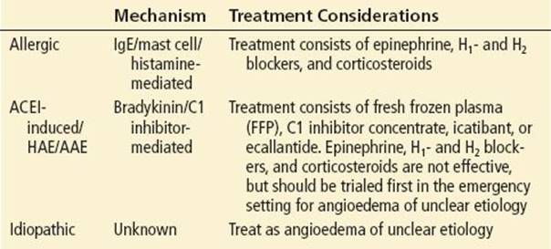
Acute allergic angioedema is mediated by a type 1 hypersensitivity reaction involving allergen binding to IgE receptors on mast cells. The resulting degranulation and histamine release can produce both angioedema as well as more traditional manifestations of allergic reactions such as urticaria, bronchospasm, and even anaphylaxis (1,2). Typical triggers include foods, drugs, contrast dye, latex, environmental allergens, and insect stings (1). This histamine-mediated mechanism is also the common pathway for most drug-related angioedema, including angioedema caused by β-lactams, sulfonamides, opiates, aspirin, and nonsteroidal anti-inflammatory drugs (NSAIDs), with the notable exception of ACEIs (1).
With approximately 40 million people taking ACEI medications worldwide, ACEI-associated angioedema is becoming an increasingly prevalent disease entity with an incidence approaching 2% according to some studies (2,6). African American patients have a four to five times greater incidence of angioedema; smoking, older age, and female sex are additional risk factors (6). The pathophysiology of ACEI-associated angioedema relates to angiotensin-converting enzyme’s dual role in inhibiting both the renin–angiotensin pathway (thereby lowering blood pressure) and the degradation of bradykinin, a powerful vasodilator (2). The initial presentation of ACEI-associated angioedema is highest during the first month of use, but about a third of cases present more than 6 months after initiation of ACEI therapy, sometimes up to several years (2,6).
Epidemiologic data regarding HAE and AAE are sparse; the incidence of HAE is reported to be between 1:50,000 and 1:100,000 persons, with AAE being even more rare (4). In both HAE and AAE, decreased C1 inhibitor activity results in upregulation of the kinin–kallikrein system, leading to increased bradykinin levels, mast cell degranulation, and vascular permeability (2). The pathophysiology of HAE is due to a genetic defect leading to decreased levels or function of the native C1 inhibitor or in some cases due to a mechanism involving estrogen (1,2,4). Features that suggest HAE include presentation at younger age, often in the first three decades of life, a positive family history, and recurrent attacks of otherwise unexplained angioedema or abdominal pain (2,4). A family history is not always present, however, due to de novo gene mutations and varying degrees of gene expression (2). Also, a history of attacks in setting of stress, trauma, or surgery, even minor surgery such as dental procedures, is suggestive of HAE (1,2). The pathophysiology of AAE is less understood, but is likely the result of either increased consumption of C1 inhibitor or an autoantibody binding to C1 inhibitor rendering it nonfunctional (1,2). The latter can occur in B-cell lymphoproliferative disorders including malignancies such as lymphoma and various autoimmune diseases (1,2). An additional feature that suggests AAE is presentation at greater age, often after the fourth decade of life (2,7).
In comparison to angioedema in adults, angioedema in the pediatric population is a rare and much less studied phenomenon (2). The allergic variant is the most common with frequent links to food and other environmental triggers (2). In one small study, angioedema was attributed to food in 40%, insect bites in 30%, upper respiratory infections in 20%, and antibiotic use in 10% of cases (8). Compared to adults, children with angioedema have better prognosis; most respond rapidly to treatment and are less likely to require airway intervention or intensive care unit (ICU) admissions (8).
CLINICAL PRESENTATION
Patients with angioedema characteristically present with complaints of swelling in:
• Face and lips
• Tongue, soft palate, and uvula
• Oropharynx and larynx
• Bowel walls manifesting as abdominal pain, nausea/vomiting, diarrhea, or ascites
• Distal extremities
• Genitals (1)
The typical time course of symptoms is onset within minutes to hours and resolution within one or several days. On physical examination, the edema is nonpitting, nonpruritic, usually does not involve gravitationally dependent areas, and can be painful owing to distension and swelling of the skin (2). Up to 15% of patients with angioedema will develop some degree of airway obstruction (2). Important clinical clues that suggest airway involvement include:
• Dyspnea and respiratory distress
• Voice change
• Hoarseness
• Stridor
• Dysphagia
• Drooling (9,10).
Also, urticaria, bronchospasm, or even anaphylaxis may present if the angioedema is allergic in nature. Abdominal wall involvement leading to abdominal complaints such as pain, nausea or vomiting, diarrhea, and ascites are a less recognized manifestation of angioedema and may be a diagnostic challenge for emergency physicians (2,4,8). They can occur in isolation without the usual facial involvement, and at times the abdominal pain can be so severe that it is mistaken for an acute surgical abdomen (2).
DIFFERENTIAL DIAGNOSIS
The differential diagnosis of angioedema is broad (Table 176.2). In the ED, important common alternative diagnoses to consider include dependent edema (for example, due to congestive heart failure), acute abdominal pathology (for example, appendicitis), and infections (for example, cellulitis and abscess) (1,7).
TABLE 176.2
Differential Diagnosis of Angioedema

ED EVALUATION
The diagnosis of angioedema in the acute setting is established primarily on clinical grounds. Laboratory studies are of limited utility in the ED. Nevertheless at follow-up, a number of diagnostic tests are available, including complement component C4, which is usually low in patients with HAE and AAE, but normal in allergy- and ACEI-induced angioedema, as well as C3, C1q, and C1 inhibitors, which may serve to further differentiate between HAE and AAE and their respective subtypes (1,2,4). Diagnostic imaging similarly has little to offer in the ED. Nevertheless, for abdominal manifestations of angioedema, ultrasound and computed tomography (CT) scans of the abdomen and pelvis may be helpful to either exclude potentially dangerous alternative diagnoses or support the diagnosis of abdominal angioedema by demonstrating characteristic findings of bowel wall edema and sometimes ascites (2).
As always, initial ED evaluation of suspected angioedema should focus on airway, breathing, and circulation. As deterioration can be rapid, timely evaluation and frequent reassessment of the airway are the first priorities for the emergency physician (9). Symptoms of dyspnea, stridor, hoarseness, and voice change suggest airway compromise and have been shown in retrospective chart reviews to correlate with immediate or eventual airway intervention and ICU care (9,11). The anatomic location of the angioedema also has important prognostic implications, although the literature is limited. Retrospective chart reviews have shown that isolated edema of the face, lips, or soft palate conferred a good prognosis with no patient requiring airway intervention or ICU admission. Evidence of laryngeal edema, on the other hand, universally resulted in either airway intervention or ICU admission, while tongue edema showed no clear tendencies (2,3,11). One chart review addressed the particular use of fiber optic nasopharyngoscopy and showed that the presence or absence of laryngeal edema on fiber optic examination was highly predictive of which patients would or would not require immediate or eventual airway intervention (11). Based on these data, the authors recommend the use of fiber optic nasopharyngoscopy if available whenever there is a clinical suspicion of laryngeal edema, especially if dyspnea, stridor, hoarseness, or voice change is present or if there is significant tongue swelling.
Next, adequate circulation should be ascertained in case of suspected allergic reaction or anaphylaxis with careful blood pressure monitoring. With the airway, breathing, and circulation assessment complete and therapy initiated if needed, a goal-directed history and physical examination should follow, focusing on finding the etiology of the angioedema, which can in turn guide the eventual medical management.
ED MANAGEMENT
The basic concepts of managing angioedema in the ED are outlined in Figure 176.1. For unstable patients who require immediate definitive airway intervention, orotracheal intubation by direct or video laryngoscopy is recommended. Concomitant preparation for cricothyrotomy is essential should direct laryngoscopy fail. If airway intervention is occurring on a less emergent basis, the safest option is nasotracheal fiber optic intubation, preferably in a controlled environment such as an operating room (2,9). Nasotracheal fiber optic intubation can be especially helpful if there is significant tongue swelling (2). Ultimately, the best choice of intubation technique will incorporate consideration of patient stability, anticipated difficulty of the airway, the provider level of comfort with the array of techniques, and local practice patterns. The risk of increasing laryngeal edema through the trauma of repeated intubation attempts should also be taken into consideration (2). Rescue airway devices such as laryngeal mask airway (LMA) and blind insertion airway devices are not effective for laryngeal edema, but may be considered if the edema is limited to the more proximal airway such as tongue and soft palate (2). Finally, any hypotension due to anaphylaxis should be treated with intramuscular or intravenous epinephrine and intravenous fluids (Fig. 176.1) (2).
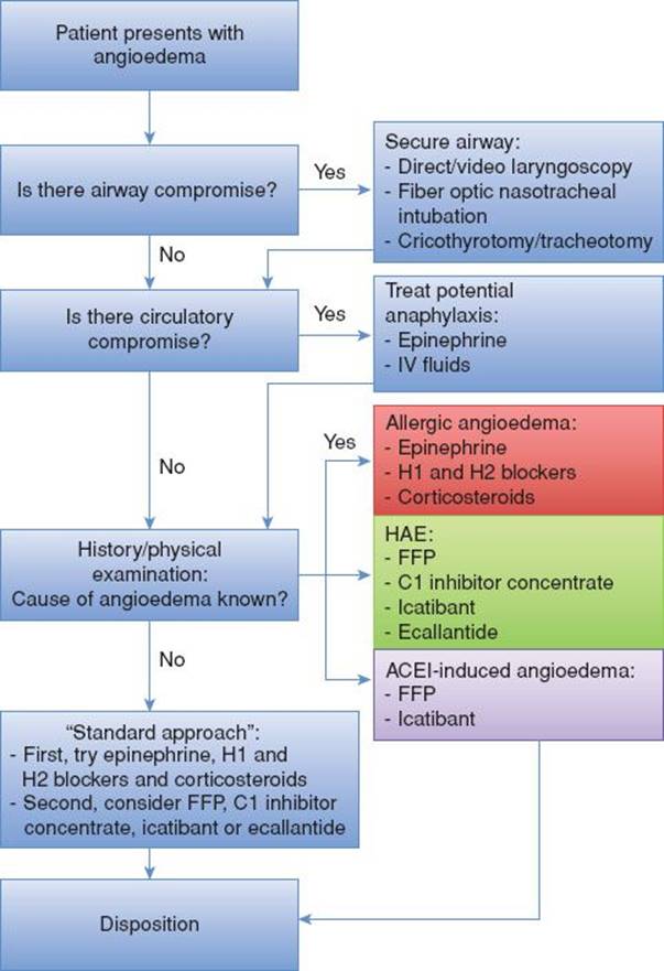
FIGURE 176.1 Clinical approach to angioedema. AAE, acquired angioedema; IV, intravenous; HAE, hereditary angioedema; FFP, fresh frozen plasma; ACEI, angiotensin-converting enzyme inhibitor. (Modified from Wilkerson RG. Angioedema in the emergency department: An evidence-based review. Emerg Med Pract. 2012;14(11):1–21.)
Once any airway or circulatory compromise has been addressed, a number of pharmacologic options are available to treat angioedema. It is important for emergency physicians to identify the key features of the various etiologies of angioedema because the best choice in medications depends on the underlying disease mechanism (Fig. 176.1; Table 176.1) (2). If the underlying mechanism is already known, for example, if the patient has a past medical history of HAE or presents after exposure to an obvious allergic trigger, every effort should be made to tailor therapy to these etiologies. Realistically, however, the cause of angioedema is difficult to ascertain in the ED based on history alone or using current diagnostic tools. In these cases of angioedema of unclear etiology, the authors propose a “standard approach.”
In this “standard approach”, all angioedema is assumed to be allergic in nature at first and treated initially with epinephrine, H1- and H2 blockers, and corticosteroids, since these agents are widely available, inexpensive, and generally safe (Fig. 176.1; Tables 176.1 and 176.3) (2). Epinephrine should be administered intramuscularly or in severe cases intravenously. H1 blockers, (primarily diphenhydramine, but second-generation antihistamines can also be considered), H2 blockers (such as cimetidine and ranitidine), and corticosteroids can be administered intravenously or orally (Table 176.3) (2). H1 blockers offer the main antihistamine effect, though the addition of H2 blockers may potentiate the effect as extrapolated from studies on urticaria (2). A short oral course of corticosteroids may be administered as a prophylactic measure against a rebound reaction (2). While these agents are effective for histamine-mediated allergic angioedema, they are ineffective for bradykinin-mediated ACEI-associated angioedema, HAE, and AAE (2).
TABLE 176.3
Medical Treatment of Adult Angioedema

For angioedema of unclear etiology refractory to these first-line therapies, as well as for known HAE, and to lesser extent, ACEI-induced angioedema or AAE, there are several alternative agents for consideration. These agents include fresh frozen plasma (FFP) and several newly developed therapies: purified C1 inhibitor concentrate, icatibant, and ecallantide (Fig. 176.1; Table 176.1) (2,4). The recommended dosing for these agents is listed in Table 176.3(2). The first of these therapies, FFP, contains exogenous C1 inhibitor and angiotensin-converting enzyme and has been shown to be effective for HAE and ACEI-induced angioedema in case reports, though not for AAE (2,12). Purified C1 inhibitor concentrate has also been shown in several clinical trials to be both effective and safe for HAE with only mild reactions reported (2,13,14). One formulation, Berinert, was recently approved in the United States for acute presentation of HAE (2). Data are currently unavailable for the effectiveness of C1 inhibitor for ACEI-induced angioedema and AAE. Icatibant and ecallantide are both agents that modify the kinin–kallikrein pathway, icatibant as a bradykinin receptor inhibitor, and ecallantide as a kallikrein inhibitor (2,4). Like C1 inhibitor concentrate, both agents have been shown in clinical trials to be effective for acute attacks of HAE and recently approved for use in the United States (2,15,16). Both medications have a favorable side effect profile; however, rare hypersensitivity reactions including anaphylaxis have been reported with ecallantide (2,15,16,17). With the exception of case reports suggesting the effectiveness of icatibant for ACEI-induced angioedema, there is little evidence that either icatibant or ecallantide is effective for ACEI-induced angioedema or AAE (2). In summary, while purified C1 inhibitor concentrate, icatibant, and ecallantide offer great potential for the management of HAE, data on their effectiveness for ACEI-induced angioedema and AAE are limited. Moreover, these newer agents are still not available at all hospitals and can be costly, ranging from $4,700 to $9,500 per dose (2). Thus, it is suggested that these therapies be considered as second-line therapy options for angioedema of unclear etiology.
For pediatric patients presenting with acute angioedema, management is similar to that of adults, with initial emphasis on airway, breathing, and circulation (14). Epinephrine, H1- and H2 blockers, and corticosteroids are particularly important first-line pharmacologic interventions in pediatric patients since allergic angioedema is more common (Table 176.4) (1). For pediatric cases of HAE, FFP, and Berinert C1 inhibitor concentrate are considered appropriate therapies (Table 176.4) (14). Icatibant and ecallantide are currently not approved for pediatric use (2).
TABLE 176.4
Medical Treatment of Pediatric Angioedema

An often overlooked component of addressing angioedema in the ED is prevention and patient education. Patients with allergic angioedema should avoid any potential triggers including discontinuing any inciting drugs. If allergic angioedema or anaphylaxis is suspected, an epinephrine autoinjector device should be prescribed (1,2). Patients who had angioedema from one kind of ACEI should avoid other drugs within the ACEI class (2,6). Whether angiotensin receptor blockers (ARBs) should be used by patients with previous ACEI-induced angioedema is controversial, and data are limited (2). The incidence of developing angioedema in this group is estimated to be 10% or less (18,19). Nevertheless, the risk of angioedema should be weighed against the indications for ARBs, particularly in cases when ARBs are thought to be more beneficial than other antihypertensive drugs.
CRITICAL INTERVENTIONS
• Perform rapid assessment and management of airway compromise and anaphylaxis.
• Treat all cases of angioedema with epinephrine, H1- and H2 blockers, and corticosteroids, before considering FFP, C1 inhibitor concentrate, icatibant, or ecallantide.
• Identify potential precipitating agents, especially allergic triggers and ACEIs.
DISPOSITION
The disposition of patients with angioedema is determined by the severity of initial symptoms, the anatomic location of the angioedema, the disease course, and response to therapy. As general guideline, patients who may warrant ICU admission include those with:
• Airway compromise and laryngeal edema requiring definitive airway
• Symptoms of dyspnea, stridor, hoarseness, or voice change
• Diffuse tongue involvement
In contrast, patients who may be admitted to a floor unit or discharged home include those with:
• Involvement limited to the face, lips, and soft palate
• Localized tongue involvement (9)
The patients who are candidates for discharge should be observed for a period of at least 4 to 6 hours in the ED to ensure no further extension of the edema (2). Outpatient referral for further work-up may be appropriate, especially in cases of recurrent angioedema. Allergy skin testing for allergic angioedema, prophylactic drug regimens for HAE, and work-up for underlying malignancy or autoimmune disease for AEE may all be addressed outpatient (1,2).
Common Pitfalls
• Failure to adequately assess and manage the airway
• Failure to adequately reassess for airway deterioration
• Failure to observe low-risk patients for an adequate period of time before discharge
• Failure to address underlying precipitants and discontinue their use
ACKNOWLEDGMENTS
The authors gratefully acknowledge the contributions of Aparajita Sohoni to the contents of this chapter.
REFERENCES
1. Krishnamurthy A, Gershwin ME, Gershwin ME. Pediatric angioedema. Clinic Rev Allerg Immunol. 2008;34:250–259.
2. Wilkerson RG. Angioedema in the emergency department: An evidence-based review. Emerg Med Pract. 2012;14(11):1–21.
3. Zirkle M, Bhattacharyya N. Predictors of airway intervention in angioedema of the head and neck. Otolaryngol Head Neck Surg. 2000;123(3):240–245.
4. Longhurst, H, Cicardi M. Hereditary angio-oedema. Lancet. 2012;379:474–481.
5. Greenberger PA. Idiopathic anaphylaxis. Immunol Allergy Clin North Am. 2007;27(2):273–293, vii–viii.
6. Byrd JB, Adam A, Brown NJ. Angiotensin-converting enzyme inhibitor-associated angioedema. Immunol Allergy Clin North Am. 2006;26(4):725–737.
7. Weldon D. Differential diagnosis of angioedema. Immunol Allergy Clin North Am. 2006;26(4):603–613.
8. Shah UK, Jacobs IN. Pediatric angioedema: Ten years’ experience. Arch Otolaryngol Head Neck Surg. 1999;125(7):791–795.
9. Ishoo E, Shah UK, Grillone GA, et al. Predicting airway risk in angioedema: Staging system based on presentation. Otolaryngol Head Neck Surg. 1999;121(3):263–268.
10. Sondhi D, Lippmann M, Murali G. Airway compromise due to angiotensin-converting enzyme inhibitor-induced angioedema: Clinical experience at a large community teaching hospital. Chest.2004;126(2):400–404.
11. Bentsianov BL, Parhiscar A, Azer M, et al. The role of fiberoptic nasopharyngoscopy in the management of the acute airway in angioneurotic edema. Laryngoscope. 2000;110(12):2016–2019.
12. Prematta M, Gibbs JG, Pratt EL, et al. Fresh frozen plasma for the treatment of hereditary angioedema. Ann Allergy Asthma Immunol. 2007;98(4):383–388.
13. Craig TJ, Bewtra AK, Bahna SL, et al. C1 esterase inhibitor concentrate in 1085 hereditary angioedema attacks–final results of the I.M.P.A.C.T.2 study. Allergy. 2011;66(12):1604–1611.
14. Farkas H, Varga L, Szeplaki G, et al. Management of hereditary angioedema in pediatric patients. Pediatrics. 2007;120(3):e713–e722.
15. Levy RJ, Lumry WR, McNeil DL, et al. EDEMA4: A phase 3, double-blind study of subcutaneous ecallantide treatment for acute attacks of hereditary angioedema. Ann Allergy Asthma Immunol.2010;104(6):523–529.
16. Sheffer AL, Macginnitie AJ, Campion M, et al. Outcomes after ecallantide treatment of laryngeal hereditary angioedema attacks. Ann Allergy Asthma Immunol. 2013;110(3):184–188.
17. Stolz LE, Sheffer AL. Prospective, double-blind, placebo-controlled trials of acute attacks of hereditary angioedema. Expert Rev Clin Immunol. 2012;8(1):25–32.
18. Beavers CJ, Dunn SP, Macaulay TE. The role of angiotensin receptor blockers in patients with angiotensin-converting enzyme inhibitor-induced angioedema. Ann Pharmacother. 2011;45(4):520–524.
19. Haymore BR, Yoon J, Mikita CP, et al. Risk of angioedema with angiotensin receptor blockers in patients with prior angioedema associated with angiotensin-converting enzyme inhibitors: A meta-analysis. Ann Allergy Asthma Immunol. 2008;101(5): 495–499.