
Paul Jhun
An adverse drug reaction (ADR) is defined by the World Health Organization as a “noxious, unintended, and undesired effect of a drug that occurs at doses used for prevention, diagnosis, or treatment” (1). ADRs occur in 10% to 20% of hospitalized patients and account for 3% to 6% of all hospital admissions (2,3), many of whom present to the emergency department for diagnosis and care.
ADRs are classified as type A or type B reactions. Type A ADRs are predictable reactions that can affect any healthy patient, given sufficient dose and duration, due to the known pharmacologic properties of the drug (drug interactions, overdose/toxicity, side effects). Type B ADRs are unpredictable hypersensitivity reactions that affect a susceptible subset of the population due to unpredictable individual immunologic response. Type B ADRs include true immunologic drug reactions, idiosyncratic drug reactions, and drug intolerance (expected side effects at subtherapeutic doses). Type A reactions constitute 80% to 90% of ADRs, while Type B reactions make up 10% to 20% of ADRs (2,3).
DRUG ALLERGY THEORY
Drug allergy or hypersensitivity accounts for 6% to 10% of ADRs. The multivalency theory proposed by Karl Landsteiner forms the current understanding of the immunologic pathophysiology of drug allergies (4). By definition, a monovalent antigen has only one site of attachment. Landsteiner proposed that drug antigens must be presented to the immune system in multivalent form in order to: (1) sensitize the immune system, inducing immunoglobulin synthesis; and (2) activate the immune system, eliciting the mast cell and basophil release of vasoactive mediators. Multivalency can be achieved through four main mechanisms: (1) the antigen is already a large macromolecule with multiple repeating epitopes, such as recombinant proteins, antisera, vaccines, or insulin; (2) the antigen is a hapten, a small molecule able to bind with larger macromolecules, that can form a multivalent hapten-carrier complex, such as β-lactam antibiotics, heavy metals, antithyroid medications, or quinidine; (3) the antigen undergoes metabolism, generating intermediate haptens capable of forming multivalent hapten-carrier complexes, such as antimicrobial sulfonamides, phenytoin, procainamide, or acetaminophen; or (4) the antigen directly binds certain T-cell receptors and activates the immune system, such as sulfamethoxazole, carbamazepine, lamotrigine, or lidocaine (5). Of note, a few small molecules have multiple recurrences of a single epitope, creating an inherently multivalent antigen, such as succinylcholine.
Risk factors predisposing a patient to drug hypersensitivity reactions include the genetic predisposition of the patient (history of atopy, history of prior drug reactions, and familial propensity), the characteristics of the drug (inherent reactivity, preparation, dosage, frequency, and duration of therapy), and the disease state (concomitant medications or diseases). Interestingly, specific drug hypersensitivity reactions have been associated with certain disease states, such as when sulfonamides are administered to patients with AIDS or when amoxicillin is administered to patients actively infected with Epstein–Barr virus (6,7).
CLINICAL PRESENTATION
Classification of Reactions
Drug hypersensitivity reactions are conceptually categorized according to the Gell and Coombs four-group classification system: type I acute IgE-mediated response, type II delayed IgG/IgM-mediated cell destruction, type III delayed immune complex reaction, and type IV delayed T-cell-mediated hypersensitivity.
Type I reactions are mediated by previously sensitized patients who have preformed drug-specific IgE antibodies attached to the surface of their mast cells and basophils. When the patient is re-exposed to the offending drug, the circulating drug antigen binds the drug-specific IgE antibodies activating the mast cells and basophils, leading to immediate vasoactive mediator release and clinical sequelae such as urticaria, flushing, pruritis, angioedema, laryngeal edema, bronchospasm, gastrointestinal distress, and hypotension.
Type II cytotoxic reactions result when an offending drug binds to certain cell types and acts as an antigen. When drug-specific IgG or IgM antibodies bind these complexes, the cells are destroyed. Almost all type II reactions involve formed elements of the blood leading to hemolytic anemia, thrombocytopenia, or neutropenia, and their respective clinical sequelae.
In type III reactions, the drug antigen binds drug-specific IgG, forming immune complexes that precipitate in various tissues, clinically presenting as serum sickness, drug fever, or vasculitis.
Type IV reactions are mediated by sensitized T lymphocytes that recognize a particular drug antigen and recruit other lymphocytes and mononuclear cells to the site of that antigen. Type IV reactions classically have prominent dermatologic findings due to the fact that there are nearly twice as many T cells residing in the skin compared to circulation (8). Clinical manifestations include contact dermatitis, morbilliform or maculopapular eruptions, exfoliative dermatitides, or drug-induced hypersensitivity syndrome. However, type IV reactions may also involve specific organs without cutaneous findings, such as isolated drug-induced hepatitis, interstitial nephritis, or pneumonitis.
The World Allergy Organization proposed a simpler, two-group clinical classification system to help practitioners identify IgE-mediated reactions, based on the timing of symptom onset: immediate and delayed reactions (9). Immediate reactions are those beginning within 1 hour of first dose administration and are likely IgE-mediated type I reactions. Delayed reactions are those appearing after 1 hour of dose administration and are not likely to be IgE-mediated.
Drug reactions that resemble type I IgE-mediated hypersensitivity reactions, but are not immune-mediated, are termed idiosyncratic or pseudoallergic reactions. Most pseudoallergic reactions are clinically indistinguishable from true allergic reactions because they often share the final common pathway of mast cell and basophil activation. Of note, the term anaphylaxis is reserved for immune-mediated type I hypersensitivity reactions, while the terms anaphylactoid and nonallergic anaphylaxis are preferred for idiosyncratic reactions. Common medications and their idiosyncratic reactions include radiocontrast media (RCM)-induced urticaria and hypotension, opiate-induced urticaria, aspirin-induced bronchospasm, nonsteroidal anti-inflammatory drug-induced urticaria, and vancomycin “red man syndrome.”
DIFFERENTIAL DIAGNOSIS
Given the broad spectrum of clinical presentations of drug adverse reactions, physicians should include ADRs as part of their differential diagnosis.
ED EVALUATION
The diagnosis of drug hypersensitivity reactions in the emergency department is largely dependent on the patient history and presentation. Particularly for the emergency physician, early recognition and diagnosis of anaphylaxis is critical, while recognition of severe exfoliative dermatitides and drug-induced hematopoietic or organ damage is important in determining appropriate management and disposition. Important historical information includes assessing risk factors such as previous exposures to the same or similar drugs, a complete list of medications to determine allergenic potentials, and temporal association of drug administration times to symptom onset to establish a causal link. Improvement of symptoms after withdrawal of the suspected drug is suggestive information. A suspicion for immune-mediated cell destruction should prompt the physician to obtain a complete blood count with a peripheral smear. An indirect Coombs test, if available, may be helpful in diagnosing immune hemolytic anemia.
While objective testing is available for certain drug allergic reactions, such tests require referral to an allergy–immunology specialist for implementation and interpretation. Skin testing is conducted primarily to evaluate suspected type I hypersensitivity reactions, although limitations include lack of standardization and validation for many drugs. Patch testing can be performed for evaluating contact dermatitis caused by topically applied medications. In vitro tests, such as radioallergosorbent or enzyme-linked immunosorbent assay tests are largely investigational and not standardized or validated, so are of limited value. Drug provocation testing by graded challenges can be performed, but should be done so by appropriate specialists after careful consideration of risks and benefits for a particular individual. The best studied drug allergen testing involves penicillin. Intradermal skin testing is the diagnostic procedure of choice for evaluating suspected IgE-dependent penicillin allergy (5,7).
ED MANAGEMENT
The standard management of drug hypersensitivity reactions includes prompt discontinuation of the medication and provision of supportive care that is specific to the type and severity of manifestations. Mild pruritis, flushing, and urticaria may be treated with antihistamines. Corticosteroids may be indicated for severe systemic or cutaneous reactions, immune complex reactions, or drug-induced hematologic diseases. Early epinephrine is the mainstay of treatment for anaphylactic reactions. Oxygen and airway support, inhaled β-agonists, intravenous fluids, and pressors also may be required.
Although type B ADRs are unpredictable, several steps can be taken to minimize the chance of developing a drug hypersensitivity reaction. Perhaps most importantly, medications should be prescribed only when necessary. A careful history of risk factors for drug reactions, including prior ADRs, should be obtained. If a drug allergy exists, crossreactive drugs should be avoided. The oral route should be used when possible (1,5,7). Preparations that are less sensitizing are preferred when available. Documentation in the medical record of any apparent reaction may help avert a similar or worse reaction in the future. Finally, when potentially immunogenic parenteral agents are given in the emergency department, patients should be observed for at least 1 hour after administration, to ensure an immediate IgE reaction does not occur.
CRITICAL INTERVENTIONS
• Recognize and administer epinephrine promptly for anaphylaxis (adult: epinephrine 0.2 to 0.5 mg [1:1,000 {1 mg/mL} solution] IM, or epinephrine 0.1 mg [1:10,000 {0.1 mg/mL} solution] IV; pediatric: epinephrine 0.01 mg/kg [1:1,000 {1 mg/mL} solution] IM, maximum single dose of 0.3 mg) (10)
• Obtain, document, and be aware of a history of prior drug hypersensitivity reactions
• Identify and withdraw offending drug(s) and provide alternative drug therapy if indicated
DISPOSITION
Disposition decisions for patients with drug reactions hinge on the nature and severity of the reaction. Patients who present with mild reactions, respond well to treatment, and are reliable with good support systems may be considered for expedited discharge to home. In borderline cases, a period of observation of several hours may be helpful. Patients with persistent or recurring airway edema, bronchospasm, hypotension, cardiovascular complications, or altered mental status require admission to the hospital in an intensive care setting. Patients who receive treatment for an initially severe reaction but who rapidly improve are typically either observed for a period of 6 to 12 hours or admitted to the hospital; in the case of anaphylaxis, this facilitates treatment if a late-phase reaction occurs. Discharged patients should receive instructions including cautions regarding possible delayed or recurrent reactions. Referral to or consultation with an allergy–immunology specialist is indicated in suspected cases of drug hypersensitivity reactions, especially suspected cases of IgE-mediated reactions. Further testing can be performed and patients can be evaluated for induction of drug tolerance or desensitization for offending drugs that are medically necessary.
PENICILLIN ALLERGY AND CROSSREACTIVITY
Penicillin and its derivatives are among the most widely prescribed antibiotics and are the most common cause of drug-induced allergic reactions. Penicillin can be responsible for any of the four types of Gell and Coombs. The overall incidence of reported adverse reactions to penicillins is estimated to be up to 10% of patients (7). Interestingly, up to 90% of these individuals are able to tolerate penicillin after complete evaluation and testing (7). This could be due to inaccurate clinical history of penicillin allergy or more importantly could be due to the gradual loss of penicillin-specific IgE that happens in patients over time. Studies have shown that approximately 50% of patients with IgE-mediated penicillin allergy lose sensitivity 5 years after their last reaction, and approximately 80% lose sensitivity after 10 years (11,12). Life-threatening anaphylaxis secondary to penicillin after parenteral administration appears to be uncommon, with an incidence of 1 to 2 per 10,000 treated patients (7). Patients with a suspected history of IgE-mediated penicillin allergic reaction should be referred to an allergy–immunology specialist to undergo skin testing, which is the most reliable method for evaluating IgE-mediated penicillin allergy. In patients who report a reaction history concerning for anaphylaxis, especially if the reaction was recent, penicillins and crossreactive drugs should generally be avoided until evaluation by a specialist.
Regarding crossreactivity risks with the use of the other β-lactam antibiotics in a patient with a confirmed penicillin allergy, crossreactivity rate is high with first-generation cephalosporins and these drugs should generally be avoided. Most second- and all third- and fourth-generation cephalosporins appear to have negligible crossreactivity (13). Carbapenems appear to have low crossreactivity rates of about 1% in limited studies (14,15). Monobactams do not appear to immunologically crossreact, likely due to its distinctly different chemical structure (5,7). Overall, recent studies have shown that cephalosporin administration in patients with a reported history of penicillin allergy, unconfirmed by skin testing, resulted in an allergic reaction in 1% of patients, while cephalosporin administration in patients with confirmed penicillin-allergy patients resulted in an allergic reaction in 2% of patients (7,13). A recent consensus allergy and immunology guideline states that patients with a complete, negative penicillin skin testing may receive cephalosporins safely (7). However, if the patient has a reported history of penicillin but has not received skin testing, this guideline states that the practitioner has the option of either (1) obtaining penicillin or cephalosporin skin testing to determine the safety of cephalosporin administration or (2) directly administering the cephalosporin (only in the absence of severe and/or recent penicillin allergy reaction history), with the knowledge that there is a very small but real risk for allergic reaction. In such cases of direct cephalosporin administration in a patient with a reported but unconfirmed history of penicillin allergy, crossreactivity risk can be further mitigated by administering a third- or fourth-generation cephalosporin.
RADIOCONTRAST MEDIA REACTIONS
More than 15 million diagnostic radiographic examinations using RCM are performed in the United States each year. Adverse reactions due to RCM are reported to have an annual incidence of 2% to 12%, with deaths estimated between 1 to 3 per 100,000 administrations (16,17). RCM adverse reactions can be categorized into hypersensitivity reactions and chemotoxic reactions.
RCM-induced hypersensitivity reactions are further divided into immediate and delayed types. Immediate hypersensitivity reactions generally present within 1 hour of administration and are believed to be idiosyncratic, nonimmune-mediated, and independent of dose and infusion rate. These acute reactions share the final common pathway of mast cell activation and vasoactive mediator release, are clinically indistinguishable from type I reactions, and are treated similarly. Delayed RCM hypersensitivity reactions are type IV reactions that can present several days after administration and are generally self-limited (18).
As newer generation RCM products have been developed, the incidence of adverse reactions has also decreased. First-generation RCMs are ionic monomers that are categorized as high-osmolar RCM (>1,400 mOsm/kg), compared with normal serum osmolality. Second-generation agents, such as iohexol, are nonionic monomers that are categorized as low-osmolar RCM (500 to 900 mOsm/kg). The newest agents are nonionic dimers, such as iodixanol, that have the same osmolality of plasma and are categorized as iso-osmolar RCM. Estimated rates of severe, immediate hypersensitivity reactions are 0.1% to 0.3% for the high-osmolar RCM and 0.01% to 0.04% for low-osmolar RCM (1,17). Of note, seafood or shellfish allergy is not an independent risk factor for RCM hypersensitivity reactions and no special precautions need to be taken for such patients (1).
Chemotoxic RCM reactions are related to the RCM chemical properties and are dependent on dose and infusion rate. These reactions include nephrotoxicity, vasovagal reactions, and iodine-induced thyroid dysfunction. Regarding contrast-induced nephropathy (CIN), there is no specific treatment once acute kidney injury develops, so management is aimed at prevention. The mechanism of CIN is thought to be related to renal vasoconstriction and/or direct renal cytotoxic effects of the contrast agent, and the diagnosis is based on the increase in the serum creatinine level beginning within 24 to 48 hours of RCM administration. Risk factors for CIN include chronic kidney disease, diabetes mellitus, multiple myeloma, and causes of reduced renal perfusion such as advanced heart failure and hypovolemia. A high total dose of RCM as well as first-generation RCM are additional risk factors. The use of low osmolar or iso-osmolar RCM in patients with pre-existing chronic kidney disease is associated with decreased incidence of CIN compared to high osmolar RCM (19,20).
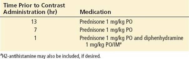
The risk-to-benefit ratio of a contrast study should be considered before it is ordered and performed. If possible, ultrasound or magnetic resonance imaging should be utilized. If RCM must be used and the patient has a history of prior anaphylactoid reaction, the patient can be pretreated with corticosteroids and antihistamines to decrease the likelihood of recurrent anaphylactoid reaction. One common protocol is shown in Table 177.1. To minimize the risk of developing CIN, recommended approaches have included infusion of isotonic sodium chloride or sodium bicarbonate before and after the administration of contrast (e.g., 3 mL/kg/hr for 1 hour before and 1 mL/kg/hr for 6 hours after) (20). Also, while evidence is conflicting, some suggest benefit from adjunctive treatment with N-acetylcysteine, 1,200 mg bid the day before, and the day of the radiocontrast administration (20). Finally, nonsteroidal anti-inflammatory drugs should be avoided as they increase the risk of nephrotoxicity.
TABLE 177.1
Premedication Protocol Prior to Administration of Radiocontrast Media to a Patient with Prior History of Anaphylactoid Reaction
Common Pitfalls
• Failure to obtain medication list and drug allergy history
• Administration of crossreactive drugs to patients with a known drug hypersensitivity reaction
• Failure to diagnosis and quickly manage anaphylaxis
REFERENCES
1. Khan DA, Solensky R. Drug allergy. J Allergy Clin Immunol. 2010;125(2 suppl 2):S126–S137.
2. Gomes ER, Demoly P. Epidemiology of hypersensitivity drug reactions. Curr Opin Allergy Clin Immunol. 2005;5:309–316.
3. Thong BY, Tan TC. Epidemiology and risk factors for drug allergy. Br J Clin Pharmacol. 2011;71(5):684–700.
4. Landsteiner K. The Specificity of Serological Reactions. New York, NY: Dover; 1962.
5. Celik G, Pichler WJ, Adkinson NF. Drug allergy Chapter 68. In: Adkinson NF, ed. Middleton’s Allergy: Principles and Practice. 7th ed. Philadelphia, PA: Elsevier; 2009.
6. Gruchalla RS, Pirmohamed M. Antibiotic allergy. New Engl J Med. 2006;354:601–609.
7. Joint Task Force on Practice Parameters, American Academy of Allergy, Asthma and Immunology, American College of Allergy, et al. Drug allergy: An updated practice parameter. Ann Allergy Asthma Immunol. 2010;105(4):259–273.
8. Clark RA, Chong B, Mirchandani N, et al. The vast majority of CLA+ T cells are resident in normal skin. J Immunol. 2006;176(7):4431–4439.
9. Johansson SG, Bieber T, Dahl R, et al. Revised nomenclature for allergy for global use: Report of the Nomenclature Review Committee of the World Allergy Organization, October 2003. J Allergy Clin Immunol. 2004;113(5):832–836.
10. Lexi-Comp OnlineTM. Lexi-Drugs OnlineTM. Hudson, OH: Lexi-Comp, Inc.; May 31, 2013.
11. Sullivan TJ, Wedner HJ, Shatz GS, et al. Skin testing to detect penicillin allergy. J Allergy Clin Immunol. 1981;68:171–180.
12. Blanca M, Torres MJ, García JJ, et al. Natural evolution of skin test sensitivity in patients allergic to beta-lactam antibiotics. J Allergy Clin Immunol. 1999;103:918–924.
13. Campagna JD, Bond MC, Schalbelman E, et al. The use of cephalosporins in penicillin-allergic patients: A literature review. J Emerg Med. 2012;42(5):612–620.
14. Romano A, Viola M, Gueant-Rodriguez RM, et al. Imipenem in patients with immediate hypersensitivity to penicillins. N Engl J Med. 2006;354(26):2835–2837.
15. Romano A, Viola M, Gueant-Rodriguez RM, et al. Brief communication: Tolerability of meropenem in patients with IgE-mediated hypersensitivity to penicillins. Ann Intern Med. 2007;146(4):266–269.
16. Christiansen C. X-ray contrast media-an overview. Toxicology. 2005;209(2):185–187.
17. Hong H, Wang HS, Liu ZP. Agents that induce pseudo-allergic reaction. Drug Discov Ther. 2011;5(5):211–219.
18. Meth MJ, Maibach HI. Current understanding of contrast media reactions and implications for clinical management. Drug Saf. 2006;29(2):133–1141.
19. Rudnick MR, Goldfarb S, Wexler L, et al. Nephrotoxicity of ionic and nonionic contrast media in 1196 patients: A randomized trial. The Iohexol Cooperative Study. Kidney Int. 1995;47(1):254–261.
20. Kidney Disease Improving Global Outcomes (KDIGO) Acute Kidney Injury Work Group. KDIGO clinical practice guideline for acute kidney injury. Kidney Inter Suppl. 2012:2(1):69–86.