
Meghan A. Cummins and Keith T. Borg
Parasitic infestations are much more widespread than many people realize. Parasites affect not only impoverished people in remote countries but can also cause important health problems for the rich and poor throughout the world, including the United States. Parasites are considered by many physicians to be the most undiagnosed infectious disease challenge in the United States today. Diagnosing parasitic infections in the emergency department (ED) can be challenging because there is a large and varied group of organisms that cause parasitic infections with a wide range of clinical symptoms. Parasitic disease, however, can usually be diagnosed through methods available to most emergency physicians, and effective treatments are available that are generally minimally toxic to the human host (1,2). A history of foreign travel or habitation in an endemic area, including certain regions of the United States, should alert the clinician to the possibility of parasitic infection. As with many disease processes, the key is considering parasitic infestation in the differential diagnosis.
Symptoms may not manifest themselves until well after the traveler has returned home. In addition, over the past two decades, the proportion of the US population born abroad has increased steadily, with newer immigrants typically coming from Latin America and Asia (3). The proper treatment of immigrants in the United States requires a familiarity with diseases common in the developing world, such as malaria, amebiasis, and neurocysticercosis. Given the constant change in the globalization of infectious disease, the clinician should always consult current references, including the Centers for Disease Control (CDC) website (http://www.cdc.gov/travel). Parasitic infestation is a global problem. Here in the United States, the CDC cites food as the catalyst behind 80% of the pathogenic outbreaks.
Four major groups of organisms infest humans. Protozoans cause malaria, amebiasis, giardiasis, and many other diseases. Helminths, or worms, including nematodes (roundworms), cestodes (tapeworms), and trematodes (flukes), are also responsible for a wide variety of human illnesses. A comprehensive review of parasitic diseases would encompass infections due to literally scores of organisms, so this chapter is limited to the parasitic diseases most common in EDs in the United States (trichomoniasis is excluded). This chapter is divided into three discussions: systemic illnesses (malaria), predominantly gastrointestinal (GI) disease, and predominantly central nervous system (CNS) disease (cysticercosis).
Malaria
CLINICAL PRESENTATION
Awareness about malaria and measures that travelers can take to avoid infective mosquito bites are more important than ever. Cases of malaria continue to rise in the United States. Thus, fever in a patient with a history of recent travel or habitation in the tropics should suggest the diagnosis of malaria. Caused by four species of the protozoan Plasmodium (P. vivax, P. ovale, P. falciparum, and P. malariae), malaria is the most common parasitic disease in the world. According to the CDC (www.cdc.gov/malaria), malaria currently infects an estimated 350 to 500 million people worldwide and on average, 1,500 cases are reported in the United States annually. Malaria causes <1 million deaths annually (most commonly children in sub-Saharan Africa). Although malaria is no longer considered endemic in the United States, increased immigration from and travel to countries where the disease is common has made malaria more frequent as a presenting complaint in ED in this country (4). The number of cases of malaria acquired by international travel is growing as a result of the increased risk of malaria transmission where malaria control has faded and also because of the spread of drug-resistant strains of malaria (5). Patients with malaria are frequently misdiagnosed on their initial presentation to the ED, thus increasing their risk for serious morbidity and mortality.
Classic symptoms of malaria:
• Fever, chills, and rigors lasting 4 to 10 hours, culminating in an episode of profuse diaphoresis followed by defervescence.
• This pattern may recur at 2- or 3-day intervals, depending on the species of Plasmodium involved.
• General malaise, headache, and, occasionally, hypotension.
• Splenomegaly and jaundice may be seen in established infections. (Physical examination is generally not helpful.)
• Laboratory abnormalities include a mild anemia, neutropenia, thrombocytopenia, and increased prothrombin time. Clinical evidence of impaired hemostasis is rare.
The periodicity of the disease can be explained by examining the life cycle of the infecting organism. In the human, the life cycle of the parasite consists of two stages. The initial stage is known as the liver or exoerythrocytic stage. This is where the parasites multiply in hepatocytes and cause them to rupture. The second stage is known as the blood or erythrocytic stage and occurs when parasites are released into the bloodstream and invade the erythrocytes. This is the cause of clinical illness. Plasmodium species may be injected into the human circulation following the bite of the Anopheles mosquito, after sharing needles or receiving a blood transfusion, and by congenital transmission. Immature sporozoites migrate to the liver, beginning the exoerythrocytic phase, the duration of which is species-dependent. Ultimately, mature parasites (merozoites) are released into the circulation and invade red blood cells, beginning the erythrocytic phase. Replication within red cells is followed 48 to 72 hours later by red cell lysis and release of further merozoites into the circulation, which again invade erythrocytes and repeat the cycle. The fever corresponds to episodes of red cell lysis.
The classic pattern of periodic fever is not always seen, however, particularly early in the course of the illness. The variability of the clinical course also depends on the infecting species and the host’s immune status. People living in endemic areas often have partial “immunity” that results in much less severe manifestations and symptoms. It is presumed to be an acquired immunity caused by frequent exposure to the infecting organism. Months after leaving an endemic area, these persons may experience relapses that resemble the acute form of the disease, probably caused by a loss of immunity and the release of exoerythrocytic parasites from the liver. P. vivax and P. ovale can persist for long periods in the exoerythrocytic state. Even patients who have received adequate antimalarial therapy may experience relapses associated with the release of exoerythrocytic organisms. Cases have been reported 2 months to 4.5 years after a traveler’s return (5).
P. falciparum causes the most severe type of malaria and is responsible for most complications and deaths related to malaria (4). Falciparum malaria has two important features that account for differences in its presentation and in the severity of the disease it produces. First, this species causes widespread capillary obstruction, resulting in end-organ hypoxia and dysfunction, with severe complications that can include CNS dysfunction, hemolytic anemia, hypoglycemia, pulmonary edema, septicemia, renal failure, or splenic rupture. A second distinguishing feature of P. falciparum is that, unlike other malarial species that infect only the most mature red cells, it infects red cells of all ages, resulting in a greater degree of hemolysis and anemia and the potential for acute tubular necrosis. Finally, unlike P. ovale and P. vivax, cases of P. falciparum malaria in patients with late presentations are caused by blood-stage parasites that were drug resistant or had survived incomplete prophylaxis.
ED EVALUATION
The diagnosis of malaria is a difficult one to make because of the disease’s nonspecific clinical features—fever, chills, headache, and malaise. Because approximately 90% of travelers who contract malaria do not become ill until returning home, the diagnosis should be entertained for anyone presenting to the ED with a febrile illness and a history of travel or habitation in an endemic area. Kain et al. (6) estimated that approximately 30,000 travelers from industrialized countries contract malaria each year. A high degree of suspicion is necessary for diagnosis by physicians who rarely encounter this disease. The most common incorrect diagnosis is a viral syndrome, but a history of recurrent fever generally distinguishes malaria from these disorders. Other disorders that cause fever and are endemic in foreign countries, such as viral hepatitis, dengue fever, typhoid fever, and amebiasis, should also be considered. Recent data suggest that a significant number of those presenting with malaria do so later than previously thought. More than 35% of malaria-infested travelers in one study developed malaria more than 2 months after their return. This suggests that we may need to modify treatment to more aggressively treat the liver phase of the disease for effective prevention (5).
The standard way to make the diagnosis of malaria is through demonstration of intracellular forms of the parasite on a thin Giemsa-stained peripheral blood smear. If malaria is strongly suspected, negative blood films should be repeated every 12 to 24 hours for total of three sets. Three negative sets make the diagnosis very unlikely. Sometimes, it is necessary to use the thick-smear technique, which usually requires the expertise of a trained technician. New rapid testing by PCR, and even real-time PCR assays for chloroquine-resistant strains of P. falciparum malaria, as well as other methods, are being developed and should aid in the rapid diagnosis of malarial infections (7).
Although admittedly rare in the United States, congenital malaria should be considered when the emergency physician is presented with the febrile infant of an immigrant mother. It has been estimated that about 15% to 20% of all cases of malaria brought back to Western countries from recent travelers are in children (8).
ED MANAGEMENT
Management of malaria in the ED is guided by the severity of the infection. Assessment of airway patency and respiratory, circulatory, and neurologic status should be performed in a timely fashion. The emergency physician should be prepared to treat potential complications such as anemia, coagulopathies, pulmonary edema, azotemia, hypoglycemia, metabolic acidosis, and hypotensive shock (9).
Volume should be restored and fever controlled. Hypotension usually responds well to crystalloid infusion. Fever usually responds to standard cooling measures; for temperatures <104°F, oral antipyretics are likely to be effective. Acetaminophen is recommended rather than aspirin because of the common occurrence of thrombocytopenia. For patients with high-density parasitemia (>10% parasitemia), altered mental status, nonvolume overload pulmonary edema, or renal failure, exchange transfusion may be helpful (10).
Pharmacologic therapy of malaria is outlined in Table 193.1. Antimalaria pharmacotherapy and prophylaxis continue to pose difficult problems. Most antimalarial agents act on the parasites at blood stage and therefore do not prevent late onset illness (5). Chloroquine remains the oral drug of choice (with hydroxychloroquine being second-line treatment) for acute treatment of all infections except those due to chloroquine-resistant P. falciparum or P. vivaxacquired in Papua New Guinea or Indonesia. For chloroquine-resistant P. falciparum infection or for severely ill patients with an unidentified malarial infection, the recommended agents include either oral quinine plus doxycycline (tetracycline or clindamycin may be substituted for doxycycline), atovaquone–proguanil, or artemether–lumefantrine. Mefloquine may be used if the other drugs are not available. Patients with P. vivax acquired within Papua New Guinea or Indonesia should use atovaquone–proguanil plus mefloquine as first-line therapy and consider quinine plus doxycycline or tetracycline as alternative treatments. Patients with severe malaria (regardless of species or geographic location where contracted) or those who are unable to tolerate oral medications, should be treated with intravenous (IV) quinidine plus tetracycline, doxycycline, or clindamycin.
TABLE 193.1
Treatment of Malaria

Critically ill patients should have access to continuous cardiac and blood pressure monitoring, while having frequent blood glucose, hemoglobin, parasitemia levels, and electrolyte levels checked. As stated previously, exchange transfusion may be indicated in patients who meet clinical criteria (10). Finally, newer artemisinin-derived drugs similar to artemether–lumefantrine, such as dihydroartemisinin–piperaquine and artesunate–mefloquine are being tested against multidrug-resistant strains of P. falciparum (11–13).
In addition to acute therapy, patients infected with P. vivax or P. ovale require more prolonged treatment to prevent reactivation of exoerythrocyte forms. These patients should be treated with primaquine phosphate, 30 mg base/d for 14 days for adults and 0.5 mg base/kg/d for 14 days for children. Patients should be screened for G-6-PD deficiency before beginning primaquine therapy, and the drug is contraindicated during pregnancy. Patients who are G-6-PD deficient and for whom primaquine is thus contraindicated should be counseled about the possibility of recurrence (10).
To prevent fatal outcomes in cases of falciparum malaria, the following are required: improved health information and preventive measures for the 30 million travelers who visit regions where malaria is endemic, improved recognition of infection by physicians, and prompt initiation of effective therapy (6).
DISPOSITION
Hospital admission is indicated for patients with severe dehydration, significant underlying medical illness, inability to tolerate oral medications, or evidence of end-organ involvement such as stroke, renal failure, or pulmonary edema. Those whose ability to comply with an outpatient regimen is questionable should also be admitted. Suspected P. falciparum infection is commonly considered to require hospitalization because of the increasing prevalence of drug resistance and the potential for life-threatening complications. Severely ill patients or those with significant parasitemia (>3% of red cells containing parasites) should be treated in an intensive care unit with IV medications.
Prophylaxis is the most important aspect of malaria control in the United States, given the frequency of foreign travel. Before going to an endemic area, travelers should consult an expert to determine whether chemical prophylaxis is indicated and whether chloroquine resistance has been reported in the area. Decisions about prophylaxis are increasingly complicated as a result of malaria drug resistance. Chloroquine is usually recommended for persons traveling to endemic areas but may not be necessary in all cases, particularly for a short trip to an urban area. When traveling to a chloroquine-resistant area, another drug should be chosen. The particular medication chosen must depend on the pattern of resistance in the area. Malarone (atovaquone/proguanil) is the prophylactic drug of choice for adults traveling to most chloroquine-resistant areas. Other options include mefloquine (Lariam), doxycycline, and hydroxychloroquine sulfate (Plaquenil). Primaquine is also an option for prophylaxis specifically against P. vivax for trips of short duration only. See Table 193.2 for prophylactic drug dosages.
TABLE 193.2
Malaria Prophylaxis Drug Regimes
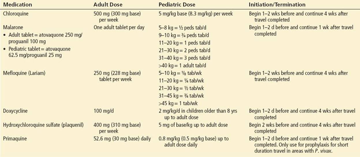
Contraindications to the use of mefloquine include serious psychiatric illness, seizures, or serious cardiac arrhythmias (14). Because no drug regimen guarantees protection against malaria, if fever develops within the first year after travel to a malarious area, travelers should be encouraged to seek medical attention (10). Consultation of current guidelines for areas of travel can be obtained from the CDC or WHO websites.
Gastrointestinal Disease
CLINICAL PRESENTATION AND DIFFERENTIAL DIAGNOSIS
The GI tract is commonly affected by parasites. This is because exposure occurs readily through fecal contamination of food and water. Protozoans and helminths are common causes of GI symptoms such as abdominal pain, malabsorption, obstruction, diarrhea, rectal prolapse, and pruritus ani. The protozoans that cause GI diseases include Giardia lamblia, Entamoeba histolytica, and Cryptosporidium.
G. lamblia, a flagellated protozoan, affects the small intestine. It is the most commonly identified intestinal parasite in the United States. This infection may be transmitted through the ingestion of contaminated food or water, by person-to-person contact, or through fecal–oral transmission (15). Patients may experience explosive, watery, foul-smelling diarrhea or may express a more insidious onset of symptoms. Cramping epigastric pain is common; bloody diarrhea is unusual. Physical examination of the abdomen often reveals diffuse upper quadrant tenderness without evidence of peritoneal irritation. Malabsorption may be responsible for the substantial weight loss that is associated with Giardia infestations in more than 50% of patients (15). Microscopic stool examination may demonstrate active trophozoites, but cysts are more commonly found. At least three stools taken at 2-day intervals should be examined for ova and parasites. Commercially available tests to detect Giardia antigen in the stool exist. The “string test” may be used in an outpatient setting to help diagnose Giardia infestation. This test involves the patient swallowing a gelatin capsule attached to a string. The string slowly works its way into the duodenum over 4 to 6 hours, at which time it is pulled up and any blood or other material from the digestive tract still attached to the string is looked at under a microscope. Parts of the parasites or eggs may be observed in a positive test. Because Giardia is most often transmitted by contaminated water or by the fecal–oral route, efforts at prevention should focus on these routes of transmission.
E. histolytica, upon invasion of the colonic mucosa, can cause amebic dysentery with abdominal pain, tenesmus, and bloody stools (16). It may result in severe dehydration. The large bowel is typically most affected. Transmission of amebiasis is linked directly to hygiene and sanitation; risk factors for amebiasis include recent travel and institutionalization. The spectrum of clinical disease ranges from mild diarrhea to fulminant rectocolitis. Massive GI bleeding may occur when amebic trophozoites invade large areas of colonic mucosa and cause ulceration; toxic megacolon, perforation, and peritonitis are other severe complications of amebic dysentery. Extraintestinal infection, most commonly amebic liver abscess (3), may occur with invasion of the trophozoites through the colonic mucosa. The diagnosis of amebic dysentery is made by stool examination for cysts and trophozoites or in biopsy specimens obtained by sigmoidoscopy or colonoscopy (16). More recently, PCR assay techniques have shown greater sensitivities and specificities for both E. histolytica and Entamoeba dispar species than stool microscopy or culture (17).
Cryptosporidium may affect any part of the GI tract. Immunocompetent patients frequently experience a clinical syndrome similar to that of giardiasis, whereas immunodeficient patients typically develop more serious disease and may die of dehydration and electrolyte abnormalities. In addition to water sources, such as drinking water, pools, lakes, and water parks, there have been several food-borne outbreaks of cryptosporidiosis in North America, including the severe outbreak in Milwaukee (18). Diagnosis is based on the microscopic identification of fecal oocysts, although newer more sensitive techniques, including molecular methods such as PCR, are being used in reference laboratories to identify Cryptosporidium at the species level. The disease is typically self-limited. Paromomycin has been used to treat cryptosporidiosis in several cases. Treatment with nitazoxanide, 500 mg twice daily (either in tablet or suspension form) for 3 days, has shown some promise in treating enteritis and diarrhea caused by Cryptosporidium (19).
Infection with the helminths Ascaris lumbricoides and Strongyloides stercoralis and the hookworms Necator americanus and Ancylostoma duodenale causes cramping upper abdominal pain when associated with large worm burdens. Patients presenting from tropical climates, particularly Central America, South America, and Southeast Asia, may be infested with more than one of these GI parasites. Hookworms are also endemic in the southeastern United States.
Infection with A. lumbricoides and S. stercoralis may present with nonspecific abdominal pain and diarrhea. Ascaris, a roundworm, can migrate into the biliary system through the ampulla of Vater and induce inflammation and fibrosis of the biliary ducts through chemical irritation. They may also cause partial or intermittent obstruction of the bile ducts (20). Midepigastric and right upper quadrant pain may also mimic peptic ulcer disease. Ascaris has also been reported to cause bowel obstruction, especially in children (21). Upper respiratory symptoms can develop due to pulmonary migration of larvae (Loeffler syndrome). S. stercoralis larvae may mature within the GI tract, as the gravid adult female releases rhabditiform larvae that rapidly evolve into the invasive filariform larval form. The filariform larvae can penetrate the GI tract or perianal skin, causing the hyperinfection syndrome characteristic of strongyloidiasis (2); this is particularly likely to appear in the immunocompromised patient. Both Necator and Ancylostoma penetrate the mucosa of the small bowel in heavy acute infestations and cause cramping epigastric pain. In addition, GI blood loss at worm attachment sites may result in a profound hypochromic microcytic anemia.
The physical examination of patients with worm infestation is often unremarkable. Minimal diffuse upper abdominal tenderness may be present, but peritoneal signs are unusual when the worms are intraluminal. Presentation as an acute abdomen does occur, however, when there is complete biliary obstruction or small bowel obstruction secondary to a massive worm burden, as may be the case with acute Ascaris infestation. A “whirlpool” pattern of intraluminal worms may be seen with plain abdominal x-rays (21). In ascariasis and hookworm disease, microscopic examination of the stool often reveals typical ova. Rhabditiform and filariform larvae of strongyloidiasis may be seen in stool samples and Papanicolaou-stained gastric aspirate or sputum smears. The larval forms of S. stercoralis are more likely than ova to be visible in the stool samples from these patients. Eosinophilia is common with all helminthic infections and should alert the clinician to this possibility.
Trichuris trichiura and Enterobius vermicularis often present as an annoyance rather than the cause of serious illness. Rectal prolapse may occur with massive infestation by T. trichiura, the whipworm, particularly in children (1). Large worm burdens with T. trichiura may cause a clinical pancolitis and a mild chronic illness (22), which is referred to as “trichuris dysentery syndrome”: chronic diarrhea, anemia, and growth retardation (22). In patients with rectal prolapse from trichuriasis, the adult worms are normally visible on the exposed rectal mucosa. Microscopic examination of stool samples demonstrates the typical barrel-shaped ova of T. trichiura.
Infestation with the pinworm E. vermicularis is the most common helminthic infection of humans, with the prevalence highest among children ages 5 to 10 (9). Pinworm infection causes severe perianal pruritus, especially at night, when female worms crawl out onto the perianal skin to lay eggs (2). This is typically the live worm seen by patients in the perianal region. Examination of the perineum may reveal secondary bacterial infection of perianal excoriated skin. On occasion, dead parasites or eggs deposited in ectopic sites may lead to the formation of granulomas or abscesses (23). For diagnosis, clear cellulose tape may be placed against the perianal skin, lifted off, and then applied, adhesive side down, to a glass slide and viewed under a microscope to identify the characteristic eggs. Family members and other close contacts should be examined and, if necessary, treated.
ED MANAGEMENT
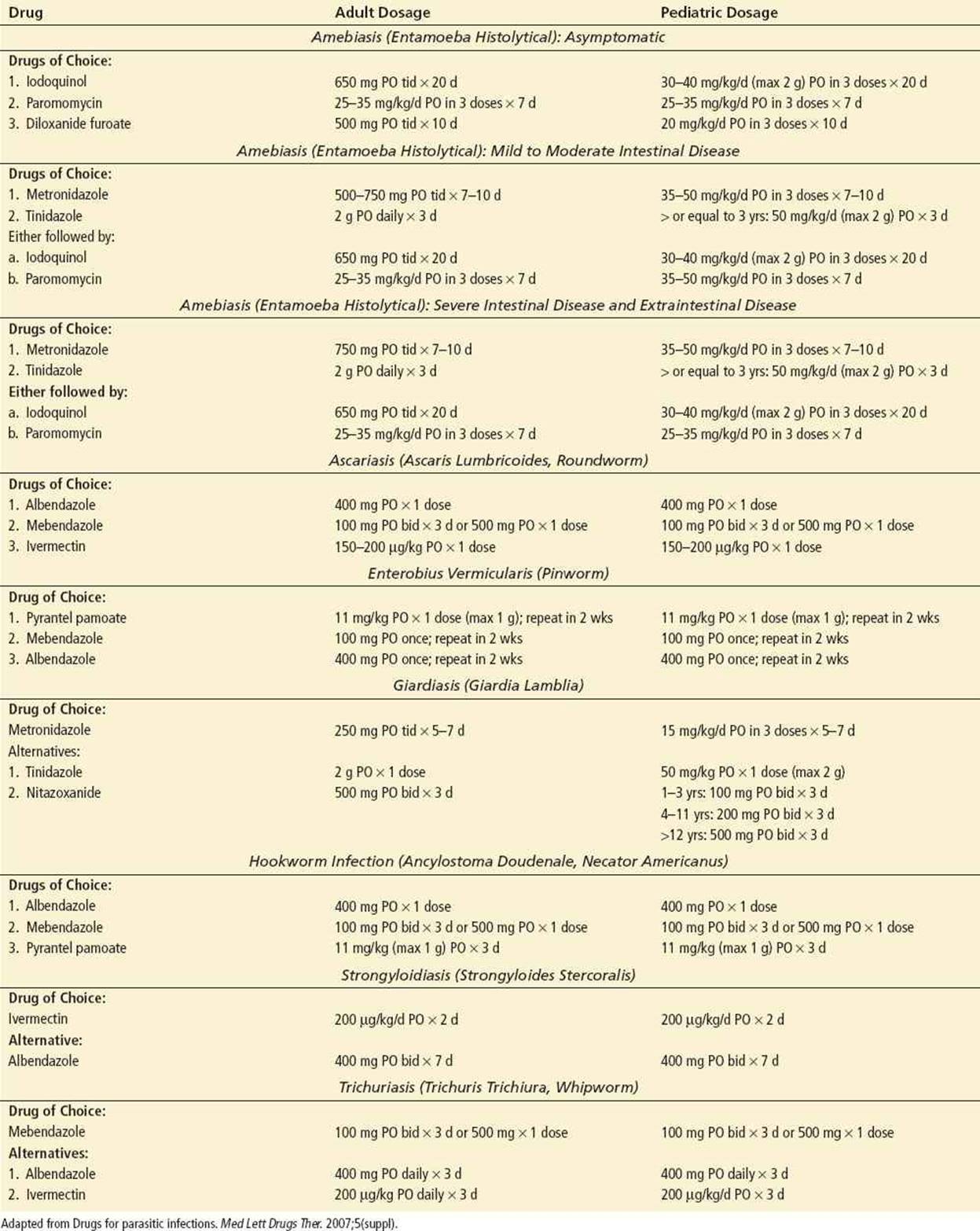
Pharmacologic therapy of GI parasitic disease is outlined in Table 193.3.
TABLE 193.3
Treatment of Gastrointestinal Parasitic Infections
DISPOSITION
Many GI parasitic illnesses can be treated with the appropriate antiparasitic agents on an outpatient basis. However, there may be specific indications for inpatient treatment, depending on the causative organism, extent of disease, concomitant medical conditions (immunocompromised patients, etc.), and the reliability of compliance with treatment and adequate follow-up.
Indications for inpatient treatment and potential complications:
• Giardia infection: abnormal electrolytes secondary to excessive diarrhea may require hospitalization.
• E. histolytica infection: fulminant rectocolitis, massive GI bleeding, perforation, peritonitis, toxic megacolon, and amebic liver abscess are potential complications.
• Cryptosporidium infection: severe diarrhea, especially in immunocompromised patients.
• A. lumbricoides infection: intestinal obstruction that requires surgery, especially in the pediatric population.
• S. stercoralis infection: hyperinfection syndrome may be fatal in the immunocompromised patient.
• Necator and Ancylostoma infections: severe hypochromic microcytic anemia in patients with heavy worm burdens.
• T. trichiura infection: severe anemia or rectal prolapse.
• E. vermicularis: rarely if ever requires inpatient care.
Cysticercosis
Parasitic disease may present with CNS pathology and requires the emergency physician to have a basic understanding of the life cycle of the organisms involved to diagnose and treat the infection.
Protozoal infestations such as malaria may present with neurologic manifestations. Falciparum malaria can cause sludging of parasitized red cells in brain capillaries, resulting in severe CNS dysfunction, which can progress to coma and death. Early recognition and treatment are essential to prevent this outcome.
Helminthic CNS disease is caused primarily by two tapeworms: Taenia solium (pork tapeworm) and Echinococcus granulosus. T. solium, an intestinal parasite in humans, can invade other tissues in its larval form, causing cysticercosis. Although 60% of reported cases of cysticercosis involve the brain, it is believed that muscular and subcutaneous encystment is more common (24). Humans may acquire the infestation by eating cured or undercooked pork meat that has been infected with the larvae of T. solium (24). In addition, infection may be acquired through the fecal–oral route by consuming contaminated food, such as raw vegetables, or water (25). Cysts formed in the brain (i.e., neurocysticercosis) can cause seizures or produce symptoms through mass effect (2). Worldwide and in the United States, cysticercosis is the most common parasitic disease of the CNS (25). Immigrants from endemic areas, such as Mexico and Central America, account for the vast majority of cases in the United States. Cysticercosis is most prevalent in communities where there is close contact between humans and pigs, and where hygiene is poor (25). E. granulosus also forms cysts in the CNS that can mimic neoplastic disease by causing compression effects or seizures.
CLINICAL PRESENTATION
Although T. solium is the most common parasitosis of the human CNS, it is symptomatic in only 50% of patients (24). Patients with cysticercosis of the brain may present with focal neurologic findings or impaired sensorium. The clinical features depend primarily on the location of the cyst. The cyst may be located in the meninges, brain parenchyma, or ventricles. The common presenting features include:
• seizures (most prevalent)
• adult-onset seizures in the Third World are most often caused by neurocysticercosis (26)
• focal neurologic deficits
• increased intracranial pressure
• obstructive hydrocephalus
• stroke
• status epilepticus and mechanical hydrocephalus (most frequent causes of death)
ED EVALUATION AND MANAGEMENT
The information obtained from laboratory examination is often limited. A mild peripheral leukocytosis and eosinophilia may be seen. Cerebrospinal fluid (CSF) findings suggestive of cysticercal arachnoiditis include an elevated protein level, eosinophilia, and a predominantly lymphocytic pleocytosis. Several immunologic tests of the serum and CSF have been developed to aid in the diagnosis of cysticercosis (25). Microscopic examination of stool preparations may reveal typical eggs, proglottids (gravid uterus segments), or scolices, which are the heads of the worm. Although the patient with neurocysticercosis may no longer have a tapeworm at the time the diagnosis is made, stools of both the patient and family members should be examined for several days (25). Serologic diagnosis is necessary to confirm E. granulosusinfection.
Laboratory personnel should be consulted to determine the most appropriate procedures for collecting and handling specimens. The laboratory should be informed of the patient’s clinical diagnosis to assist in sample analysis. Consultation with an infectious disease expert may be helpful if the clinician is unfamiliar with the potential manifestations of a particular disease.
Computed tomography (CT) scanning of the head may show both calcified and noncalcified cysts (20,25). Contrast-enhanced CT scanning may demonstrate ring-enhancing lesions in both cysticercosis and echinococcal cyst disease. Magnetic resonance imaging (MRI) has been shown to be more sensitive than CT scan for the detection of active neurocysticercosis, because of the superior ability of MRI to image cysts in the brainstem, subarachnoid space, and ventricles. CT, however, is a better modality for imaging inactive, calcified cysts (20).
Treatment of parasitic CNS disease may involve medical or surgical interventions and depends on the clinical signs and symptoms, the presence of active noncalcified or inactive calcified cysts, and the occurrence of complications. In addition to symptomatic treatment, options for the treatment of neurocysticercosis include anticysticercal drugs, corticosteroids, CSF shunting, and surgical cyst removal (25). Most parasitic infections in the active phase with viable cysts are readily treated with safe and effective agents. Patients can commonly be treated as outpatients, with appropriate follow-up.
If active neurocysticercosis is suspected, praziquantel (50 mg/kg/d in three divided doses for 15 days) or albendazole (400 mg two times a day for 8 to 30 days) should be initiated (10). Albendazole has shown to be superior to praziquantel in reducing the number of live cysts and in clinically relevant outcomes (27). Corticosteroids should be given for 2 to 3 days before and during drug therapy to suppress the inflammatory response induced by destruction and death of the live cysts (10). Albendazole (400 mg two times a day for 28 days) has been used to treat echinococcal cysts. Surgical removal of cysts remains the most effective treatment, especially when obstructive hydrocephalus is a complicating factor (14). Patients who present with inactive disease (e.g., seizures secondary to parasitic calcifications) are not candidates for treatment with antihelminthic agents. Seizures are treated no differently than seizures from other causes. Neurologic or neurosurgical consultation is appropriate for these patients.
DISPOSITION
Most patients with cysticercosis will either remain asymptomatic without treatment or will be able to receive antihelminthic treatment as an outpatient. However, the clinician should be aware of the need for inpatient treatment in patients who present with signs of severe neurocysticercosis infection; patients with status epilepticus, mechanical hydrocephalus, or stroke require inpatient care and definitive neurosurgery.
CRITICAL INTERVENTIONS
• Diagnose malaria using a thin Giemsa-stained peripheral blood smear to demonstrate intracellular forms of the parasite. Repeat negative blood films every 12 to 24 hours to obtain three negative sets if diagnosis is strongly suspected.
• Diagnose infestation with the pinworm E. vermicularis using clear cellulose tape placed against the perianal skin, lifted off, and then applied (adhesive side down) to a glass slide and viewed under a microscope to identify the characteristic eggs.
• Diagnose cysticercosis with CT and/or brain MRI in addition to serologic testing.
Common Pitfalls
• Failure to consider parasitic disease as a potential etiology for a variety of systemic, GI, CNS, and skin complaints. Parasitic disease should be considered in any patient presenting from an endemic area or with a travel history to an endemic area.
• Failure to consider parasitic infestation in travelers months after their return.
• Failure to consult reference textbooks and current therapeutic guidelines concerning the appropriate diagnosis and treatment for these diseases.
REFERENCES
1. Becker BM, Cahill JD, Gibler WB. Parasites. In: Marx JA, Hockberger RS, Walls RM, eds. Emergency Medicine—Concepts and Clinical Practice. St. Louis, MO: Mosby; 2002.
2. Garg RK. Neurocysticercosis. Postgrad Med J. 1998;74:321–326.
3. Li E, Stanley SL Jr. Protozoa: Amebiasis. Gastroenterol Clin North Am. 1996;25: 471–492.
4. Kain KC, Keystone JS. Malaria in travelers: Epidemiology, disease, and prevention. Infect Dis Clin North Am. 1998;120:267–284.
5. Schwartz E, Parise M, Kozarsky P, et al. Delayed onset of malaria—implications for chemoprophylaxsis in travelers. N Engl J Med. 2003;49:1510–1516.
6. Hasugian AR, Purba HL, Kenangalem E, et al. Dihydroartemisinin-piperaquine versus artesunate-amiodaquine: superior efficacy and posttreatment prophylaxis against multidrug-resistant Plasmodium falciparum and Plasmodium vivax malaria. Clin Infect Dis. 2007;44(8):1067–1074.
7. Farcas GA, Soeller R, Zhong K, et al. Real-time polymerase chain reaction assay for the rapid detection and characterization of chloroquine-resistant Plasmodium falciparum malaria in returned travelers. Clin Infect Dis.2006;42(5):622–627.
8. Yamashita P, Kelsey J, Henderson SO. Subcutaneous cysticercosis. J Emerg Med. 1997;16:583–586.
9. Griffith KS, Lewis LS, Mali S, et al. Treatment of malaria in the United States: A systematic review. JAMA. 2007;297(20):2264–2277.
10. Drugs for parasitic infections. Med Lett Drugs Ther. 2007;5(suppl).
11. Shandera WX, Bollam P, Hashmey RH, et al. Hepatic amebiasis among patients in a public teaching hospital. South Med J. 1998;91:829–837.
12. Villamazir E, Mizrahinn M. Ascaris lumbricoides infestation as a cause of intestinal obstruction in children: Experience with 87 cases. J Pediatr Surg. 1996;31:201–204.
13. Sincalir D, Zani B, Donegan S, et al. Arttemisinin-based combination therapy for treating uncomplicated malaria. Cochrane Database Syst Rev. 2009;3: CD007483.
14. Smithius F, Kyaw MK, Phe O, et al. Efficacy and effectiveness of dihydroartemisinin- piperaquine versus artesunate-mefloquine in falciparum malaria: An open-label randomized comparison. Lancet.2006;367(9528):2075–2085.
15. Lamont EB, Sayah A. An occult cause of persistent nausea and vomiting. J Emerg Med. 1996;15:633–635.
16. Kyriacou DN, Spira AM, Talan DA, et al. Emergency department presentation and misdiagnosis of imported falciparum malaria. Ann Emerg Med. 1996;27: 696–699.
17. Beaver PC, Jung RC, Cupp EW. Clinical Parasitology. Philadelphia, PA: Lea & Febiger; 1984:240–245.
18. Jotte RS, Scott J. Malaria: Review of features pertinent to the emergency physician. J Emerg Med. 1993;11:729–736.
19. Blessmann J, Buss H, Nu PA, et al. Real-time PCR for detection and differentiation of Entamoeba histolytica and Entamoeba dispar in fecal samples. J Clin Microbiol. 2002;40(12):4413–4417.
20. Kramer MH, Sorhaqe FE, Goldstein ST, et al. First reported outbreak in the United States of cryptosporidiosis associated with a recreational lake. Clin Infect Dis. 1998;26:27–33.
21. Ortega YR, Adam RD. Giardia: overview and update. Clin Infect Dis. 1998;25: 545–549.
22. Grencis RK, Cooper ES. Enterobius, trichuris, capillaria, and hookworm including Ancylostoma caninum.Gastroenterol Clin North Am. 1996;25:579–597.
23. Avolio L, Avoltini V, Ceffa F, et al. Perianal granuloma caused by Enterobius vermicularis: Report of a new observation and review of the literature. J Pediatr. 1998;132:1055–1056.
24. Rossignol JF, Kabil SM, El-Gohary Y, et al. Effect of nitazoxanide in diarrhea and enteritis caused by cryptosporidium species. Clin Gastroenterol Hepatol. 2006;4(3):320–324.
25. Garcia HH, Pretell EJ, Gilman RH, et al. A trial of antiparasitic treatment to reduce the rate of seizures due to cerebral cysticercosis. N Engl J Med. 2004; 350(3):249–258.
26. Ladhani S, Aibara RJ, Riordan FA, et al. Imported malaria in children: A review of clinical studies. Lancet Infect Dis. 2007;7(5):349–357.
27. Matthaiou DK, Panos G, Adamidi ES, et al. Albendazole versus praziquantel in the treatment of neurocysticercosis: A Meta-analysis of comparative trials. PLoS Negl Trop Dis. 2008:2(3):e194.