
Tarlan Hedayati and Michael F. Murphy
Airway evaluation and management to ensure continuing and adequate gas exchange remain the first priority of resuscitation, taking precedence over other interventions. Further, it is widely recognized that a conceptual framework focused on rapid airway evaluation, critical action analysis and performance, and facility with an array of airway management techniques minimizes the risk of failure and improves outcome (1).
The failure to maintain a patent airway and the failure to affect adequate gas exchange are the cornerstones of emergency airway management. Decisions regarding how and when the airway will be managed depend on the integration of numerous factors including the skills and knowledge of the physician, the equipment available, the condition of the patient, and the anatomy of the airway.
There are only four ways to provide gas exchange for the patient who is not breathing:
1. Bag–mask ventilation (BMV)
2. Extraglottic device (EGD)
3. Endotracheal intubation (ETI)
4. Surgical airway (usually cricothyrotomy in emergency medicine)
Emergency ETI performed by emergency physicians has been the most discussed, researched, and published over the years (2).
By the early 1970s, ETI had become a skill integral to the practice of emergency medicine. It was generally accomplished either nasally or orally employing varying degrees of brute force. By the mid to late 1990s, most emergency practitioners employed neuromuscular blockade in some form most of the time to facilitate oral ETI. It had become evident that neuromuscular blockade not only made the technical task of intubation easier and faster but that the complication rates were lower and the success rates higher. However, for many practitioners, the need to evaluate the airway for difficulty and the development of a systematic method of doing so lagged behind the clinical introduction of rapid sequence intubation (RSI). There was also the need to expand the rescue options beyond cricothyrotomy.
BMV, long considered to be the cornerstone of airway management (Chapter 2), is a critically important skill in emergency medicine. However, it has become apparent that BMV is a difficult skill to teach and maintain (3). In recent years, EGDs (e.g., laryngeal mask airways [LMA], King LT Airway), which are easily inserted and highly successful at achieving adequate gas exchange, have increasingly emerged as potential first-line gas exchange techniques.
The current challenges facing emergency airway managers include the following:
• Should EGDs supplant BMV as the first-line airway management technique?
• Which patients should not receive paralytics?
• Is cricoid pressure still recommended to prevent passive regurgitation and aspiration of gastric contents?
• How is the airway best rescued in the event intubation and/or ventilation is impossible or has failed?
THE DECISION TO INTUBATE
The following are the five generally accepted indications to secure an airway by ETI in emergency medicine practice:
1. Failure of the patient to maintain a patent airway
2. Failure of the patient to adequately protect the airway from aspiration
3. Failure of the patient to maintain adequate gas exchange (oxygen and carbon dioxide)
4. Need for the initiation of paralytic agents or therapeutic hyperventilation
5. Inability of the patient to adequately clear secretions
A sixth indication that is frequently invoked is intubation prior to transport, when it is reasonably anticipated that although an indication to intubate does not exist now, one may develop during the transport. This risk-to-benefit analysis recognizes the practicalities and challenges of intubating in a transport vehicle.
There are probably few other situations in emergency practice when judgment and knowledge of the anticipated clinical course of a disorder are as crucial to the emergency physician as the decision to intubate a patient (4).
EVALUATION OF THE PATIENT
Human beings protect their airway at all costs, triggering an extreme autonomic response when threatened. ETI stimulates a similar autonomic response. As such, potent medications are generally employed to attenuate the adverse responses to laryngoscopy and intubation such as hypertension, tachycardia, increased intracranial pressure (ICP), and bronchospasm. The emergency physician must attempt to balance the anticipated aggression of the response to intubation by selecting appropriate classes and dosages of medication to mitigate them. Underdosing may fail to mitigate an aggressive response; overdosing may produce hypotension or premature apnea.
The evaluation of the patient to be intubated is focused on the three crucial organ systems: The central nervous system (CNS), the cardiovascular system, and the respiratory system. The patient’s hemodynamic stability, physiologic reserve of vital organ systems, and the anticipated response to laryngoscopy and intubation dictate which medications and what doses are required to maintain stability and mitigate adverse effects during the procedure.
EVALUATION OF THE AIRWAY
Airways that are difficult to manage are fairly common in emergency medicine with some estimates being as great as 20% of all emergency intubations. However, the incidence of intubation failure is quite uncommon (0.5% to 2.5%). Moreover, the disastrous situation of being able to neither intubate nor ventilate rarely occurs (0.1% to 0.5%) (2).
The most important way to avoid airway management failure is to predict when difficulty is likely to be encountered with BMV, ETI, EGD, or cricothyrotomy, particularly if one is relying on the latter two techniques to rescue the airway in the event of ETI or BMV failure.
The distinction among the terms difficult laryngoscopy, difficult ETI, difficult BMV, difficult EGD, difficult cricothyrotomy, and difficult airway is important. For instance, difficulty with BMV or an EGD when ETI is easily performed would not be described as a difficult airway. Rather, the narrative would describe each independently.
The failed airway is clearly defined as three failed attempts at intubation and/or failure to maintain oxygen saturations. The difficult airway is not so easily defined. Rather than a strict definition, the concept of the difficult airway has the following five “dimensions.”
1. Difficult BMV
2. Difficult laryngoscopy
3. Difficult intubation
4. Difficult EGD
5. Difficult cricothyrotomy
These five dimensions can be reduced to the following four technical operations:
1. Difficult BMV
2. Difficult EGD
3. Difficult laryngoscopy and intubation
4. Difficult cricothyrotomy
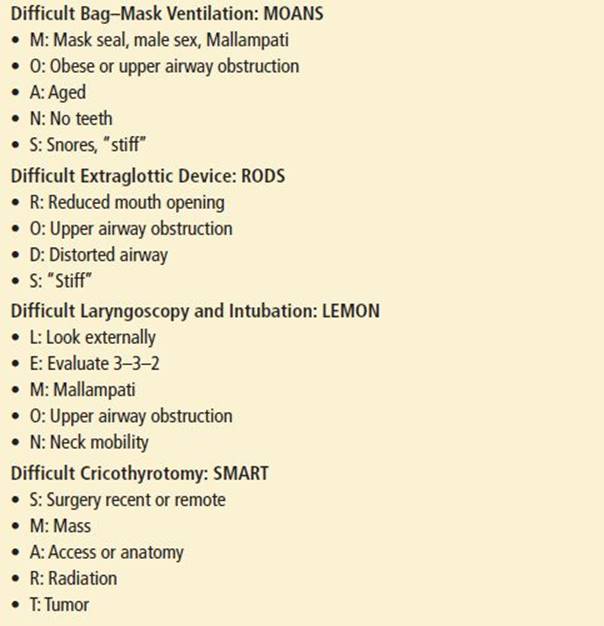
The evaluation of the airway for difficulty in an emergency must be done quickly, with care taken not to omit anything important. Mnemonics are efficient memory aid strategies, so one for each technical operation has been crafted to permit a rapid and complete evaluation (Table 1.1).
TABLE 1.1
Mnemonics Useful in Evaluating the Airway for Difficulty
Difficult Bag–Mask Ventilation
If the emergency physician is uncertain that neuromuscular blockade-facilitated orotracheal intubation (RSI) will be successful, he or she must be confident that BMV or EGD-facilitated ventilation is possible or at the very least that a cricothyrotomy can be performed rapidly.
The five indicators of difficult BMV (5,6) can be easily recalled for rapid use in the emergency setting by using the mnemonic MOANS (Table 1.1) (7).
• Mask seal, Male sex, Mallampati. Bushy beards, crusted blood on the face, or a disruption of lower facial continuity are the most common examples of conditions that may make an adequate mask seal difficult. Male sex and Mallampati scores of 3 or 4 have also been associated with difficult mask ventilation.
• Obese or obstructed. Patients who are obese (body mass index [BMI] >26 to 28 kg/m2) are often difficult to ventilate adequately by bag and mask. Parturients at term; patients with angioedema, Ludwig angina, upper airway abscesses (e.g., peritonsillar), or epiglottitis; and others ought to be considered at this juncture.
• Advanced age. Patient age older than 55 to 57 years is associated with a higher risk of difficult BMV, perhaps because of a loss of muscle and tissue tone in the upper airway with age.
• No teeth. An adequate mask seal may be difficult in the edentulous patient, perioral tissues tend to cave into the structurally unsupported mouth.
• Stiff or snores. Patients with high airway resistance or diminished pulmonary compliance are exceedingly difficult to adequately manage with a bag and mask, as are patients who snore.
Difficult Extraglottic Devices
Four factors may indicate that EGDs may be unsuccessful at rescuing the airway and may be remembered using the RODS mnemonic (Table 1.1) (8):
• Restricted mouth opening. Depending on the EGD to be employed, a certain amount of oral access is critical to employ the device.
• Obstruction. Upper airway obstruction at the level of the larynx or lower will not be bypassed through the use of an EGD.
• Disrupted or distorted anatomy. The “seat and seal” of the EGD may be compromised by a disrupted or distorted airway. In addition, a seal may be exceedingly difficult or impossible to achieve in the face of a fixed flexion deformity of the neck (9).
• Stiff lungs or cervical spine. Ventilation with an EGD may be difficult or impossible in the face of substantial increases in airway resistance (e.g., severe asthma) or decreases in pulmonary compliance (e.g., pulmonary edema). In addition, patients unable to fully extend the neck may prove to be difficult to ventilate with EGDs due to improper positioning of the EGD.
Difficult Laryngoscopy and Intubation
Difficult laryngoscopy and intubation ordinarily implies that the operator had a poor view of the glottis. Cormack and Lehane (10) provided some clarity to the way physicians think of the “difficult airway” by parsing the act of intubation into its two subcomponents: Laryngoscopy and intubation. They also introduced the most widely used system of categorizing the degree of visualization of the larynx during laryngoscopy (Fig. 1.1) (7). Cormack–Lehane view grades 3 (epiglottis only visible) and 4 (no glottic structures at all visible) are often used as surrogates to represent difficult laryngoscopy and to predict difficult intubation. View grades 1 (visualization of the entire laryngeal aperture) and 2 (visualization of the posterior cords and arytenoids) are not typically associated with difficult intubation, although some grade 2’s may be very anterior and difficult or impossible to intubate.

FIGURE 1.1 Cormack and Lehane system of categorizing the degree of visualization of the larynx during laryngoscopy.
The mnemonic LEMON (Table 1.1) is a useful guide to identifying as many of the risks as quickly as possible to meet the demands of an emergency situation.
• Look externally. If the airway looks difficult, it probably is! A litany of physical features is associated with difficult laryngoscopy and intubation, making the glottic aperture difficult to visualize. A small mandible may indicate that the tongue is “retrofitted” over the larynx; a large mandible elongates the pharyngeal axis, serving to extend the distance to the larynx beyond the visible horizon. Buck teeth block access to the oral cavity and also elongate the length of the oral axis. A high, arched palate is often associated with a long narrow oral cavity, making access a problem. A short neck may mean the larynx is positioned higher in the neck relative to the base of the tongue, making it more difficult to bring the glottis into view. Lower facial disruption interferes with adequate mask seal and may make the glottis impossible to find.
• Evaluate 3–3–2. This step recognizes the importance of the geometric relationships of various parts of the airway to successful intubation (11). One ought to be able to open one’s mouth with the width of three of their own fingers, accommodate three of their own fingers between the tip of the mentum and the hyoid bone, and fit two fingers between the hyoid bone and the thyroid notch. The first “three” assesses the adequacy of oral access. The second “three” addresses the capacity of the mandibular space to accommodate the tongue on laryngoscopy. More than or less than three fingers are both associated with greater degrees of difficulty in visualizing the larynx at laryngoscopy. The former because the length of the oral axis is elongated; the latter because the mandibular space may be too small to accommodate the tongue, leaving it to obscure the view of the glottis. The final “two” identifies the location of the larynx in relation to the base of the tongue. If more than two fingers are accommodated, the larynx is further below the base of the tongue beyond the visible horizon. Fewer than two fingers may mean that the larynx is tucked up under the base of the tongue and may be difficult to expose. This condition is often called anterior larynx.
• Mallampati score. Mallampati determined that the degree to which the posterior oropharyngeal structures are visible is loosely associated with intubation success (12). He had patients sit on the side of the bed, open their mouths as widely as possible, and protrude their tongues as far as possible, without phonating. Figure 1.2 depicts how the scale is constructed. Although classes I and II patients are associated with low intubation failure rates, the importance with respect to the wisdom of using neuromuscular blockade rests with those patients in classes III and IV, particularly those in class IV where intubation failure rates may exceed 10%. By itself, the scale is neither sensitive nor specific; however, it is easily performed in an emergency and may reveal important information about access to the oral cavity and the potential for difficult glottic visualization.

FIGURE 1.2 Mallampati score.
• Obstruction. Upper airway obstruction is always a difficult airway. The four cardinal signs of upper airway obstruction are muffled voice (“hot potato voice”), difficulty swallowing secretions (either because of pain or obstruction), dyspnea, and stridor. The first two signs do not ordinarily herald imminent total upper airway obstruction; dyspnea and stridor are much more sinister. The presence of stridor generally indicates that the airway has been reduced to 4.5 mm or less (13). Upper airway obstruction should always be managed with extreme care. The administration of small doses of opioids and benzodiazepines to manage anxiety may induce total obstruction due to diminished upper airway musculature tone.
• Neck mobility. The ability to position the head and neck is one of the factors employed to achieve an optimal view of the larynx. Although cervical spine immobilization, of and by itself, does not automatically lead to one, it should be considered in addition to other features indicating difficulty.
Difficult Cricothyrotomy
There are no absolute contraindications to performing an emergency cricothyrotomy. However, some conditions may make it difficult or impossible to perform the procedure. It is imperative to identify those conditions up front, particularly if one is relying on a rapidly performed cricothyrotomy as a rescue technique. The mnemonic SMART (Table 1.1) is used to quickly assess the patient for features that may indicate a difficult cricothyrotomy.
• Surgery—Recent or remote: The anatomy may be distorted making the airway difficult to find or access.
• Mass: A hematoma or infective process in the direct path of the cricothyrotomy produces a mass effect that may make the procedure technically difficult, but should never be considered a contraindication in a life-threatening situation.
• Access or anatomy: Obesity should be considered a surrogate for any problem that makes percutaneous access to the anterior neck problematic. A fixed flexion deformity of the cervical spine and halo traction make cricothyrotomy more difficult.
• Radiation: Past radiation therapy may distort tissues, making the procedure difficult.
• Tumor: Tumor in or around the airway may present difficulty from an access perspective as well as from bleeding.
No single indicator, combination of indicators, or weighted scoring system of indicators can be relied on to assure success or predict failure when it comes to oral intubation. Thus, emergency physicians must evaluate the airway for difficulty, identify those factors known to increase the degree of difficulty, match them to the skill, experience, and judgment of the individual performing the intubation, and make the following decision: Does this airway meet the threshold of being sufficiently difficult to warrant using the difficult airway algorithm or is it safe to proceed directly to RSI?
RAPID SEQUENCE INTUBATION
“RSI is the administration, after preoxygenation, of a potent induction agent followed immediately by a rapidly acting neuromuscular blocking agent (NMBA) to induce unconsciousness and motor paralysis for tracheal intubation” (14). Attenuating the adverse responses to intubation, maintaining adequate oxygen saturations, and protecting the airway from aspiration are integral to the procedure.
The adage in anesthesia with respect to neuromuscular blockade to facilitate the orotracheal intubation of a patient who has some effective spontaneous ventilation has always been “don’t take anything away from the patient that you cannot replace.” Although such a rigid principle is not always consistent with the realities of emergency airway management, it is useful to remember.
The use of NMBAs assumes that the operator is entirely versed in BMV, the use of extraglottic rescue devices, airway evaluation for difficulty, and the performance of a surgical airway.
Walls and Murphy (14) have referred to the steps of RSI as the “Seven P’s of RSI.” It is a useful framework for discussing the procedure.
• Preparation. Establish IV access, place the patient on a cardiac monitor and pulse oximetry, and prepare intubation equipment.
• Preoxygenation. This is a crucial step, permitting oxygen saturations to remain acceptable during the period of apnea as the NMBA becomes fully effective. The goal is to replace the nitrogen in the patient’s functional residual capacity (FRC, normally 30 mL/kg) with oxygen. The ability to do so varies with the time available, the ability of the oxygen delivery apparatus to deliver the high concentrations of oxygen used, and the patient’s condition.
• Pretreatment. The goal of pretreatment is to attenuate the adverse responses to intubation, particularly the surges in heart rate, blood pressure, ICP, and airway resistance. Ideally, the pretreatment medications ought to precede the induction agent by 3 minutes to be optimally effective. Not all patients are equally at risk from these responses. Based on the best available evidence and expert recommendations at least the following patients should be pretreated (15).
• Those suspected of having elevated ICP and imperfect autoregulation may benefit from lidocaine 1.5 mg/kg and fentanyl 3 to 13 μ/kg (ordinarily 3 to 5 μ/kg). In addition, a defasciculating dose of a nondepolarizing paralytic decreases fasciculations and reduces ICP. It has been taught that typically 1/10 of the intubating dose of any available paralytic agent will achieve this.
• Those with significant ischemic heart disease or major vessel dissection or rupture may benefit from fentanyl 3 to 13 &b.mu;/kg (ordinarily 3 to 5 &b.mu;/kg). Caution ought to be exercised in administering fentanyl to patients with compensated or uncompensated shock.
• Adults with significant existing reactive airway disease may benefit from lidocaine 1.5 mg/kg.
• Children up to the age of 10 years, or any patient with hemodynamically significant bradycardia and receiving succinylcholine may receive atropine 0.01 to 0.02 mg/kg (minimum 0.1 mg; maximum 0.5 mg). This recommendation is currently considered to be controversial and the administration of atropine is optional.
• Paralysis with induction. Induction and paralytic agents are rapidly administered intravenously. Induction agents have profound effects on all three vital organ systems: The CNS, cardiovascular system, and ventilation. These effects depend on the particular drug, the patient’s underlying physiologic condition, and the dose and speed of injection of the drug. Because RSI requires rapid administration of the sedative induction agent, the choice of drug and the dose must be tailored to capitalize on desired effects, while minimizing adverse side effects (Table 1.2). In some cases, an amnestic dose may be selected, rather than an induction dose. While the sedative induction dose might be tailored, the dose of the NMBA is not (e.g., succinylcholine 1 to 2 mg/kg; rocuronium 1 to 1.5 mg/kg).
• Protection and positioning. Though controversial, the use of cricoid pressure, or the Sellick maneuver, to prevent regurgitation and aspiration is maintained until proper placement of the endotracheal tube (ETT) is confirmed. Premature release of cricoid pressure is a common error and may place the patient at risk of aspiration, particularly if an inadvertent esophageal intubation has occurred. Cricoid pressure must be released immediately should active vomiting occur; otherwise, there is danger of esophageal rupture. Most experts on airway management agree that positioning the head and neck is an important step in gaining the best view of the larynx with conventional laryngoscopy. It has long been taught that the “sniffing the morning air” or “sipping English tea” positioning of the head and neck, when possible, is best. Although there is some dissension as to whether this positioning does indeed deliver the best view of the larynx, most agree that it is a reasonable place to start (16).
• Placement and proof. The tube is passed through the laryngeal cords. Proper ETT placement is confirmed and proven using physical examination as well as devices such as the end-tidal CO2 detector.
• Postintubation management (PIM). The tube should be secured using commercially available devices or basic tape to prevent accidental extubation. If tape is used, then a “bite block” or oral airway should also be placed to avoid accidental cutting of the tube should the patient bite down. A postintubation chest radiograph is the standard practice to confirm placement of the tube and assess the depth of intubation. Finally, postintubation sedation with or without paralytics should be provided.
TABLE 1.2
Induction Agents and Dosage Selection

APPROACH TO THE AIRWAY: HOW TO PROCEED
Once one has decided that intubation is indicated, one progresses rapidly to identifying exactly what kind of airway the patient has and how the practitioner will proceed:
• Is this a “crash” airway where the patient is unconscious, unresponsive, and near death (17)?
• Is this a difficult airway where one anticipates difficulty with BMV, laryngoscopy and intubation, EGD rescue, or cricothyrotomy?
• If neither of these exists, RSI is reasonable.
• Has a failed airway supervened?
This systematic approach to airway management in the emergency department encompasses all possible presentations. Identifying the type of airway to be managed permits the practitioner to select an appropriate course of action. Algorithms are presented below for each of these clinical presentations. Figure 1.3 depicts this conceptual framework (17).

FIGURE 1.3 Universal emergency airway algorithm.
Crash Airway Algorithm
The patient who presents in an unresponsive state or is deemed unlikely to respond in any way to direct laryngoscopy is said to have a crash airway, and the crash airway algorithm (Fig. 1.4) is employed (17). Noteworthy points in the crash airway algorithm include the following.
• BMV is occurring as preparations are made to intubate.
• The initial action is to attempt oral intubation immediately via direct laryngoscopy without pharmacologic assist.
• If the first attempt at orotracheal intubation is unsuccessful, it is assumed that the patient is not optimally relaxed, and succinylcholine is administered in an increased dose.
• If BMV is unsuccessful at any time and if intubation has failed, a failed airway is present, and one proceeds to the failed airway algorithm (Fig. 1.5).
• Once three attempts have failed, one proceeds to the failed airway algorithm (Fig. 1.5).

FIGURE 1.4 The crash airway algorithm.

FIGURE 1.5 The failed airway algorithm.
Difficult Airway Algorithm
Should the situation not be a crash airway, one must determine whether it is a difficult airway. Although all emergency intubations are difficult to some degree, the evaluation of the airway for those features that reliably predict difficult BMV, difficult laryngoscopy, difficult EGD rescue, or difficult cricothyrotomy is extremely important. In the event the airway is judged to be difficult, the difficult airway algorithm (Fig. 1.6) is used (17). The evaluation for difficulty is presented in the next section.
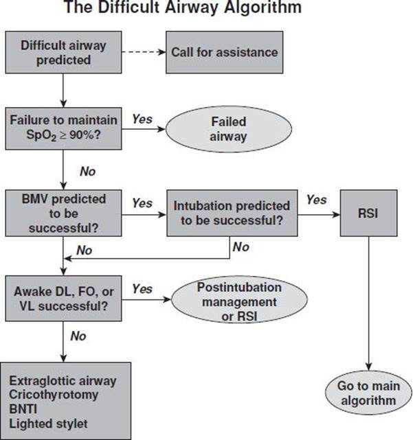
FIGURE 1.6 The difficult airway algorithm.
Noteworthy points in the difficult airway algorithm include the following:
• This iteration of the difficult airway algorithm recognizes that the emergency physician may be forced to act before a detailed evaluation of the airway can be performed. RSI is recommended, though this action must be accompanied by preparations to perform a surgical airway (“double set up”) and to ensure the immediate availability of an EGD in the event of failure (18).
• Determine how much time is available. If ventilation and oxygenation are adequate, then a careful assessment and a planned approach can be undertaken. If not, does BMV provide adequate ventilation and oxygenation? If not, the situation is equivalent to a “can’t intubate; can’t oxygenate” failed airway, and one proceeds to the failed airway algorithm (Fig. 1.5) (17).
• If the patient can be adequately oxygenated, the next step is to consider RSI. If BMV or gas exchange rescue with EGD are deemed unlikely to succeed or if success at laryngoscopy and intubation is felt to be questionable, RSI is not recommended.
• Awake laryngoscopy is the cornerstone of difficult airway management. An “awake look” is intended to either intubate the nonparalyzed patient orally or identify whether the patient can be safely paralyzed and intubated using an RSI technique. An awake look in an emergency airway situation relies virtually entirely on the intravenous titration of systemically active sedation and topical anesthesia.
• If at any time oxygenation and ventilation become inadequate, the situation has become a failed airway. If oxygenation remains adequate, several options remain, all of which place a cuffed (in adults) ETT in the trachea.
In the absence of an identified crash or difficult airway, RSI is the method of choice for airway management in the emergency department.
Failed Airway Algorithm
The failed airway is easily defined as follows:
1. Three failed attempts at orotracheal intubation by a skilled intubator, or
2. Failure to maintain acceptable oxygen saturations.
The practical problem in day-to-day practice is recognizing failure when it occurs and changing gears rapidly to deal with it appropriately. Clinically, the failed airway presents itself in two ways.
1. Time is available: “Can’t intubate; can oxygenate.”
2. Immediate action is required: “Can’t intubate; can’t oxygenate.”
Noteworthy points in the failed airway algorithm (Fig. 1.5) include the following (17):
• Unlike the difficult airway, when the placement of a cuffed tube in the trachea is the goal, the failed airway calls for immediate action to provide emergency oxygenation by whatever appropriate maneuvers are available, regardless of whether this action results in a secure, protected airway. The devices considered for the failed airway are somewhat different from, but inclusive of, the devices used for the difficult airway.
• When a failed airway has been determined to be present, the response is dictated by whether BMV is possible and adequate.
• A “can’t intubate; can’t oxygenate” situation mandates immediate cricothyrotomy. The airway manager may elect to attempt to rapidly place an alternative device, usually an EGD (17,19), simultaneously with the preparation for a cricothyrotomy, in the hope that a “can’t intubate; can’t oxygenate” situation can be converted into a “can’t intubate; can oxygenate” situation.
• If at any time during the management of the failed airway adequate oxygenation and ventilation cannot be assured, immediate performance of a cricothyrotomy is mandated.
RESCUE DEVICES
Airway rescue devices can be divided into the following two groups, depending on the situation.
• “Can’t intubate; can’t oxygenate.” Cricothyrotomy is the standard method of airway management in this situation. However, devices that can be inserted rapidly and are associated with high success rates include the Combitube and the intubating laryngeal mask airway (Fastrack). The advantage of the Fastrack compared to the Combitube is the ability to intubate through the Fastrack.
• “Can’t intubate; can oxygenate.” Videolaryngoscopes, flexible and rigid endoscopic devices, and nasotracheal intubation require time to perform and are inappropriate in an emergency situation where ventilation and oxygenation must not be delayed.
SELLICK MANEUVER
Brian Sellick introduced cricoid pressure in 1961 as a protective measure, reasoning that backward pressure on the cricoid cartilage would occlude the upper esophagus and further reduce the incidence of aspiration. With widespread use of the technique in all arenas of airway management by the late 1980s, it assumed the status of “Standard of Care” in emergency patients and others at risk of aspiration.
However, scientific evidence that the maneuver actually reduces the incidence of aspiration remains lacking due to the rarity of clinically evident or significant aspiration, and it will likely remain that way. In the meantime, investigators have shown that the esophagus is not entirely obstructed by the Sellick maneuver. In addition, the maneuver is often not performed correctly, it attenuates lower esophageal sphincter tone, and it hinders placement of and ventilation through an LMA. It has also been found to produce partial or total airway obstruction that may hinder mask ventilation and ETI. Furthermore, cricoid pressure may lend a false sense of security if done incorrectly and some anesthetists have seen regurgitation in spite of its application. Three reviews on rapid sequence induction and cricoid pressure have pointed out that there have been no randomized controlled trials comparing the incidence of regurgitation on induction with and without cricoid pressure in patients at high risk of regurgitation (20).
On the positive side, there is evidence that the position of the esophagus during cricoid pressure is irrelevant, as the maneuver leads to compression of the upper esophagus and hypopharynx, providing some degree of protection from the passage of alimentary tract contents into the hypopharynx (21).
CONFIRMATION TECHNIQUES
Endoscopic bronchoscopy remains the gold standard of verifying correct ETT placement by permitting direct visualization of tracheal rings. Visualization of the ETT entering the larynx also provides a highly reliable method of verifying intratracheal placement. Detection of expired carbon dioxide provides reliable evidence of tracheal rather than esophageal intubation and is the standard of care in emergency medicine. In nonarrested patients, carbon dioxide detection is highly reliable, indicating correct placement 99% to 100% of the time (22). Soft drinks in the stomach that contain carbon dioxide may mimic the exhaled carbon dioxide from the lungs for a couple of breaths, the so-called “cola complication,” although this confounding result ought not to persist beyond six breaths.
The migration of an ETT from the trachea to the esophagus during the tosses and turns of transport is an ever present hazard. It has been demonstrated that the continuous monitoring of exhaled carbon dioxide during the prehospital phase of care minimizes the risk of such displacement going unrecognized (23).
As might be expected, carbon dioxide detection techniques tend to be less accurate in identifying correct placement of the ETT in patients with circulatory arrest, with reported false-negative rates (carbon dioxide not detected, tube in the trachea) as high as 30% to 35% (24). In this circumstance, the endotracheal placement of the tube can be evaluated by an esophageal detector device (EDD) that consists of a self-inflating suction bulb or syringe armed with an adapter to fit a standard ETT connector. The collapsed bulb or syringe rapidly fills with air if the ETT is in the trachea; it does not inflate if the tube is in the esophagus, with a specificity of approximately 99%. Use of this device instead of carbon dioxide detection is not recommended because of its failure to detect esophageal intubation in as many as 20% of cases (24).
Physical examination techniques used to confirm intratracheal placement of an ETT, although neither sensitive nor specific, remain important adjuncts to more elaborate techniques, particularly in the cardiopulmonary arrested patient. Auscultation of the chest for breath sounds and of the epigastrium for absence of air entry into the stomach, observation of chest motion during ventilation, and condensation inside the ETT during exhalation are common, but notoriously unreliable, methods of ascertaining proper ETT placement.
In summary, although carbon dioxide detection remains the most reliable method of verifying tracheal placement of the ETT in prehospital care, the incorporation of multiple methods of confirmation is superior to any single method.
SUMMARY
RSI by emergency practitioners is associated with high success rates and low complication rates. The challenge is to recognize those patients who should not be paralyzed and to develop alternative skills to successfully manage those airways. There is no “recipe” of RSI pretreatment and induction medications that can be applied in every case. A careful assessment of risk and benefit is required in each patient to select the appropriate medication and its dose. An array of airway management skills is required to appropriately manage difficult airways and rescue failed airways.
CRITICAL INTERVENTIONS
• Evaluate the airway prior to airway intervention
• Preoxygenate patients prior to RSI
• Perform cricothyrotomy or insert ILMA in patients who cannot be ventilated or intubated
• Know when not to paralyze a patient
• Recognize airway management failure immediately
REFERENCES
1. Benumof JL. The ASA difficult airway algorithm: New thoughts and considerations. In: Fifty-first Annual Refresher Course Lectures and Clinical Update Program, No. 235. Philadelphia, PA: American Society of Anesthesiologists; 2000.
2. Walls RM, Brown CA 3rd, Bair AE, et al. Emergency airway management: A multi-center report of 8937 emergency department intubations. J Emerg Med. 2011;41:347–354.
3. Komatsu R, Kasuya Y, Yogo H, et al. Learning curves for bag and mask ventilation and orotracheal intubation: An application of the cumulative sum method. Anesthesiology. 2010;112:1525–1531.
4. Walls RM. The decision to intubate. In: Walls RM, Murphy MF, et al., eds. Manual of Emergency Airway Management. 4th ed. Philadelphia, PA: Lippincott Williams & Wilkins; 2012:22–34.
5. Langeron O, Masso E, Huraux C, et al. Prediction of difficult mask ventilation. Anesthesiology. 2000;92:1229–1236.
6. Kheterpal S, Martin L, Shanks AM, et al. Prediction and outcomes of impossible mask ventilation: 50,000 anesthetics. Anesthesiology. 2009;119:891–897.
7. Walls RM, Murphy MF. Identification of the difficult and failed airway. In: Walls RM, Murphy MF, et al., eds. Manual of Emergency Airway Management. 4th ed. Philadelphia, PA: Lippincott Williams & Wilkins; 2012:8–21.
8. Ishimura H, Minami K, Sata T, et al. Impossible insertion of the laryngeal mask airway and oropharyngeal axes. Anesthesiology. 1995;83:867–869.
9. Buckham M, Brooker M, Brimacombe J, et al. A comparison of the reinforced and standard laryngeal mask airway: Ease of insertion and the influence of head and neck position on oropharyngeal leak pressure and intracuff pressure. Anaesth Intensive Care. 1999;27:628–631.
10. Cormack RS, Lehane J. Difficult tracheal intubation in obstetrics. Anaesthesia. 1984;39:1105–1111.
11. Murphy MF, Doyle J. Identification of the difficult airway. In: Hung OR, Murphy MF. Difficult and Failed Airway Management. New York, NY: McGraw Hill; 2011:3–14.
12. Mallampati SR, Gatt SP, Gugino LD, et al. A clinical sign to predict difficult intubation: A prospective study. Can Anaesth Soc J. 1985;32:429–434.
13. Donlon JV. Anesthetic and airway management of laryngoscopy and bronchoscopy. In: Benumof JL, ed. Airway Management: Principles and Practice. St. Louis, MO: Mosby; 1996:666–685.
14. Walls RM, Murphy MF. Rapid sequence intubation. In: Walls RM, Murphy MF, et al., eds. Manual of Emergency Airway Management. 4th ed. Philadelphia, PA: Lippincott Williams & Wilkins; 2012:220–232.
15. Caro DA, Bush S. Pretreatment agents. In: Walls RM, Murphy MF, et al., eds. Manual of Emergency Airway Management. 4th ed. Philadelphia, PA: Lippincott Williams & Wilkins; 2012:233–240.
16. Adnet F, Baillard C, Borron SW, et al. Randomized study comparing the “sniffing position” with simple head extension for laryngoscopic view in elective surgery patients. Anesthesiology. 2001;95:836–841.
17. Walls RM. The emergency airway algorithms. In: Walls RM, Murphy MF, et al., eds. Manual of Emergency Airway Management. 4th ed. Philadelphia, PA: Lippincott Williams & Wilkins; 2012:1–7.
18. Cook TM, Woodall TM, Harper J, et al. Major complications of airway management in the UK: Results of the fourth national audit project of the royal college of anesthetists and the difficult airway society: Parts 1 and 2. Br J Anesth. 2011; 106:616–642.
19. Apfelbaum J, Hagberg C, Caplan RA, et al. Practice guidelines for the management of the difficult airway: Updated report by the ASA task force. Anesthesiology. 2013;118:251–270.
20. Law JA, Broemling N, Cooper RM, et al. The difficult airway with recommendations for management–part 1–difficult tracheal intubation encountered in an unconscious/induced patient. Can J Anesth.2013;60(11):1089–1118.
21. Ovassapian A, Salem MR. Sellick’s maneuver: To do or not do. Anesth Analg. 2009;109:1360–1362.
22. Grmec S. Comparison of three different methods to confirm tracheal tube placement in emergency intubation. Intensive Care Med. 2002;28:701–704.
23. Silvestri S, Ralls GA, Krauss B, et al. The effectiveness of out-of-hospital use of continuous end-tidal carbon dioxide monitoring on the rate of unrecognized misplaced intubation within a regional emergency medical services system. Ann Emerg Med. 2005;45:497–503.
24. Li J. Capnography alone is imperfect for endotracheal tube placement confirmation during emergency intubation. J Emerg Med. 2001;20:223–229.