
Judd E. Hollander and Adam J. Singer
Nearly 9 million patients with traumatic lacerations are treated annually in emergency departments (EDs) in the United States (1). The ultimate goals are to restore the physical integrity and function of the injured tissue in a cosmetically pleasing manner and to reduce the risk of infection. Treatment of these wounds involves a series of decisions that determine the methods of evaluation, wound preparation, wound closure, and postoperative care most likely to help attain these goals.
CLINICAL PRESENTATION
Lacerations occur predominantly in young adults; the majority of patients with lacerations are males (2). Most wounds are located on either the head or neck (50%) or the upper extremity (35%), usually involving the fingers or hands. The most common mechanism of injury is application of a blunt force, such as bumping the head against a coffee table. Such contact crushes the skin against an underlying bone, causing the skin to split. Other causes of injury include sharp instruments, glass, and wooden objects (2). While mammalian bites continue to receive much attention, they are a relatively infrequent cause of puncture wounds and lacerations (2).
ED EVALUATION
Evaluation of the patient with a traumatic wound begins with an expeditious, comprehensive assessment of the patient. This assessment can be divided into primary and secondary surveys, following advanced trauma life support algorithms. Unless the wound compromises the ABCs, formal wound evaluation and management occur during the secondary survey and management phase of the trauma evaluation.
Proper wound management begins with a thorough patient history. It is important to determine hand dominance and occupation. Host factors such as the extremes of age, diabetes mellitus, chronic renal failure, obesity, malnutrition, and the use of immunosuppressive medications all increase the risk of wound infection and can impair wound healing. Wound healing can also be impaired in the presence of inherited and acquired connective tissue disorders. The tendency of patients to form keloids should be ascertained because the development of keloids can result in a scar with less than acceptable cosmesis. Black and Asian populations are more prone to keloid formation (3).
Identification of the mechanism of injury is essential to help ascertain the presence of potential wound contaminants and foreign bodies that might result in chronic infection and delayed healing (3). Failure to diagnose foreign bodies in wounds is a leading cause of litigation against emergency physicians. Missed tendon and nerve injuries and failure to prevent infection are other common wound-related causes of litigation. In patients with scalp lacerations, a history of loss of consciousness and any neurologic complaints should be obtained. In patients with hand injuries, a history of neurologic deficits should be obtained to help predict the presence of tendon or nerve injury.
A careful history can also predict the likelihood of foreign bodies. Most organic and inorganic components of soil potentiate infection. However, sand grains are relatively innocuous. The black dirt on the surface of highways appears to have minimal chemical reactivity.
Crush injuries that tend to cause greater devitalization of tissue are more susceptible to infection than are wounds resulting from the more commonly seen shearing forces. Impact injuries with low energy levels may not result in division of the skin; they can, however, disrupt vessels and produce a hematoma and ecchymosis.
The presence of allergies to local anesthetics, latex, and antibiotics should be determined. The tetanus status of all patients should be assessed, and patients should receive immunization in accordance with current Centers for Disease Control and Prevention recommendations (Table 21.1). Before inspecting the wound, the emergency physician should question the patient regarding the time and mechanism of injury. The amount of time elapsed since the accident may influence treatment decisions.
TABLE 21.1
Recommendations for Tetanus Prophylaxis

Adequate wound examination should always be conducted in a field where bleeding has been controlled. One way to minimize the possibility of missing an injury to a vital structure is to start the wound examination with a careful neurovascular assessment of pulses, motor function, and sensation distal to the laceration. Finger tourniquets or a blood pressure cuff may then be used to obtain a bloodless field, but they should not be used for more than 30 to 60 minutes.
Moist areas of the body, such as the axillae, perineum, toe webs, and intertriginous areas, harbor millions of bacteria per square centimeter. Lacerations of the oral cavity are usually heavily contaminated with facultative species and obligate anaerobes. Wounds with fecal contaminants run a high risk of infection despite therapeutic intervention.
Wounds located on highly vascular areas, such as the face and scalp, are less likely to be infected than are wounds located in less vascular areas, such as the extremities (2). Lacerations of the scalp and face have a very low infection rate regardless of the intensity of cleansing (4).
Wound location also contributes to the cosmetic appearance of the scar by affecting static and dynamic skin tensions (eFig. 21.1). Lacerations over joints are subject to large, dynamic skin tensions and will have a wider scar than will similar lacerations subject to less tension. Wounds that run perpendicular to the lines of minimal skin tension will also be prone to the development of wider and more visible scars.

eFIGURE 21.1 Langer lines of least skin tension.
eTABLE 21.1
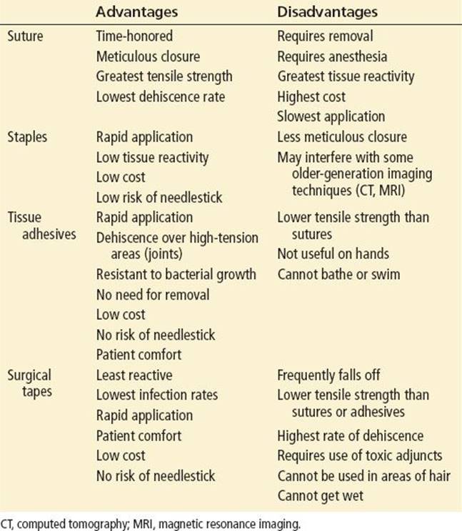
Advantages and Disadvantages of the Common Wound-Closure Techniques
eTABLE 21.2
Characteristics of Nonabsorbable Sutures
TABLE 21.2
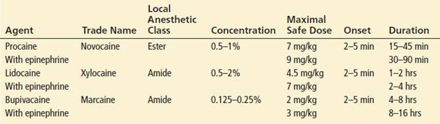
Properties of Commonly Used Local Anesthetics

eTABLE 21.3
Characteristics of Absorbable Sutures
TABLE 21.3
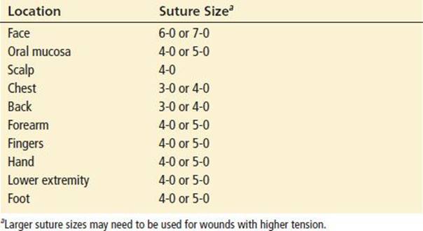
Suture Selection Based on Anatomic Location

KEY TESTING
• The majority of lacerations require no diagnostic testing besides a history and physical examination.
• Depending upon the likelihood of a retained foreign body or fracture from the injury, radiography, ultrasound, or computerized tomography may be necessary.
ED MANAGEMENT
Aseptic Technique
The use of sterile gloves makes common sense, despite the lack of clinical evidence supporting their use (5,6). Though the practice of universal precautions is recommended in all patients, double gloving is recommended when caring for patients who can transmit serious infections (human immunodeficiency virus or hepatitis) to the healthcare provider.
Latex Allergy
Severe reactions in latex-sensitive individuals have resulted in fatalities. Populations that appear to be at increased risk for allergic reactions to latex include patients who have undergone multiple operations, patients with atopic disease, children with spina bifida, and medical personnel with frequent latex exposures.
Wound Examination
Wound examination should always be conducted under optimal lighting conditions and with bleeding in the field controlled. Wound examination should begin with a neurovascular assessment of pulses, motor function, and sensation distal to the laceration.
Bleeding should be controlled with direct pressure. When lacerations continue to bleed despite reasonable pressure, placement of a sphygmomanometer cuff proximal to the injury, with inflation to a pressure greater than the patient’s systolic blood pressure, will help contain bleeding to allow proper examination. Palpation of the bones adjacent to the injured site may detect tenderness, a defect, or instability consistent with an underlying injury. Radiography should be used to detect fractures. Detection of an open fracture will dictate a change from usual management because it will require the administration of systemic antibiotics and may require irrigation in the operating room. Wounds occurring adjacent to a joint should be carefully evaluated for joint violation. If in doubt, sterile fluid should be injected into the joint, and the physician should look for communication to verify violation of the joint space.
Amputated parts should be cared for as outlined in the chapters on hand injuries and replantation emergencies (Chapters 41 and 47). Digits should be covered with a protective saline-soaked dressing, placed within a waterproof bag, and then placed in a container of ice water for preservation and consideration of future reattachment.
Complicated injuries involving most open fractures, joint penetration, flexor tendon injuries, nerve injuries, or arterial injuries are often treated in the operating room. In most other cases, wound repair will occur in the ED.
The ultimate appearance and function of a scar can be predicted by the static and dynamic skin tensions on the surrounding skin (see eFig. 21.1). The most aesthetically pleasing scar occurs when the long axis of the scar is parallel to the direction of minimal tension, because the static skin tensions continually pull on the wound edges even after wound closure. The ultimate width of the scar is proportional to the magnitude of the static skin tensions.
An appreciation of the effects of various dynamic tensions can be used to determine the need for tension-relieving absorbable sutures in addition to the nonabsorbable skin closure.
Wound Anesthesia
Most lacerations will require some form of anesthesia prior to wound closure. All visibly contaminated wounds and wounds that will require anesthesia for closure should receive anesthesia prior to cleansing so that adequate mechanical removal of bacteria, soil, and other debris can be more readily accomplished.
Local Infiltration
The most common form of anesthesia for traumatic injuries is local infiltration. Unfortunately, local anesthesia is painful. To reduce the pain associated with local infiltration, several strategies can be employed. The addition of sodium bicarbonate to lidocaine (pKa, 7.9) in a 1:9 ratio increases the ratio of uncharged to charged ions and results in more rapid diffusion of anesthetic agent into nerve endings. It reduces the pain associated with local infiltration without altering the ability of host defenses to prevent infection (7). The use of warm anesthetic solutions, small needles, slow rates of infiltration, injection through the wound edges in noncontaminated wounds, and pretreatment with topical 4% tetracaine or lidocaine, epinephrine, and tetracaine (LET) are additional strategies that can be used to decrease the pain of infiltration (3,8).
For injection of local anesthesia, a 27- to 30-gauge needle attached to a 10-mL syringe should be used. The needle should be passed into the wound edge rather than the intact skin because wound edge injections are less painful. Full anesthesia to pinprick is present 5 to 6 minutes after subdermal injection. The local anesthetic agent should be instilled slowly to decrease the pain that accompanies tissue infiltration. The needle should be passed through the dermal tissue initially; then the anesthetic agent can be injected as the needle is slowly withdrawn. This technique minimizes distention and pain by providing a potential space for the anesthetic solution. Aspiration of the syringe before injection is recommended to prevent inadvertent intravascular injection.
Because the duration of local anesthesia induced by bupivacaine is nearly four times longer than that of lidocaine, bupivacaine (0.25%) should be used when longer durations of anesthesia are desirable (Table 21.2).
Vasoconstrictors such as epinephrine can be used as adjuncts to the local anesthetic agents. They will reduce bleeding and oozing within the laceration, which will make exploration and closure easier to accomplish. The addition of epinephrine also delays systemic absorption and increases the therapeutic window. Clinical studies have not found that the vasoconstriction associated with epinephrine use results in differences in infection rates. While traditionally these agents have been avoided in fingers, toes, and the penis owing to the risk of vasospasm and subsequent necrosis, more recent evidence suggest that their use is safe (9).
Regional Anesthesia
When the nerve supply to a wound is easily accessible, a regional nerve block is a valuable alternative to local anesthesia. The use of regional nerve blocks will prevent distortion of the wound edges, facilitating approximation and increasing the likelihood of an excellent cosmetic result. Regional nerve blocks can be accomplished by only one or two needle passages through skin proximal to the injury site. Regional nerve blocks of the supraorbital, infraorbital, and mental nerves are useful for facial lacerations; however, they are more painful and less reliable than local infiltration (10). Median, radial, and ulnar nerve blocks may be useful for lacerations of the hand. Digital or metacarpal nerve blocks can be used for finger lacerations. Regional nerve blocks can also be used for lacerations of the feet. Field blocks can be considered for more proximal extremity injuries.
A 27- or 25-gauge needle is preferred to a 30-gauge needle for a regional nerve block, because either one is more resistant to deflection during passage through tissue. The duration of sensory analgesia can be significantly prolonged when epinephrine (1%) is added to the anesthetic solution. When the needle puncture site is the mucous membrane, anesthetizing the mucous membrane with a topical anesthetic agent makes the introduction of the needle painless.
Topical Anesthesia
Topical anesthesia eliminates needle use and the risk of inadvertent needle sticks while allowing the application of painless anesthesia. Various combinations of lidocaine (1% to 4%), adrenaline (1:1,000 to 1:2,000), and tetracaine (0.5% to 2.0%) are as effective as eutectic mixture of local anesthetic cream (EMLA) (11,12). An EMLA has a slower onset of action, is more expensive than LET, and is not approved for use on injured skin; therefore, it is not recommended for use in the ED.
Preparation of Skin
Removal of the hair surrounding a laceration may facilitate meticulous wound closure. Because many bacteria normally reside in hair follicles, shaving the hair prior to repair may increase wound infection rates. Reduced damage to hair follicles may be achieved by using hair clippers instead of a razor. Removal of the eyebrow hair should be avoided because it does not always grow back. Additionally, the presence of hair serves as a guide to approximation of wound edges during laceration repair.
The skin surrounding the wound should either be cleansed with normal saline or disinfected with an iodophor (such as Betadine) or chlorhexidine; the physician should avoid contacting the wound itself with disinfectant. A recent study in the operating room demonstrated significantly reduced wound infection rates after skin preparation with chlorhexidine (13). Although these agents can reduce the bacterial concentration on intact skin, they appear to damage wound defenses, making the wound more susceptible to development of infection. Consequently, any contact of these agents with the wound should be avoided.
Debridement
Appropriate debridement is an important factor in the management of contaminated wounds. Retained devitalized soft tissue, fat, muscle, and skin can damage wound defenses and increase the likelihood of infection through the promotion of bacterial growth and the inhibition of phagocytosis. Identification of the exact limit of devitalized tissue in wounds may be difficult. Evaluation of the color, consistency, contraction, and circulation is useful.
In some anatomic sites, such as the trunk, debridement is best accomplished by complete excision of the skin and deep tissues. Heavily contaminated wounds with serpiginous defects in these regions can be converted to clean wounds by more generous tissue excision. When a heavily contaminated wound contains specialized tissues, such as the nerves or tendons that perform important physical functions, complete excision often is not feasible. In such instances, mechanical wound cleansing followed by excision of all fragments of tissue that are not clearly viable is indicated.
The benefits of debridement must be weighed against the consequences of excision of the tissue. Debridement of skin and underlying tissue leaves a significant soft tissue defect that makes reapproximation more difficult.
Assessment for Foreign Bodies
Metal, bone, teeth, pencil graphite, certain plastics, glass, gravel, sand, some fish bones, some wood, and some types of aluminum are visible on plain radiographs. Almost all glass fragments 2 mm or larger are visible on radiographs (14). If the wound was caused by metal or glass and no foreign body is found on wound exploration or on plain films, it is unlikely that a foreign body exists. Computed tomography (CT) and magnetic resonance imaging (MRI) are useful for identifying and locating objects that have densities similar to soft tissue. Sonography may also be useful, particularly for wooden foreign bodies.
Cleansing the Wound
The two basic methods of wound cleansing are hydraulic forces (irrigation) and direct contact (scrubbing).
Irrigation
Some debate exists over both the optimal method of irrigation and the preferred solution. The efficacy of wound irrigation is correlated with the pressure at which the irrigant is delivered to the wound. High-pressure irrigation (>7 pounds per square inch) successfully cleans wounds of small particulate matter, such as bacteria and soil. Such cleansing has reduced the infection rate of experimentally contaminated wounds. In contrast, low-pressure syringe irrigation, even with large volumes of fluid, demonstrates negligible capability for removing small particles (bacteria) but removes large particulate matter, such as detached devitalized tissue.
High wound impact pressures can be easily obtained using a 30- to 60-mL syringe and a 19-gauge needle or Zerowet splash shield (15). However, it must be noted that sustained high- pressure irrigation can result in tissue damage. As a result, at very high pressures, infection rates may actually increase.
For relatively clean wounds in highly vascularized areas containing loose areolar tissue, such as the eyelid, high pressures should be avoided. In fact, high-pressure irrigation has not been found to offer any advantages for cleansing clean, noncontaminated facial lacerations (4). Conversely, high-pressure irrigation is clearly indicated for contaminated wounds of the lower extremity. The choice of irrigation solution is relatively straightforward. Normal saline compares favorably with more expensive, less readily available alternatives. Because of their tissue toxicity, detergents, hydrogen peroxide, and concentrated forms of povidone–iodine should not be used to irrigate wounds. Irrigation of wounds with tap water results in comparable infection rates versus irrigation with sterile saline (16). Irrigation volume should be individualized based on patient characteristics and such wound characteristics as location and etiology. Use of a device designed to reduce splatter will minimize the healthcare provider’s risk of exposure to potentially infectious materials.
Scrubbing
Direct scrubbing of the wound with gauze or a sterile surgical brush helps remove both bacteria and particulate matter, which potentiate the risk of wound infection. However, scrubbing also contributes to the tissue damage and reduces the ability of the wound to resist infection. Therefore scrubbing should be reserved for highly contaminated wounds.
Use of Drains
The potential benefits of the use of surgical drains in a clinical setting must be weighed against its harmful effects. Drains evacuate potentially harmful collections of pus and blood from wounds. When no definite localized fluid collection exists, drains must be considered prophylactic, and their potentially harmful effects become more important. The general use of drains is not indicated in the management of most wounds in the ED.
Open Wound Management
The technique of wound closure largely depends on whether the wound has lost tissue and the risk of wound infection. With primary closure, the wound is immediately closed. Primary closure results in a reduction in healing time in comparison with other closure methods. It also may reduce the bleeding, inconvenience, and discomfort often associated with open wounds. Secondary wound closure, in which the wound is left open and allowed to close on its own, is particularly well suited for highly contaminated or infected wounds. Though this method may reduce the risk of infection, it is relatively slow and uncomfortable. Delayed primary (or tertiary) closure combines the advantages of both primary and secondary closures. The wound is left open for a period of 3 to 5 days, after which it may be closed if no infection supervenes. For wounds with associated significant tissue loss, grafts or flaps are often required to close the defect. These procedures are usually performed in the operating room.
Though there is a direct relationship between the time interval from injury to laceration closure and the risk of subsequent infection, the length of this “golden period” is highly variable (3). A study of forearm lacerations found that closure within 4 hours had a lower infection rate than later closure (17). A large series of pediatric patients (2,834 patients) with lacerations did not find a difference in infection rate for lacerations closed within or >6 hours from the time of injury (18). Facial lacerations healed well regardless of the time to closure. In contrast, trunk and extremity lacerations exhibited lower rates of healing if they were closed more than 19 hours from the time of injury (63% to 75%) as compared to earlier (75% to 91%) (19).
On the basis of these data, it seems appropriate to consider each laceration separately, taking the time from injury until presentation into account, along with laceration location, degree of contamination, risk of infection, and importance of cosmetic appearance, before deciding whether to perform primary wound closure. Wounds that are not closed primarily owing to a high risk of infection should be considered for delayed primary closure after 3 to 5 days, when the risk of infection decreases.
Wound Closure
Careful handling of tissues and judicious use of electrocautery are necessary to reduce further trauma that may lead to poor cosmetic outcome (20). The optimal wound-closure technique varies with the clinical situation. Sutures are the most commonly employed wound-closure technique. Tissue adhesives can be used in up to one-third of ED laceration repairs. Staples and surgical tapes can be used in selected situations. The advantages and disadvantages of various wound-closure techniques are summarized in Table 21.4.
TABLE 21.4
Advantages and Disadvantages of the Various Suture Techniques

Sutures
The choice of suture technique should be determined by the configuration and biomechanical properties of the wound, in combination with an assessment of its risk of infection. Percutaneous sutures pass through the epidermal and dermal layers of the skin. They are used alone for low-tension lacerations and in combination with deep dermal sutures for higher tension lacerations. The dermal suture reapproximates the divided edges of the dermis without penetrating the epidermis. Often, dermal and percutaneous sutures are used together.
Most often, percutaneous sutures are performed with nonabsorbable sutures, such as nylon and polypropylene. Both nylon and polypropylene retain most of their tensile strength for more than 60 days and are relatively nonreactive (3). Removal of nonabsorbable sutures is required. Percutaneous sutures of either monofilament nylon or polypropylene exert the least damage to the wound defenses.
Nonabsorbable sutures made from natural fibers potentiate infection more than synthetic nonabsorbable sutures, correlating with the reaction of tissue to these sutures in clean wounds. The incidence of infection in contaminated tissue containing monofilament sutures is lower than in those containing multifilament (braided) sutures. Although use of absorbable sutures is generally reserved for dermal closures, more rapidly absorbing forms can be used to close skin in children and avoid the discomfort and inconvenience of suture removal.
Dermal closures are generally accomplished with absorbable sutures. Chromic gut lasts for up to 2 weeks, but it is associated with increased tissue reactivity. Polyglactin (Vicryl) and polyglycolic acid (Dexon) maintain tensile strength for 20 to 28 days and have less tissue reactivity. Some synthetic absorbable sutures, such as polydioxanone (PDS) and polyglyconate (Maxon), retain their tensile strength for as long as 2 months. They are particularly useful in areas with high static and dynamic tensions. These sutures should be used only in the deeper areas, because they can become extruded after long periods of time. Antibiotic-impregnated absorbable sutures (such as Vicryl Plus and Monocryl Plus) have in vitro antibacterial actions against a wide variety of bacteria. They should be considered especially in contaminated wounds.
The use of dermal sutures relieves skin tension, decreases dead space and hematoma formation and, theoretically, should improve cosmetic outcome. Research in experimental animal models has found that deep sutures increase the risk of infection in highly contaminated wounds, but they do not do so in clean, noncontaminated lacerations (3). Deep sutures should not be placed in adipose tissue and in contaminated wounds. No studies of traumatic lacerations have demonstrated any benefit to the use of deep sutures (21). Many practitioners prefer to use deep sutures in gaping lacerations and in cosmetically important lacerations despite a lack of evidence. Deep sutures should not be used in the hand as they may inadvertently cause damage to important structures such as nerves and tendons.
Undermining the wound margin decreases the forces required for wound closure. Theoretically, it should limit the width of the ultimate scar. However, this benefit must be weighed against its potential damage to the skin blood supply, which may increase the likelihood of infection. Consequently, undermining should be reserved for clean, noncontaminated wounds subjected to strong static and dynamic tensions.
The magnitude of the suture’s damage to the local tissue defenses is related to the quantity of the suture within the wound. As a result, the narrowest diameter suture (5-0 or 6-0) that is strong enough to resist disruption of the skin should be used (Table 21.3). Approximation of the midportion of the laceration first, with subsequent bisection of the remaining portion, will enable the use of the least amount of suture. Interrupted dermal sutures placed in each quadrant of the wound subjected to strong static and dynamic skin tensions may provide sufficient strength to permit early suture removal.
Careful attention to the following details may help ensure proper wound edge eversion and an optimal cosmetic outcome after suturing. The needle should enter the wound at an angle of 90 degrees. This will result in a wider “bite” at the depth of the wound than at its surface, helping to achieve wound eversion. This is best accomplished by maximally pronating the hand that is holding the needle holder. It is also important to match corresponding layers of the skin on either side of the wound by taking equally sized bites.
The choice of a particular suture technique is based upon the specific wound, its location, tension, and advantages and disadvantages of the various suture techniques (Table 21.4). Simple interrupted sutures are commonly used for low-to-moderate tension wounds. For long linear lacerations, continuous sutures may be used to save time and distribute the tension more evenly. Continuous subcuticular (or intradermal ) sutures can be used instead of percutaneous sutures to avoid the need for suture removal. However, this method is more time-consuming and technically challenging. A half-buried horizontal mattress suture (or corner stitch) is most appropriate for narrow corners and flaps, as it does not compromise tissue perfusion. Vertical mattress sutures help achieve maximal wound edge eversion, but they may result in tissue strangulation. They are best reserved for closure of high-tension wounds in thin skin where placement of deep dermal sutures is difficult. Whatever method is used, the sutures should be tied tightly enough to achieve complete apposition of the wound edges. However, care should be taken not to tighten the suture too tightly, as this may compromise perfusion and result in necrosis and sloughing of the wound edges.
Topical Skin Adhesives
The only topical skin adhesives that are strong enough to close wounds are the cyanoacrylate polymers. A growing number of butylcyanoacrylate, octylcyanoacrylate, or combination topical skin adhesives are now available. The topical skin adhesives also have the advantage of forming a microbial barrier to prevent wound contamination (22).
Several clinical studies have compared the use of 2-octylcyanoacrylate to 5-0 and 6-0 sutures. Quinn et al. (23) found the long-term cosmetic outcomes to be equivalent. The use of 2-octylcyanoacrylate was faster and less painful than the use of sutures. The largest study to date enrolled 818 patients who were randomly assigned to receive skin closure with either 5-0 or 6-0 sutures or 2-octylcyanoacrylate (24). Of the 818 patients enrolled, 333 had subcuticular or subcutaneous sutures placed prior to laceration closure. Comparing the group of patients who received 2-octylcyanoacrylate with the group of patients who received skin closure with sutures, the 3-month cosmetic outcomes, short-term infection rates, and wound dehiscence rates were all similar. The time to wound closure was reduced by more than 50% in the group treated with 2-octylcyanoacrylate. Subgroup analysis of patients from both adult and pediatric EDs found equivalent 3-month cosmetic outcomes (25,26).
Although in vitro and in vivo studies have found that cyanoacrylate tissue adhesives possess gram-positive antimicrobial properties, it is imperative that clinicians adhere to standard wound preparation and wound-cleansing procedures. Meticulous wound cleansing is essential regardless of wound closure method. Although wound cleansing can sometimes be accomplished without the use of local anesthesia, the decision to close wounds with tissue adhesives should not, in and of itself, lead the clinician to avoid the use of anesthesia when needed for wound cleansing.
The application of tissue adhesives is rapid and relatively painless. Adhesives also have the advantage that they do not require later removal, as do sutures. They will usually slough off in 5 to 10 days as the keratinized layer of epithelium sloughs. They should be used only topically, not placed within the wound margins. The topical skin adhesives are needleless alternatives to sutures for the closure of most facial lacerations, providing an excellent cosmetic appearance, comparable to that provided by sutures.
Topical skin adhesives can be used in locations that could otherwise be closed with 5-0 or 6-0 nonabsorbable sutures. Topical skin adhesives should only be used in higher-tension areas if subcutaneous or subcuticular absorbable sutures are placed to relieve tension on the skin. They should not be used over high-tension areas or areas involved in repetitive movement, such as joints, unless the wound is splinted and tension-relieving sutures are used. When tissue adhesive application leads to a suboptimal wound edge apposition, antibiotic ointment, petroleum jelly, or bathing can be used to accelerate removal.
For the repair of very small facial lacerations, the butylcyanoacrylates are equivalent to 5-0 and 6-0 sutures or octylcyanoacrylates. In general, the butylcyanoacrylates are more brittle and friable than octylcyanoacrylates and do not allow the same degree of movement and flexibility of the skin.
Staples
Staples can be applied more rapidly than sutures. Staples are associated with a lower rate of foreign body reaction and a lower infection rate. In general, staples are considered particularly useful for scalp, trunk, and extremity wounds (3). They can also be used when time is limited, such as in situations of mass casualties or multiple trauma. On the other hand, they do not allow as meticulous a skin closure as sutures, and their use is limited when cosmetic appearance is of utmost importance. They are slightly more painful to remove than sutures. In experimental animal models, staples have shown lower rates of bacterial growth and lower infection rates than sutures, although these effects appear to have limited clinical significance.
Adhesive Tapes
Linear wounds in skin subjected to minimal static and dynamic tensions are easily approximated by tape. The relatively lax skin of the face and abdomen is amenable to wound closure by tape. With tape closures, patients are spared the discomfort of suture removal and the development of suture puncture scars. An additional benefit of this technique is that patients need not be subjected to the painful injection of the local anesthetic agent required for suturing.
Surgical tapes are intrinsically less reactive than staples. However, adhesive tapes require the use of adhesive adjuncts (e.g., tincture of benzoin), which are tissue toxic. Although the various surgical tapes have different degrees of adhesion, porosity, breaking strength, and elasticity, tapes alone will not maintain wound integrity in areas subject to tension. They are seldom recommended for primary wound closure in the ED but are often used after suture removal to decrease tension on the wound until they fall off. The outcome of highly select, small, short, linear, low-tension facial lacerations that are closed with surgical tape is comparable to closure with a tissue adhesive (27).
Complex Issues in Wound Care
Bite Wounds
An estimated 1 to 2 million animal bites are treated each year in the United States. The most frequent complication from bites is infection. Dog bites have an infection rate of 1.4% to 30%, cat bites 15.6% to 50%, and human bites 9% to 18% (14). Most bite wounds are minor. Local wound care always begins with a thorough evaluation of the injury. The location, number, type, and depth of wound are noted. The wound is also assessed for signs of infection. Bite wounds are frequently puncture wounds. Meticulous examination is of utmost importance, as they are notoriously deceptive and may be more extensive than they initially appear to be. The physician should evaluate all bite wounds for injuries to deep structures such as tendons, joint capsules, blood vessels, nerves, and bone. Local or regional anesthetic should be used to facilitate wound exploration, and a proximal tourniquet may be used when necessary to create a bloodless field. If bony involvement or foreign body is suspected, a plain radiograph should be obtained. Meticulous cleansing, irrigation, and debridement are the essential components of all bite wound care. Irrigation of puncture wounds is controversial. Their small openings do not allow the irrigant solution to drain out appropriately. This may result in infiltration of the tissue. Surgical debridement may be necessary to remove compromised or necrotic tissue that may contain embedded organisms, soil, and clots that cannot be removed by irrigation alone.
Primary closure generally should not be performed for human or extremity bites. Primary closure of dog bite wounds is controversial. Recent studies support primary closure of selected dog bite wounds (28). In one study, 145 head and neck dog bite wounds underwent primary closure without the use of antibiotics, and the wound infection rate was only 1.4% (29). Dog bite wounds to the head and neck that are more than 1.5 cm long can be closed primarily with sutures. Small bite wounds (<1.5 cm) behave like puncture wounds and should be left open to allow for drainage. Patients with bite wounds that require reconstructive surgery should be immediately referred to the appropriate specialist. Hand wounds should be managed without primary closure. However, for cosmetic reasons, facial wounds should be repaired. Other bite lacerations can be closed using delayed primary closure techniques. All injured extremities should be immobilized and elevated. Bites are tetanus-prone wounds, and the recommendations for immunoprophylaxis should be followed (Table 21.1). Domestic and wild animal bite victims should be assessed for the need for rabies postexposure immunoprophylaxis (see Chapter 375).
Puncture Wounds
The feet are the most common site for puncture wounds. There are many different views on the management of plantar puncture wounds (14,30). Some physicians prefer expectant therapy, reacting to complications as they develop. Others pursue invasive exploration for and removal of foreign bodies with subsequent antibiotics in the hope of preventing unusual but devastating infections that can develop.
Infections of plantar wounds can often have serious consequences and lead to disability or amputation. Osteomyelitis, osteochondritis, and septic arthritis are the worst of the complications. Staphylococcus aureus and Pseudomonas aeruginosa are the most common isolates from plantar puncture wound infections. Pseudomonas infections are associated with puncture wounds through tennis shoes.
Exploration of plantar puncture wounds for foreign bodies is technically difficult. Foreign bodies that penetrate the plantar fascia are almost impossible to locate through a narrow puncture wound. Irrigation of deep puncture wounds may not be beneficial because the irrigant does not completely drain out of the wound. Some authors recommend enlarging the puncture wound to allow for deeper irrigation and exploration, especially if bone or joint involvement is suspected. Others believe that removing a block of tissue down to the subcutaneous layer allows adequate visualization of accessible foreign bodies and removal of most of the contaminated tissue. Still others simply trim jagged epidermal skin edges. Blind probing and grasping have unknown false-negative rates and may force foreign bodies deeper into the wound. If a wound infection is present, the risk of foreign body is very high, and imaging or exploration is mandated.
Another controversy involves the use of prophylactic antibiotics for this type of injury. Many experts do not recommend the use of antibiotics for new plantar puncture wounds. They argue that antibiotics will not compensate for inadequate initial wound care, they are ineffective in wounds with retained foreign bodies, they may contribute to a gram-negative infection, and wound infections that do not have a retained foreign body respond quickly to initiation of antibiotics. Owing to the difficulty of cleaning and exploring plantar puncture wounds, physicians should reevaluate these wounds for infection within 2 to 3 days of initial presentation. If infection develops, they should be aggressively explored for a foreign body.
Facial Lacerations
Owing to their cosmetic significance and proximity to important structures, several points concerning the repair of facial lacerations are noteworthy. Because of their rich vascular supply, facial lacerations have a relatively low rate of infection. The risks of high-pressure irrigation (edema making it more difficult to perfectly approximate wound edges) should be weighed against the benefits (cleansing), and individual decisions regarding the use of wound irrigation should be made accordingly. For example, an upper eyelid laceration resulting from a blunt impact with the corner of a table requires minimal irrigation, whereas a contused cheek laceration resulting from a fall onto a rough and dirty surface requires larger volume irrigation.
Whether nongaping facial lacerations require deep sutures is a subject of ongoing debate, but at least one study suggests that nongaping lacerations do not benefit from layered closure (21). Gaping lacerations as well as those in which underlying muscle are involved should be repaired in multiple layers. Meticulous wound approximation should follow important anatomic landmarks such as the forehead or nasolabial creases, the eyebrow, and the vermilion border. When these structures are within a laceration, initial placement of key sutures aligning the landmarks is crucial. Eyebrows should never be removed because they may not grow back.
With lip lacerations, it is important to exclude any underlying dental injuries. If a fractured tooth is not found, careful exploration of the lip wound should be performed to rule out an embedded tooth fragment. Through-and-through lacerations of the lips should be closed in three layers. The first layer should be placed within the oral labial mucosa using a 4-0 or 5-0 absorbable suture. The laceration should then be recleansed now that it is sealed off from the oral flora. The second layer should be placed in the muscle using a 5-0 absorbable suture, and the third layer should be placed in the skin using a 6-0 nonabsorbable suture. The first suture in the skin should be placed at the vermillion border. Patients should be advised to eat a soft diet and carefully rinse out their mouth after each meal. Prophylactic use of oral antibiotics for lacerations of the oral mucosa that cover oral anaerobes is recommended.
With ear lacerations, it is important to identify any underlying injuries to the cartilage. A field block performed at the base of the ear is helpful with extensive ear injuries. With simple lacerations, primary repair of the overlying skin, ensuring complete coverage of the cartilage, suffices using nonabsorbable 6-0 sutures. Large or gaping lacerations of the cartilage may be closed with a 6-0 monofilament nonabsorbable suture. Any hematoma beneath the skin needs to be evacuated, and a compressive dressing should be placed to ensure adequate contact between the skin and the cartilage. Due to its relative avascularity, necrosis of the cartilage with subsequent formation of a “cauliflower” ear may result if the cartilage is deprived of its vascular supply. With exposure of cartilage, prophylactic oral antibiotic coverage for staphylococcus and pseudomonas is recommended.
With eyelid lacerations, it is crucial to exclude any injury to the lacrimal duct or the tarsal plate. These lacerations, as well as those involving the lid margins and inner surface, should be repaired by a specialist. Bulging of subcutaneous fat suggests violation of the tarsal plate and, along with the presence of ptosis, should always prompt referral. See Chapter 25.
Post Repair Wound Care
Post repair wound care should optimize healing. It must be tailored to both the type of wound and method of wound closure. Sutured or stapled lacerations should be covered with a protective nonadherent dressing for 24 to 48 hours until enough epithelization takes place to protect the wound from gross contamination. Maintenance of a moist wound environment increases the rate of reepithelization. When possible, the site of injury should be elevated above the patient’s heart to limit the accumulation of fluid in the wound interstitial spaces.
Topical antibiotic ointments may help reduce infection rates and prevent scab formation. Patients whose lacerations are closed with topical skin adhesives should not have topical ointments applied. They will loosen the adhesive and result in dehiscence.
Prophylactic systemic antibiotic administration should not be used as a regular adjunct to wound care. Several studies and a meta-analysis have found no benefit in prophylactic antibiotics for routine laceration repair (31). Use of antibiotics should be individualized on the basis of the degree of bacterial contamination, the presence of infection-potentiating factors (e.g., soil), the mechanism of injury, and presence or absence of host predisposition to infection. In general, decontamination is far more important than antibiotics. Antibiotics should be used in most human, and extremity bites; intraoral lacerations; open fractures and open joints; or tendon lacerations (3). Additionally, patients with soft tissue lacerations who are prone to the development of infective endocarditis, patients with prosthetic joints and other permanent “hardware,” and patients with lymphedema should receive antimicrobial therapy. Intravenous antibiotics can be given prior to wound care in patients at high risk for systemic complications of infected soft tissue injuries.
Sutured or stapled wounds should be kept clean but can be washed (14). Patients with tissue adhesives may shower, but they should avoid bathing and swimming; prolonged moisture may loosen the adhesive bond. Gentle blotting should be used to dry the area, as wiping could result in dehiscence. Splinting may prove helpful in areas subject to high tension (e.g., in lacerations across joints). Patients should be instructed to observe the wound for erythema, warmth, swelling, and drainage, as these findings, as well as fever, may indicate infection. Use of standardized wound care instructions improves patient compliance and understanding.
Sutures or staples in most locations should be removed after approximately 7 days (Table 21.5). Facial sutures should be removed sooner (within 3 to 5 days) to avoid the formation of unsightly sinus tracts. Sutures subject to high tension (e.g., in joints, hands) should be left in for 10 to 14 days. When tissue adhesives are used, patients should be advised to avoid picking or scrubbing the area and to avoid exposure of the wound to water for more than brief periods until the previously noted times. When tissue adhesives remain on the skin for prolonged periods, antibiotic ointment, petroleum jelly, or bathing can accelerate removal. Acetone can be used when more rapid removal is required. Patients should be told when and with whom to follow up for suture removal or wound examination. Most wounds do not require scheduled wound checks as patients can be told to watch for the signs and symptoms of infections.
TABLE 21.5
Optimal Time from Placement of Sutures Until Suture Removal

Abraded skin and wounds may develop permanent hyperpigmentation after exposure to the sun. Consequently, they should be protected with a sun-blocking agent for at least 6 to 12 months after injury.
Prevention of Tetanus
Two-thirds of the recent tetanus cases in the United States have followed lacerations, puncture wounds, and crush injuries. Proper immunization plays the most important role in tetanus prophylaxis. Recommendations on tetanus prophylaxis are based on the condition of the wound and the patient’s immunization history. A summary guide to tetanus prophylaxis of the wounded patient, as recommended by the Centers for Disease Control, is outlined in Table 21.1. As noted, passive immunization with tetanus immunoglobulin is indicated only under specific conditions.
The only contraindication to tetanus and diphtheria toxoids is a history of neurologic or severe hypersensitivity reaction after a previous dose. Local side effects do not preclude repeated use. Local reactions, generally erythema and induration with or without tenderness, are common after the administration of vaccines containing diphtheria, tetanus, and pertussis antigens. These reactions are usually selflimited and require no therapy. If a systemic reaction is suspected to represent allergic hypersensitivity, immunization should be postponed until appropriate skin testing is undertaken. If the use of a tetanus toxoid is contraindicated, passive immunization against tetanus should be considered in a tetanus-prone wound. In those patients who have not completed a primary tetanus immunization series, appropriate follow-up should include referral to a physician who can complete active immunization.
CRITICAL INTERVENTIONS
• Obtain a tetanus immunization history and administer tetanus toxoid if it has not been given within the past 10 years (5 years if the wound is tetanus prone).
• Perform and document a neurovascular assessment distal to the laceration.
• Perform and document a careful wound exploration to exclude foreign bodies and tendon lacerations.
DISPOSITION
The appropriate surgical specialist should be consulted for patients with open fractures of long bones, nerve or vascular injuries, flexor tendon disruptions, repair of specialized structures such as the parotid or lacrimal duct, replacement of skin loss by a flap or graft, or extensive debridement.
Few patients with lacerations will require admission to the hospital. Even those patients requiring surgical intervention can often be discharged with appropriate follow-up arrangements. Often, definitive repair can be accomplished via same-day surgery.
If the initial receiving hospital does not have the appropriate emergency or surgical specialist, transfer is necessary. The patient should be transferred in the most expeditious manner possible.
Common Pitfalls
• Failure to detect sensory, motor, and vascular complications or injuries to specialized tissues.
• Failure to detect and remove foreign bodies.
• Removing hair with a razor, because it may increase the infection rate.
• Using antiseptic agents such that there is contact between the agent and the wound.
• Using drain placement as a replacement for meticulous hemostasis; drains should be reserved for removal of harmful collections of fluid.
• Using tissue adhesives without appropriate cleansing and exploration.
• Using tissue adhesives in high-tension wounds or over joints.
REFERENCES
1. Singer AJ, Thode HC Jr, Hollander JE. National trends in ED lacerations between 1992 and 2002. Am J Emerg Med. 2006;24:183–188.
2. Hollander JE, Singer AJ, Valentine S, et al. Wound registry: Development and validation. Ann Emerg Med. 1995;25:675–685.
3. Singer AJ, Hollander JE, Quinn JV. Evaluation and management of traumatic lacerations. N Engl J Med. 1997;337:1142–1148.
4. Hollander JE, Richman PB, Werblud M, et al. Irrigation in facial and scalp lacerations: Does it alter outcome? Ann Emerg Med. 1998;31:73–77.
5. Ruthman JC, Hendricksen D, Miller RF, et al. Effect of cap and mask on infection rates. Ill Med J. 1984;165:397–399.
6. Whorl GJ. Repairing skin lacerations: Does sterile technique matter? Can Fam Physician. 1987;33:1185–1187.
7. Bartfield JM, Gennis P, Barbera J, et al. Buffered versus plain lidocaine as a local anesthetic for simple laceration repair. Ann Emerg Med. 1990;19:1387–1389.
8. Hollander JE, Singer AJ. State of the art laceration management. Ann Emerg Med. 1999;34:356–367.
9. Chowdry S, Seidenstricker L, Cooney DS, et al. Do not use epinephrine in digital blocks: Myth or Truth? Part II. A retrospective review of 1111 cases. Plast Reconstr Surg. 2010;126:2031–2034.
10. Tarsia V, Singer AJ. Comparison of regional and local anesthesia for facial lacerations. Acad Emerg Med. 2002;9:449.
11. Ernst AA, Marvez-Valls E, Nick TG, et al. LAT versus TAC for topical anesthesia in face and scalp lacerations. Am J Emerg Med. 1995;13:151–154.
12. Singer AJ, Stark MJ. LET vs. EMLA for pretreating lacerations—a randomized trial. Acad Emerg Med. 2001;8:223–230.
13. Darouiche RO, Wall MJ, Jr, Itani KM, et al. Chlorhexidine-alcohol versus povidone-iodine for surgical-site antisepsis. N Engl J Med. 2010;362:18–26.
14. Capellan O, Hollander JE. Management of lacerations in the emergency department. In: Peth HA, ed. High Risk Presentations. Emergency Medicine Clinics of North America. Vol 21. Philadelphia, PA: WB Saunders; 2003:205–231.
15. Mittra ES, Singer AJ, Bluestein D, et al. Simulated wound irrigation impact pressures. Israeli J Emerg Med. 2003;3:9–16.
16. Moscati RM, Mayrose J, Reardon RF, et al. A multicenter comparison of tap water versus sterile saline for wound irrigation. Acad Emerg Med. 2007;14:404–409.
17. Morgan WJ, Hutchison D, Johnson HM. The delayed treatment of wounds of the hand and forearm under antibiotic cover. Br J Surg. 1980;67:140–141.
18. Baker MD, Lanuti M. The management and outcome of lacerations in urban children. Ann Emerg Med. 1990;19:1001–1005.
19. Berk WA, Osbourne DD, Taylor DD. Evaluation of the “golden period” for wound repair: 204 cases from a third world emergency department. Ann Emerg Med. 1988;17:496–500.
20. Singer AJ, Quinn JV, Thode HC, Jr, et al. Determinants of poor outcome after laceration and surgical incision repair. Plast Reconstr Surg. 2002;110:429–435.
21. Singer AJ, Gulla J, Hein M, et al. Single vs double layer closure of facial lacerations: A randomized control trial. Plast Reconstr Surg. 2005;116:363–368.
22. Singer AJ, Giordano P, Fitch JL, et al. Evaluation of a new high viscosity octylcyanoacrylate tissue adhesive for laceration repair. A randomized control trial. Acad Emerg Med. 2003;10:1134–1137.
23. Quinn JV, Wells GA, Sutcliffe T, et al. Tissue adhesive vs. suture wound repair at one year: Randomized clinical trial correlating early, three-month, and one-year cosmetic outcome. Ann Emerg Med.1998;32:645–649.
24. Singer AJ, Quinn JV, Hollander JE, et al. Closure of lacerations and incisions with octylcyanoacrylate: A multi-center randomized clinical trial. Surgery. 2002; 131:270–276.
25. Singer AJ, Hollander JE, Valentine SM, et al. Prospective randomized controlled trial of tissue adhesive (2-octylcyanoacrylate) vs standard wound closure techniques for laceration repair. Acad Emerg Med. 1998;5:94–99.
26. Bruns TB, Robinson BS, Smith RJ, et al. A new tissue adhesive for laceration repair in children. J Pediatr. 1998;132:1067–1070.
27. Mattick A, Clegg G, Beattie T, et al. A randomised, controlled trial comparing a tissue adhesive (2-octylcyanoacrylate) with adhesive strips (Steristrips) for paediatric laceration repair. Emerg Med J.2002;19:405–407.
28. Chen E, Hornig S, Shepherd SM, et al. Primary closure of mammalian bites. Acad Emerg Med. 2000;7(2):157–161.
29. Guy RJ, Zook EG. Successful treatment of acute head and neck dog bite wounds without antibiotics. Ann Plastic Surg. 1986;17:45–48.
30. Lammers R. Plantar puncture wounds. In: Singer AJ, Hollander JE, eds. Lacerations and Acute Wounds. An Evidence-based Guide. Philadelphia, PA: FA Davis; 2003:157–160.
31. Cummings P, Del Beccaro MA. Antibiotics to prevent infection of simple wounds: A meta-analysis of randomized studies. Am J Emerg Med. 1995;13(4): 396–400.