
Margaret Wolff
Jaundice is defined as a yellow discoloration of the skin and sclerae indicative of excessive bilirubin production or disordered bilirubin metabolism or excretion. When the serum concentration of bilirubin exceeds 2 mg/dL, jaundice usually becomes apparent and is best appreciated on the palms and soles (1). The etiologies of neonatal jaundice range from physiologic jaundice to potentially life-threatening conditions. These can be differentiated by a thorough history, physical examination, and laboratory evaluation. This chapter discusses the diagnostic approach to a young infant with jaundice. For the approach to patients greater than 8 weeks of age, see Chapter 102 (jaundice).
Bilirubin is created from the breakdown of hemoglobin and other heme-containing proteins. Bilirubin produced by this degradation process is unconjugated and poorly water soluble. Unconjugated bilirubin is conjugated in the liver with glucuronic acid (which is water soluble) and excreted into the biliary tract and in the stool (Fig. 221.1).
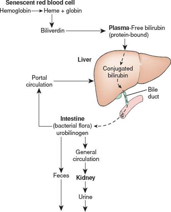
FIGURE 221.1 The process of bilirubin formation, circulation, and elimination. (Image from Porth CM. Pathophysiology: Concepts of Altered Health States. 7th ed. Philadelphia, PA: Lippincott Williams & Wilkins; 2005.)
CLINICAL PRESENTATION
Almost all neonates will have some degree of jaundice in the first week of life but are otherwise well appearing (2). Some of these neonates may appear dehydrated or have significant reduction in weight from birth weight suggesting insufficient oral intake. Other findings may suggest a more concerning cause of neonatal jaundice.
• Temperature instability, lethargy, apnea, or poor feeding suggest a serious bacterial infection (3)
• Pallor may indicate anemia
• Jaundice and vomiting suggest upper gastrointestinal obstruction
• Acholic stools suggest biliary tract obstruction
• Hepatomegaly may be associated with liver dysfunction
• Splenomegaly may indicate a hypersplenic state
• Neonates with acute bilirubin encephalopathy (kernicterus) may present with lethargy, hypotonia, and poor suck in the initial stages, and irritability and hypertonia in more advanced stages (4)
• Infants who present after the first 2 weeks of life with jaundice have pathologic jaundice and must be fully evaluated to determine the cause. These infants may be otherwise well appearing without any associated symptoms or they may exhibit any of the above signs and symptoms
DIFFERENTIAL DIAGNOSIS
Jaundice can be grouped based on underlying pathophysiology into the following categories: (1) excessive bilirubin production, (2) disordered bilirubin metabolism, or (3) disordered excretion (cholestasis) (eFig. 221.1). The majority of neonates develop some degree of jaundice due to a relative deficiency of glucuronic acid in the liver until approximately 14 weeks of age (5). In addition, neonates have more red blood cells and a shorter red cell half-life, resulting in increased bilirubin production compared to older children (6). This benign jaundice is termed physiologic neonatal hyperbilirubinemia and results in peak unconjugated bilirubin levels between 3 and 5 days that resolves without treatment. Breastfed neonates may develop breastfeeding-associated jaundice in the first week due to mild dehydration because of a delay in the let-down of breast milk as it transits from colostrum. Breastfed infants may also develop mild jaundice after the first week of life termed breast-milk jaundice due to effects of breast milk on intestinal excretion and resorption of bile (7).
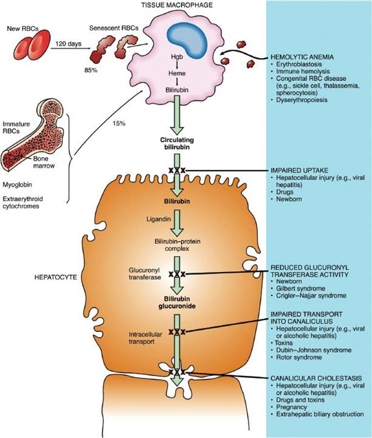
eFIGURE 221.1 Mechanisms of jaundice at the level of the hepatocyte. Bilirubin is derived principally from the senescence of circulating red blood cells, with a smaller contribution from the degradation of erythropoietic elements in the bone marrow, myoglobin, and extraerythroid cytochromes. Jaundice results from overproduction of bilirubin (hemolytic anemia) or defects in its hepatic metabolism. The locations of specific blocks in the metabolic pathway of bilirubin in the hepatocyte are illustrated. (Image from Rubin E, Farber JL. Pathology. 3rd ed. Philadelphia, PA: Lippincott Williams & Wilkins; 1999.)
Nonphysiologic hyperbilirubinemia is defined as jaundice in the first 24 hours of life, a rapidly rising bilirubin level, a bilirubin level exceeding phototherapy thresholds, jaundice that persists after 2 weeks, or conjugated hyperbilirubinemia (Table 221.1). Nonphysiologic jaundice may be caused by an exaggeration of the mechanisms that produce physiologic jaundice, or may be due to pathologic conditions leading to excessive bilirubin production, disordered bilirubin metabolism, or disordered excretion (cholestasis). After the neonatal period, excessive production from hemolysis is the most common cause of unconjugated hyperbilirubinemia (Table 221.2).
TABLE 221.1
Causes of Jaundice in Neonates

ED EVALUATION AND MANAGEMENT
The emergency physician must distinguish neonates with nonphysiologic jaundice, who require treatment, from neonates who have benign self-limited causes of jaundice. If left untreated, high levels of bilirubin are associated with kernicterus, a chronic form of bilirubin encephalopathy characterized by permanent neurologic damage (8). Although kernicterus is rare, recent cases demonstrate the need for vigilance when evaluating these infants (9,10). Unfortunately, visual determination of bilirubin levels in neonates is unreliable, making laboratory testing necessary (11,12).
Since the majority of well-appearing neonates will have physiologic jaundice that does not require further treatment (eFig. 221.2), it is reasonable to obtain an initial fractionated bilirubin level from a heelstick capillary sample (13). If this demonstrates hyperbilirubinemia with predominantly unconjugated bilirubin, the next step is to determine if phototherapy treatment is required, based on AAP Guidelines (Fig. 221.2). Patients requiring phototherapy should have more formal laboratory studies ordered, including a complete blood count, peripheral blood smear, blood type, and Coombs test. Neonates with bilirubin levels below the phototherapy threshold should be risk stratified to determine the near-term risk of hyperbilirubinemia (Fig. 221.3). Follow-up and repeat testing should be arranged as needed (4). In the rare instance that bilirubin levels indicate the need for an exchange transfusion, the decision should be made expeditiously after consultation with a neonatologist (Fig. 221.4). Patients who ultimately require exchange transfusion should be hospitalized and have phototherapy initiated promptly.

eFIGURE 221.2 Approach to the patient with unconjugated hyperbilirubinemia. TORCH, toxoplasmosis, other (infections), rubella, cytomegalovirus (infection), and herpes (simplex) (titer). (From Fleisher GR, Ludwig S, Henretig FM, et al., eds. Textbook of Pediatric Emergency Medicine. 5th ed. Philadelphia, PA: Lippincott Williams & Wilkins; 2005.)
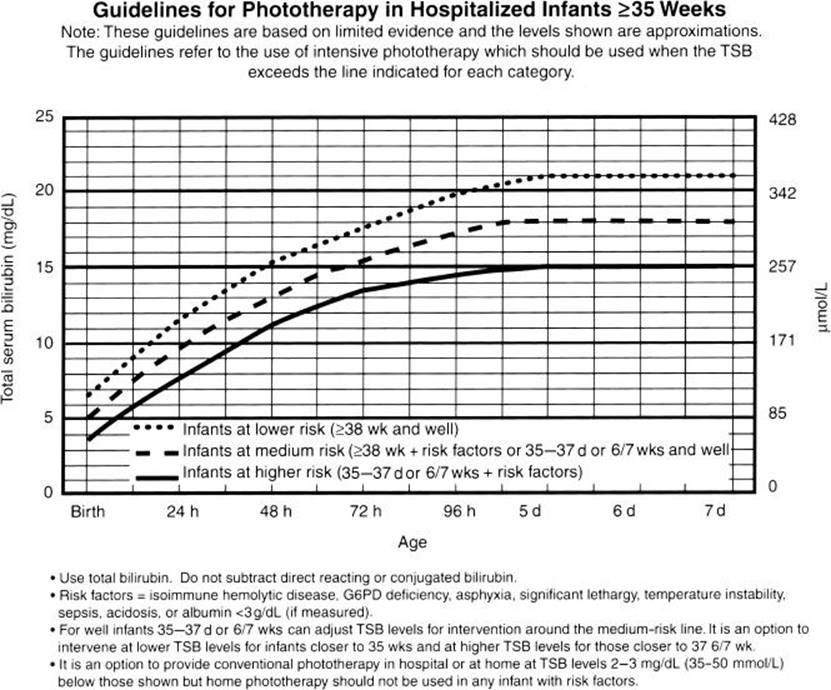
FIGURE 221.2 Management of hyperbilirubinemia in the newborn infant of 35 or more weeks of gestation. AAP guidelines for phototherapy in hospitalized infants of 35 or more weeks of gestation. Note: These guidelines are based on limited evidence and the levels shown are approximations. The guidelines refer to the use of intensive phototherapy which should be used when the TSB exceeds the line indicated for each category. Infants are designated as “higher risk” because of the potential negative effects of the conditions listed on albumin binding of bilirubin the blood–brain barrier, and the susceptibility of the brain cells to damage by bilirubin. (From MacDonald MG, Mullett MD, Seshia MMK. Avery’s Neonatology: Pathophysiology and Management of the Newborn. 6th ed. Philadelphia, PA: Lippincott Williams & Wilkins; 2005.)
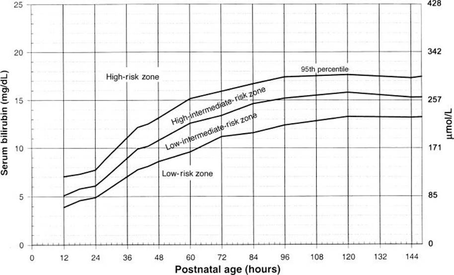
FIGURE 221.3 Nomogram for designation of risk in 2,840 well newborns ≤36 weeks of gestational age with birth weight ≤2,000 g or ≤35 weeks of gestational age and a birth weight ≤2,500 g or more based on the hour-specific serum bilirubin values. The serum bilirubin level was obtained before discharge, and the zone in which the value fell predicted the likelihood of a subsequent bilirubin level exceeding the 95th percentile (high-risk zone). (From MacDonald MG, Mullett MD, Seshia MMK. Avery’s Neonatology: Pathophysiology and Management of the Newborn. 6th ed. Philadelphia, PA: Lippincott Williams & Wilkins; 2005.)
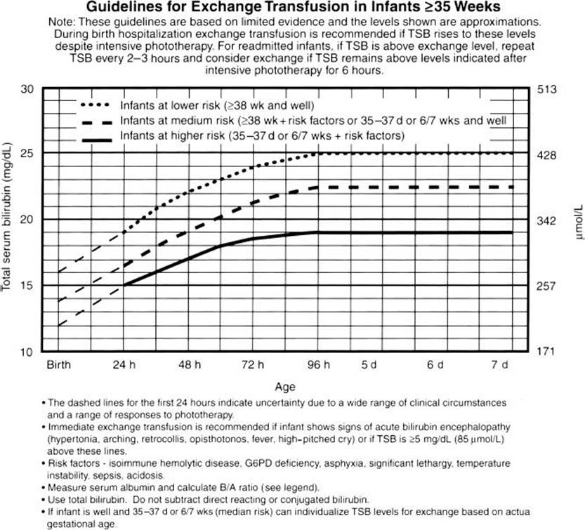
FIGURE 221.4 AAP guidelines for exchange transfusion in infants who are 35 or more weeks gestation. Note that these suggested levels are based on limited evidence, and the levels shown are approximations. During birth hospitalization, exchange transfusion is recommended if the TSB rises to these levels despite intensive phototherapy. For readmitted infants, if the TSB level is above the exchange level, repeat TSB measurement every 2 to 3 hours and consider exchange if the TSB remains above the levels indicated after intensive phototherapy for 6 hours. (From MacDonald MG, Mullett MD, Seshia MMK. Avery’s Neonatology: Pathophysiology and Management of the Newborn. 6th ed. Philadelphia, PA: Lippincott Williams & Wilkins; 2005.)
Laboratory evaluation consistent with conjugated hyperbilirubinemia (conjugated bilirubin >1 mg/dL when total bilirubin is <5 mg/dL or more than 20% of total bilirubin if the total bilirubin is ≤5 mg/dL) should prompt an evaluation for an underlying cause (eFig. 221.3) (14). If the onset was abrupt, consider perinatal TORCHS infections (i.e., toxoplasmosis, other infections, rubella, cytomegalovirus, herpes simplex, and syphilis), bacterial sepsis, and urinary tract infections (15). If the neonate appears well and does not have evidence of perinatal or bacterial infection, a urinalysis and urine culture should be ordered since a conjugated hyperbilirubinemia may be an early sign of neonatal urinary tract infection.
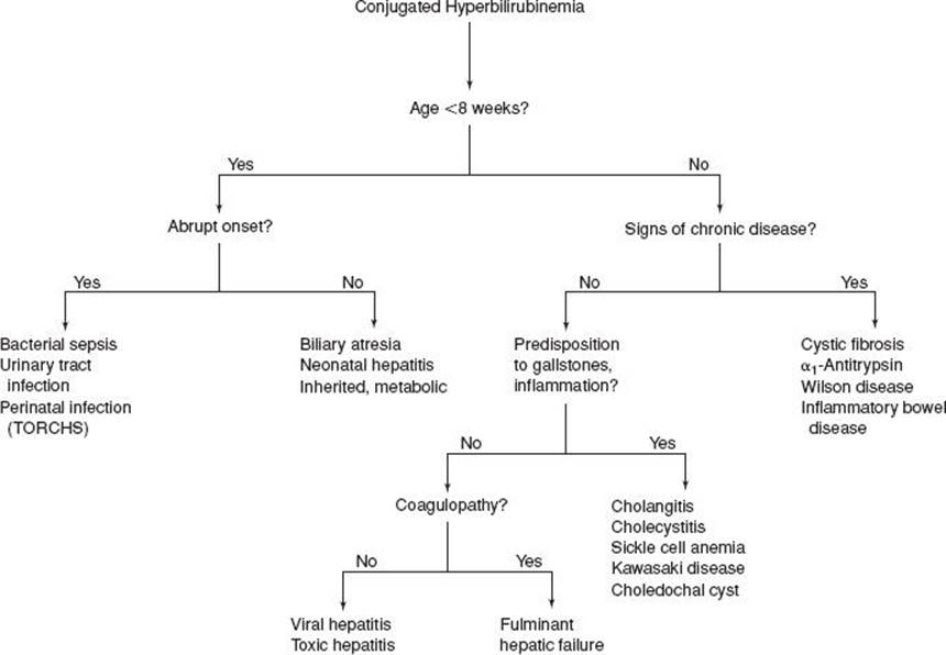
eFIGURE 221.3 Evaluation of the pediatric patient with unconjugated hyperbilirubinemia. (From Fleisher GR, Ludwig S, Henretig FM, et al., eds. Textbook of Pediatric Emergency Medicine. 5th ed. Philadelphia, PA: Lippincott Williams & Wilkins; 2005.)
Infants with biliary atresia usually present with gradual onset of jaundice and acholic stools. However, it is important to maintain a high index of suspicion for biliary atresia in any neonate presenting with conjugated hyperbilirubinemia (16). Other disorders to consider when there is a more insidious onset of conjugated hyperbilirubinemia include metabolic disorders, and viral hepatitis. Laboratory studies will assist in assessing the degree of hepatocyte injury and biliary dysfunction (serum alanine aminotransferase and aspartate aminotransferase, serum alkaline phosphatase, and gamma-glutamyl transpeptidase) as well as the biosynthetic function of the liver (prothrombin and partial thromboplastin time, serum albumin, glucose, and ammonia). Newborn screening for thyroid disease and galactosemia should be reviewed and an ultrasound of the liver and biliary tract ordered. An extensive evaluation is often required in these patients and consultation with a pediatric gastroenterologist can guide additional testing and treatment. Phototherapy is relatively contraindicated in neonates with conjugated hyperbilirubinemia due to its association with bronze baby syndrome (4).
For infants who present after the first 2 weeks of life with jaundice, measure the total and conjugated bilirubin level to assess for evidence of cholestasis. For exclusively breastfed infants with no history of dark urine or acholic stools, a normal physical examination and reliable follow-up can have this evaluation deferred until 3 weeks of age. If the conjugated bilirubin is elevated, check the results of the newborn screen and evaluate for causes of cholestasis (see above) (4,17). Infants with elevated unconjugated bilirubin levels may be diagnosed with breast-milk jaundice, if they are exclusively breastfed and are otherwise well appearing. For these patients close follow-up should be ensured, however (Table 221.2).
TABLE 221.2
Causes of Jaundice After the Neonatal Period

CRITICAL INTERVENTIONS
• Neonates presenting with temperature instability, lethargy, apnea, or poor feeding require prompt evaluation for possible sepsis and initiation of antibiotics and resuscitative efforts, as indicated.
• For neonates who meet phototherapy threshold criteria, intensive phototherapy should be initiated promptly.
• Neonates with severe hyperbilirubinemia who meet criteria for exchange transfusion require emergent transfer to a critical care setting where exchange transfusion can be initiated.
DISPOSITION
Neonates who are ill appearing or meet criteria for phototherapy or exchange transfusion require admission for treatment. Well-appearing neonates with unconjugated (physiologic) hyperbilirubinemia that do not meet the threshold for phototherapy can be discharged home if close follow-up and repeat testing as indicated by risk assessment can be ensured. The majority of young infants with conjugated hyperbilirubinemia will require hospitalization for further diagnostic evaluation.
Common Pitfalls
• Visual determination of bilirubin level in neonates is unreliable; laboratory evaluation should always be performed to determine bilirubin level.
• Jaundice may be the initial presentation of serious bacterial infections in neonates.
• If a neonate’s bilirubin level does not meet the phototherapy threshold, determine risk for subsequent significant hyperbilirubinemia and ensure necessary follow-up prior to discharge.
REFERENCES
1. Mandl KD. Jaundice–unconjugated hyperbilirubinemia. In: Fleisher GR, Ludwig S, eds. Textbook of Pediatric Emergency Medicine. Philadelphia, PA: Lippincott Williams & Wilkins; 2010.
2. Dennery PA, Seidman DS, Stevenson DK. Neonatal hyperbilirubinemia. N Engl J Med. 2001;344(8):581–590.
3. Takci S, Hanoglu D, Hascelik G, et al. Neonatal cholestasis in gram-negative septicaemia. J Pediatr Gastroenterol Nutr. 2012;55(6):e153; author reply e153.
4. American Academy of Pediatrics Subcommittee on Hyperbilirubinemia. Management of hyperbilirubinemia in the newborn infant 35 or more weeks of gestation. Pediatrics. 2004;114(1):297–316.
5. Kawade N, Onishi S, The prenatal and postnatal development of UDP-glucuronyltransferase activity towards bilirubin and the effect of premature birth on this activity in the human liver. Biochem J.1981;196:257–260.
6. Kaplan M, Muraca M, Hammerman C, et al. Imbalance between production and conjugation of bilirubin: A fundamental concept in the mechanism of neonatal jaundice. Pediatrics. 2002;110(4):e47.
7. Soldi A, Tonetto P, Varalda A, et al. Neonatal jaundice and human milk. J Matern Fetal Neonatal Med. 2011;24(suppl 1):85–87.
8. Koziol LF, Budding DE, Chidekel D. Hyperbilirubinemia: Subcortical mechanisms of cognitive and behavioral dysfunction. Pediatr Neurol. 2013;48(1):3–13.
9. Sgro M, Campbell DM, Kandasamy S, et al. Incidence of chronic bilirubin encephalopathy in Canada, 2007–2008. Pediatrics. 2012;130(4):e886–e890.
10. Selbst SM, Baghdassarian A. Pediatric emergency medicine: Legal briefs. Pediatr Emerg Care. 2011;27(12):1213–1215.
11. Keren R, Tremont K, Luan X, et al. Visual assessment of jaundice in term and late preterm infants. Arch Dis Child Fetal Neonatal Ed. 2009;94(5):F317–F322.
12. Riskin A, Tamir A, Kugelman A, et al. Is visual assessment of jaundice reliable as a screening tool to detect significant neonatal hyperbilirubinemia? J Pediatr. 2008;152(6):782–787, 787.e1–787.e2.
13. Wolff M, Schinasi DA, Lavelle J, et al. Management of neonates with hyperbilirubinemia: Improving timeliness of care using a clinical pathway. Pediatrics. 2012;130(6):e1688–e1694.
14. Brumbaugh D, Mack C. Conjugated hyperbilirubinemia in children. Pediatr Rev. 2012;33(7):291–302.
15. Bazlul Karim AS, Kamal M. Cholestatic jaundice during infancy: Experience at a tertiary-care center in Bangladesh. Indian J Gastroenterol. 2005;24(2):52–54.
16. Muraji T. Early detection of biliary atresia: Past, present & future. Expert Rev Gastroenterol Hepatol. 2012;6(5):583–589.
17. Moyer V, Freese DK, Whitington PF, et al. Guideline for the evaluation of cholestatic jaundice in infants: Recommendations of the North American Society for Pediatric Gastroenterology, Hepatology and Nutrition. J Pediatr Gastroenterol Nutr. 2004;39(2):115–128.