
Therese L. Canares and Dale Steele
Pediatric oral lesions are primarily the result of infectious etiologies including herpes, Coxsackie and group A Streptococcus (GAS), among others. Complaints of oral bleeding may arise from post-tonsillectomy eschar. Autoimmune causes of oral lesions in children include Kawasaki disease (KD) and Stevens–Johnson syndrome (SJS).
CLINICAL PRESENTATION
Herpes simplex virus (HSV) causes primary herpetic gingivostomatitis and is characterized by fever, gingivitis, and a vesicular rash that leaves shallow, painful ulcers (Fig. 240.1).
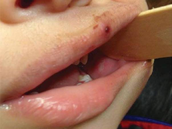
FIGURE 240.1 Infant with herpetic gingivostomatitis. An infant with a healing vesicle on the upper lip and a shallow ulcer in the buccal mucosa. Vesicles are seen in the anterior oral mucosa, periorally or, particularly in a thumb-sucking child in the periungual area (i.e., herpetic whitlow).
Coxsackievirus causes herpangina, a summertime condition characterized by small, tender vesicles with an erythematous halo that may be similar to herpes gingivostomatitis, however appear primarily in the posterior pharynx. Coxsackievirus is also the infectious etiology associated with hand-foot-and-mouth disease (Fig. 240.2).
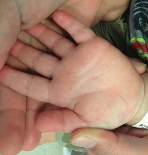
FIGURE 240.2 Hand, foot and mouth disease: This is caused by coxsackie virus, and manifests as herpangina, with fever, and a papulovesicular rash in the palms, soles or buttocks.
Infectious mononucleosis and GAS pharyngitis both present with exudative pharyngitis, palatal petechiae, cervical lymphadenopathy, and fever. GAS pharyngitis patients commonly complain of fever and sore throat without a cough. Mononucleosis is distinguished by profound fatigue and posterior cervical lymphadenopathy. Oral candidiasis causes a white, friable pseudomembrane, frequently seen in infants or immunosuppressed patients. The clinician should evaluate for erythematous satellite papules of candidal diaper dermatitis. Measles is a clinical diagnosis based on whitish-blue papules on an erythematous base in the buccal mucosa (Koplik spots), and associated cough, coryza, conjunctivitis, and rash. As declining vaccination rates threaten herd immunity, measles outbreaks in the United States are reaching their highest rates since 1996 and are increasingly relevant for the emergency clinician (1).
Surgical consultation is occasionally needed for intraoral emergencies. Delayed post-tonsillectomy hemorrhage is uncommon (1% to 5%) but potentially life-threatening and should be thoroughly evaluated (Fig. 240.3) (2).
Many autoimmune conditions have associated oral findings, however the clinician must be diligent to identify high-acuity conditions such as KD and SJS. Oral lesions of KD may include cracked red lips, strawberry tongue, crusting of the lips and mouth, or a nonexudative pharyngitis. Similarly to KD, SJS causes severe mucositis with painful erosions, crusting, and sloughing of mucous membranes (Fig. 240.4).

FIGURE 240.3 Post-operative tonsillectomy eschar and hemorrhage: During healing, a whitish-yellow fibrin clot or eschar (left) develops in the tonsillar fossae, which may be confused as exudate or thrush. The eschar sloughs from underlying granulomatous tissue 7-10 days post-operatively, which may cause bloody sputum, hematemesis, or hemodynamic instability. Clot is visible in the right tonsillar pillar.

FIGURE 240.4 Oral findings in Stevens–Johnson syndrome. A 9-year-old with ulcerated, crusted, macerated lips.
DIFFERENTIAL DIAGNOSIS
Beyond the aforementioned pathologic conditions, a number of benign oral lesions may be seen in the ED. These include aphthous ulcers, geographic tongue, angular cheilitis, gum boil, ranula, Bohn nodules, Epstein pearls, mucocele, papillomas, fibromas, hemangiomas, pyogenic granulomas, lichen planus, and dental eruption cysts.
ED EVALUATION
Oral lesions can often be diagnosed clinically. Confirmation of HSV is made by viral culture, polymerase chain reaction, or immunofluorescence staining (DFA) on cells scraped from the base of unroofed vesicles (3). GAS pharyngitis is diagnosed by rapid antigen testing and confirmatory culture (4). Mononucleosis frequently causes an atypical lymphocytosis on complete blood count (CBC). To better characterize the diagnosis of mononucleosis EBV serology should be performed, since the heterophile antibody (Monospot) test has poor sensitivity in children. Heterophile antibodies are positive in only 25% to 50% of children under 12 years of age (5). A CBC and type and screen should be obtained on those with post-tonsillectomy hemorrhage. Autoimmune disorders show nonspecific elevation of acute-phase reactants. Patients with concern for KD should have CBC, chemistry, urinalysis, and liver function tests to evaluate for anemia, sterile pyuria, and transaminitis (6).
KEY TESTING
• CBC with type and cross for post-tonsillectomy hemorrhage
• Serum electrolytes in dehydrated children with oral lesions and limited PO intake
ED MANAGEMENT
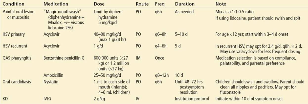
Management of oral lesions consists of supportive care and treatment of the underlying disease (Table 240.1). Assessment of dehydration and pain control are absolutely essential and may justify inpatient admission. Painful oral lesions are initially approached with analgesics such as ibuprofen or acetaminophen. “Magic mouthwash” is often used for painful lesions, with care not to overdose diphenhydramine. Avoid toxicity from topical lidocaine by using cotton-tipped applicator in infants, or have the child swish and spit (7). Narcotics are indicated for failure of over-the-counter analgesics or in severe cases. Tolerance of oral fluids must be demonstrated prior to discharge. Inability to tolerate liquids or severely dehydrated patients with tachycardia, dry mucous membranes and decreased urination may require intravenous hydration and perhaps admission. Patients with mononucleosis should be advised to avoid contact sports for at least 3 weeks to reduce the risk of splenic rupture (5). Subspecialty consultation should be made for post-tonsillectomy hemorrhage and potential intraoperative cauterization or for autoimmune conditions that require systemic treatment.
TABLE 240.1
Suggested Therapy for Pediatric Oral Lesions (8)
CRITICAL INTERVENTIONS
• Assess for dehydration in infants and toddlers with poor oral intake or decreased urine output
• Use topical lidocaine with caution for systemic toxicity (arrhythmias or seizures) (7)
• In post-tonsillectomy bleeds ensure a patent airway, obtain a CBC, T&S, and consult the surgeon
• Treat KD with IVIG within the first 10 days of illness to prevent coronary artery aneurysms (6)
DISPOSITION
Disposition is case dependent. Patients may be discharged home with over-the-counter, or rarely narcotic pain medication. Patients are admitted if unable to tolerate oral intake, there is evidence of sepsis or systemic disease warrants admission.
Common Pitfalls
• Ensure adequate pain control and tolerance of oral liquids prior to discharge.
• Consider child abuse if oral HSV coincides with genitourinary lesions in a prepubertal child.
• Maintain an index of suspicion for KD in children with prolonged fever.
• Consider mononucleosis with splenic rupture in fever, fatigue, tonsillar lesions, and left upper quadrant abdominal pain.
REFERENCES
1. Moss WJ, Griffin DE. Measles. Lancet. 2012;379:153–164.
2. Isaacson G. Tonsillectomy care for the pediatrician. Pediatrics. 2012;130:324–334.
3. Arduino PG, Porter SR. Herpes simplex virus type 1 infection: Overview on relevant clinico-pathological features. J Oral Pathol Med. 2008;37:107–121.
4. Shaikh N, Swaminathan N, Hooper EG. Accuracy and precision of the signs and symptoms of streptococcal pharyngitis in children: A systematic review. J Pediatr. 2012;160:487–493.e3.
5. Luzuriaga K, Sullivan JL. Infectious mononucleosis. N Engl J Med. 2010;362:1993–2000.
6. Harnden A, Takahashi M, Burgner D. Kawasaki disease. BMJ. 2009;338:b1514.
7. Faden H. Management of primary herpetic gingivostomatitis in young children. Pediatr Emerg Care. 2006;22:268–269.