
Bonnie Mackenzie and Siraj Amanullah
Neck masses present frequently in pediatric practice and constitute a wide range of diagnostic possibilities. The ED is often the frontline for diagnosis of congenital cysts and malignancies. A basic understanding of anatomy and embryology can simplify the approach to evaluating nontraumatic neck masses of infectious, inflammatory, or malignant origin. While the vast majority of patients have benign diagnoses related to lymphadenitis, clinicians must always ensure that potential airway problems are addressed in a timely fashion.
CLINICAL PRESENTATION
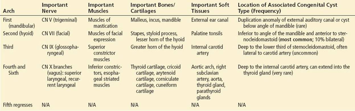
The head and neck develop from a branchial apparatus which, at the mesodermal level, is comprised of the first through fourth and sixth branchial arches (the fifth arch regresses and is not clinically relevant). The branchial arches, derived structures, and associated congenital cyst types (congenital neck masses) are summarized in Table 241.1.
TABLE 241.1
Branchial Arches, Derived Structures, and Associated Congenital Neck Masses
Anatomically, the neck can be divided into an anterior and posterior triangle separated by the sternocleidomastoid muscle. The boundaries of these triangles, and the important contents of each, are outlined in Table 241.2 and Figure 241.1 (2).
TABLE 241.2
Boundaries and Contents of the Anterior and Posterior Triangles of the Neck

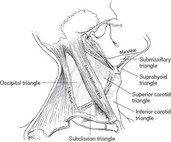
FIGURE 241.1 The anterior and posterior triangles of the neck, lithograph plate from Gray’s Anatomy.
The clinical presentation of a neck mass can include diffuse or localized swelling, fever, poor oral intake, neck stiffness, torticollis, drooling, and various other symptoms; see Table 241.3 (3).
TABLE 241.3
Common Presenting Symptoms Associated with Neck Masses
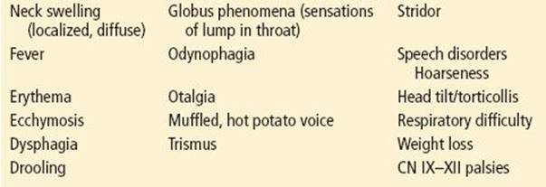
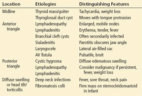
Table 241.4 lists common presentations of neck masses by anatomic location, with likely etiologies and distinguishing clinical features.
TABLE 241.4
Anatomic Neck Mass Presentations
Ill-Appearing Child with Fever
Fever and localized neck swelling that is tender and warm to the touch (with or without skin changes) suggests a superficial or deep neck infection. This may originate from local structures including tonsils, middle ear, lymph or salivary glands, a congenital cyst or, rarely, from a malignancy.
Deep neck infections include peritonsillar abscesses (with possible extension in neck planes), retropharyngeal abscesses (primarily seen in children ≤5 years old due to regression of involved lymph nodes with age), parapharyngeal abscesses, sialadenitis, supramylohyoid infections, and superinfected congenital malformations. Congenital malformations can become clinically evident at any point in life, and often present as a result of super-infection (1).
Diagnosing potentially life-threatening infections can be challenging due to the anatomic complexity and the subtle or overlapping presentations of more benign conditions such as cervical lymphadenitis. In addition, relative to adults, pediatric patients have the potential for retropharyngeal or parapharyngeal infections of lymphoid tissue in spaces that generally atrophy by adulthood. Such infections present with neck pain, swelling, or stiffness, associated with sore throat, poor oral intake, drooling, stridor, or lymphadenopathy. Drooling is an especially worrisome marker for airway compromise. In very young infants (<9 months), the most common presenting symptoms of deep neck infections include neck swelling, lymphadenopathy, fever, rhinorrhea, poor oral intake, and cough (3).
Potentially life-threatening complications of deep neck infections include mediastinitis, Ludwig angina, and Lemierre syndrome. Ludwig angina is a rapidly progressive, firm cellulitis of the floor of the mouth usually originating from a diseased tooth, but it may also originate from a peritonsillar or parapharyngeal abscess, epiglottitis, or a penetrating injury to the floor of the mouth. Lemierre syndrome, usually seen in older children, is an infected thrombosis of the internal jugular vein that is associated with parapharyngeal infections such as pharyngitis, tonsillitis, cervical lymphadenitis, and parotitis. Lemierre syndrome should be considered in a sick-appearing child with significant neck pain (3,4).
Neoplasm should be considered in a child with a firm, nonmobile, enlarging neck mass, and a history of fevers, weight loss, bone pain, or night sweats. Other concerning features include a persistently enlarged lymph node that has been present for several weeks, enlarged nodes in the posterior triangle or supraclavicular space, or lymphadenopathy that is associated with recurrent epistaxis, hoarseness, cranial nerve palsies, or otorrhea, which may indicate local tissue invasion by a mass (5,6). A list of benign and malignant head and neck neoplasms is included in the differential diagnosis in Table 241.4.
Well-Appearing Child with Fever
The most likely diagnoses include reactive lymphadenopathy, lymphadenitis due to viral or bacterial pathogens, and salivary gland infections. If fever and adenopathy are persistent (i.e., for more than a few weeks), etiologies to be considered should include mycobacterial infections, granulomatous processes, and Bartonella henselae infection (catscratch fever) (6).
Salivary gland infections should be considered as a cause of neck swelling because the majority of the parotid gland tissue is below the angle of the mandible. Parotitis often presents as an acute neck swelling that obscures the angle of the jaw Figure 241.2. In pediatric patients, acute tender enlargement of the parotid gland is usually infectious, or due to salivary duct stones (7). The differential diagnosis of parotitis includes viral infections, including mumps due to paramyxovirus, rarely seen in immunized patients, Coxsackieviruses, CMV, HIV-related EBV infection, Staphylococcus aureus infection, mycobacteriumtuberculosis, juvenile recurrent parotitis (a chronic condition without a well-defined etiology that develops between age 3 and 6 years), and bulimia nervosa sialadenosis (8).
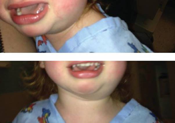
FIGURE 241.2 Parotitis in a toddler. Note how the enlarged parotid gland obscures the angle of the mandible.
Well-Appearing, Afebrile Child
This category includes mostly noninfected congenital lesions and vascular anomalies. Congenital malformations are common in the pediatric population, but may not become evident until adolescence or adulthood; they should therefore be considered in the differential of any patient presenting with a neck mass (1,9,10). The most common include branchial cleft anomalies (cysts being most common), thyroglossal duct cysts, cystic hygromas, laryngoceles, and epidermoid and dermoid cysts (see Chapter 261).
While patients with vascular malformations may not appear toxic, these lesions can potentially cause life-threatening complications due to airway compromise, bleeding, or high-output cardiac failure. Hemangioma, a very common pediatric vascular tumor, may present as a neck mass and cause subglottic obstruction or high-output cardiac failure. Hemangiomas are generally enlarging red or blue masses that typically proliferate before involuting. Cutaneous lesions may indicate the presence of hemangiomas in other organs, and one-half of infants with subglottic hemangiomas also have a cutaneous hemangioma. Any hemangioma found in the “beard distribution” (the preauricular areas, chin, anterior neck, and lower lip) should trigger further evaluation for associated airway hemangiomas (1,11). For children without stridor or drooling, this evaluation may take place as an outpatient. Cavernous hemangiomas are venous malformations due to thin-walled, dilated veins that grow with the child, are usually soft with a blue hue, and carry a risk of thrombosis or bleeding. Arteriovenous malformations can be distinguished by their palpable thrill, audible bruit, warmth, and redness. Most importantly, these lesions carry a risk of high-output cardiac failure (12).
DIFFERENTIAL DIAGNOSIS
The differential diagnosis of neck mass can be organized according to suspected etiologies (Table 241.5).
TABLE 241.5
Etiologies of Pediatric Neck Masses

ED EVALUATION
Diagnostic evaluation begins with a thorough history and physical examination. The child’s overall appearance, the location of the neck mass, the appearance of the pharynx, and the patency of the airway all help guide the clinician’s next steps. In most well-appearing children, infectious and inflammatory causes predominate and little further diagnostic testing is required. When reactive lymphadenopathy or lymphadenitis is suspected, a thorough investigation of the head and neck, including the tympanic membranes, pharynx, and skin, should be performed. In patients with fever, tonsillar erythema, and exudate, a rapid streptococcal antigen test may be diagnostic. In young children with suspected infectious mononucleosis (lymphadenopathy, fever, pharyngeal erythema, enlarged spleen), a CBC may reveal atypical lymphocytes, which support the diagnosis. In adolescents, heterophile antibody testing (monospot) can be helpful, but in younger children, EBV-specific serologies must be obtained because heterophile antibodies are often falsely negative in children less than 12 years of age (up to 50% false-negative rate) (13). Erythrocyte sedimentation rate and C-reactive protein measurements can be helpful in tracking infections, but are not necessary in well-appearing children. If there is a draining mass, a Gram stain and culture may guide therapy. Well neonates with a firm mass overlying the lower third of the sternocleidomastoid muscle (with or without torticollis) are likely to have fibromatosis colli, a clinical diagnosis confirmed with ultrasound (US). Treatment is physical therapy.
In the ill-appearing child without airway compromise a complete blood count, blood cultures, CRP, ESR, and x-rays of the neck and chest may help to initially guide diagnosis. Further testing should be based on history (exposures, duration of illness, and review of systems) and physical examination findings. In the ill-appearing child with evidence of airway compromise, x-rays of the neck (AP and lateral soft tissue) and chest can help assess the patency of the airway and lung fields (14). If there is respiratory failure, the airway should be secured by the most experienced operator, with equipment available for an emergent surgical airway (15).
In children with suspected malignancy, a complete and differential blood count is indicated, as well as a screening chest x-ray for the presence of a mediastinal mass. For suspected thyroid masses, thyroid function testing is recommended.
In addition to plain films (particularly the lateral neck film, which can quickly assess for potential airway compromise), US is the initial imaging modality of choice in children (14,16). US is readily available, can be interpreted quickly, does not involve radiation exposure, and is noninvasive. US is particularly useful in children, given their smaller neck size and less prominent subcutaneous fat, allowing for improved sonographic penetration and resolution compared to adults (14). When additional imaging is needed, magnetic resonance imaging and computed tomography (CT) are both useful for defining anatomy. CT is usually readily available, has a short study time that may obviate the need for sedation in younger children, and allows for excellent assessment of bone involvement. However, these benefits must be balanced against the risks of ionizing radiation. MRI is excellent for assessment of soft tissues and carries no radiation risks, but may have limited availability and usually requires sedation.
CRITICAL INTERVENTIONS
• Most well-appearing children with neck masses have lymphadenopathy or lymphadenitis that requires little intervention beyond consideration of antibiotic therapy if a bacterial origin is suspected.
• Antibiotics providing coverage of common head and neck pathogens should be instituted for neck masses that appear infected or for children who appear systemically ill.
• Cultures of neck drainage or blood cultures in systemically ill patients should be obtained prior to antibiotic therapy to tailor the antibiotic choice.
• Incision and drainage of neck masses is not recommended without a definitive diagnosis having been made; this may lead to fistula formation in the case of congenital malformation, or may seed the incision tract in the case of tumor.
• Lymphadenopathy that has persisted beyond several weeks or that is unresponsive to treatment should be considered a potential malignancy or atypical infection, and warrants imaging and consultation.
DISPOSITION
Well-appearing children without airway compromise may be managed as outpatients with appropriate follow-up. Most children with lymphadenitis can be prescribed outpatient antibiotics with primary care follow-up. In the stable child with evidence of a congenital malformation, referral to a pediatric surgeon or otolaryngologist is recommended. Ill-appearing children should be admitted to the hospital with airway support and antibiotics as indicated.
Common Pitfalls
• Failure to recognize impending respiratory failure from airway obstruction due to a neck mass.
• Ordering a CT scan of a neck mass when US or MRI is available and appropriate.
• Incision and drainage of a neck mass in the absence of a definitive diagnosis.
• Failure to recognize possible superinfected congenital malformations.
REFERENCES
1. Geddes G, Butterly MM, Patel SM, et al. Pediatric neck masses. Pediatr Rev. 2013;34:115–124; quiz 125.
2. Drake RL, Vogl AW, Mitchell AWM, et al. Gray’s Anatomy for Students. Philadelphia, PA: Churchill Livingstone/Elsevier; 2010.
3. Vieira F, Allen SM, Stocks RM, et al. Deep neck infection. Otolaryngol Clin North Am. 2008;41:459–483, vii.
4. Chirinos JA, Lichtstein DM, Garcia J, et al. The evolution of Lemierre syndrome: Report of 2 cases and review of the literature. Medicine (Baltimore). 2002;81:458–465.
5. Dickson PV, Davidoff AM. Malignant neoplasms of the head and neck. Semin Pediatr Surg. 2006;15:92–98.
6. Tracy TF Jr, Muratore CS. Management of common head and neck masses. Semin Pediatr Surg. 2007;16:3–13.
7. Cummings CW. Cummings Otolaryngology Head & Neck Surgery. 4th ed. Philadelphia, PA: Elsevier Mosby; 2005.
8. Patel A, Karlis V. Diagnosis and management of pediatric salivary gland infections. Oral Maxillofac Surg Clin North Am. 2009;21:345–352.
9. LaRiviere CA, Waldhausen JH. Congenital cervical cysts, sinuses, and fistulae in pediatric surgery. Surg Clin North Am. 2012;92:583–597, viii.
10. Rosa PA, Hirsch DL, Dierks EJ. Congenital neck masses. Oral Maxillofac Surg Clin North Am. 2008;20:339–352.
11. Orlow SJ, Isakoff MS, Blei F. Increased risk of symptomatic hemangiomas of the airway in association with cutaneous hemangiomas in a “beard” distribution. J Pediatr. 1997;131:643–646.
12. Higuera S, Gordley K, Metry DW, et al. Management of hemangiomas and pediatric vascular malformations. J Craniofac Surg. 2006;17:783–789.
13. Luzuriaga K, Sullivan JL. Infectious mononucleosis. N Engl J Med. 2010;362:1993–2000.
14. Friedman ER, John SD. Imaging of pediatric neck masses. Radiol Clin North Am. 2011;49:617–632, v.
15. Liess BD, Scheidt TD, Templer JW. The difficult airway. Otolaryngol Clin North Am. 2008;41:567–580, ix.
16. Fefferman NR, Milla SS. Ultrasound imaging of the neck in children. Ultrasound Clinics. 2009;4:553–569.