
Rick C. Place and Thom A. Mayer
Unintentional injury is the leading cause of death in the United States for children aged 1 to 19 years of age. Accidents, homicide, and suicide combined are responsible for over 12,000 deaths a year and are, respectively, the three leading causes of death. In children over the age of 10, motor vehicle crashes (MVCs) are by far the leading cause of death, while under 10, drowning and burns or smoke inhalation figure prominently (1). These figures contrast with the first year of life, during which only 4.4% of deaths are related to unintentional injury. During this same time period, there were over 9 million nonfatal injuries in children under 20 years of age (1).
CLINICAL PRESENTATION
Physiologic and Anatomic Differences
Just as the octogenarian is treated differently than the athletic university student, the approach to an injured infant differs greatly from that of the adolescent. While the transition is gradual, the changes are extraordinary. Pediatric physiology evolves into more adult physiology during the school-age years, between 7 and 9 years of age. This chapter focuses on the unique issues of the pediatric patient under the age of 7 and assumes an understanding of adult trauma care (Fig. 245.1) (see Chapter 18).

FIGURE 245.1 Pediatric anatomic differences.
One of the most important differences in children is the size of the head relative to the rest of the body. The infant’s prominent occiput is comparatively heavy relative to the adult’s, comprising 25% of body weight versus 10% in adults. This increased mass, coupled with weaker cervical musculature and hence less overall head control, makes infants and children highly vulnerable to injury. The cranial sutures, open at birth, gradually fuse by 12 to 18 months of age; open bulging fontanels may indicate increased intracranial pressure, but whether this results in meaningful decompression of the injured brain is debatable.
A thin and fragile skull, particularly in the first several months of life, is more liable to fracture with even a minor mechanism. In younger children, particularly those under the age of 2, a boggy lesion over the site of impact may indicate a cephalohematoma from an underlying skull fracture.
The infant’s brain has higher water content, incomplete myelinization, and greater biomechanical vulnerability to impact and shearing injuries relative to adults. This results in a higher rate of diffuse brain injury in children.
Physiologic and anatomic characteristics of the cervical spine, as well as differences in age-related activities, predispose children to different injury patterns. A relatively larger and heavier head results in a fulcrum of flexion at C2/C3, instead of C5/C6 in the fully grown adult. Weaker neck musculature, lax ligaments, anterior wedging of the vertebral bodies, and more-horizontal facets predispose to upper spinal cord injury (SCI). A predominance of upper cervical spine injuries and a greater incidence of SCI without radiographic abnormality (SCIWORA) have been noted in younger children (2,3).
The rate of cervical spine injury in children (approximately 0.7% to 1.6%) is substantially lower than in adults (2.4%). The risk of injury, however, approximately doubles (3.2%) for children involved in MVCs.
The cardiopulmonary response to stress in infants differs from older children and adults. The myocardium is stiffer, less contractile, and less able to generate ventricular wall tension. The infant heart is therefore more rate dependent, without as much ability to increase stroke volume. Similar to the heart, pulmonary reserve is more restricted by anatomic structure; minute volume is thus more dependent on rate rather than tidal volume. Children also have reduced functional residual capacity (i.e., oxygen storage) coupled with metabolic demands that are twice those of adults; consequently, desaturation may occur more rapidly and precipitously. All told, differences in baseline cardiac and respiratory rates, as well as differences in age-related norms, make recognizing compensated shock challenging for providers.
Certain structural and anatomic differences also place young children at risk for increased morbidity. Their relatively increased body surface area (BSA) increases the risk of hypothermia and leads to necessary modifications of the Rule of Nines chart used for burn care resuscitation. The less mineralized pediatric rib cage is more likely to bend rather than break, transmitting forces internally without concomitant rib fracture, resulting in higher rates of pulmonary contusion. The pediatric liver and spleen are relatively larger, in closer proximity to one another without surrounding fat and connective tissue, and exist within a less developed, weaker abdominal musculature. This makes these organs, like the lung within the pediatric rib cage, more vulnerable to transmitted blunt forces.
Children’s bones are softer and more porous, have increased ligamentous laxity and a greater percentage of unossified cartilage, which improves their ability to absorb blunt metaphyseal impact. However, this leads to a higher rate of epiphyseal injuries in children. While increased laxity serves to protect children from bony cervical spine injury, these same characteristics increase the vulnerability to cord injury, demonstrated in its purest form as SCIWORA.
General Approach to the Pediatric Trauma Patient
The fundamental primary survey–secondary survey format of the Advanced Trauma Life Support (ATLS) course remains the most effective and efficient manner of evaluating injured children of all ages.
This structured approach to major trauma is no different from that used in the evaluation of the adult trauma patient, but there are some critical differences. These include (1) the requirement for all team members to agree upon the patient’s body weight in order to facilitate error-free weight-based administration of fluids and medications; (2) the need to maintain core body temperature; (3) the need to be especially concerned about the recognition of cervical spine injuries; (4) the need to establish an airway and use appropriate noninvasive ventilation techniques; (5) the need to recognize circulatory stability and shock; (6) the importance of promptly establishing adequate vascular access; and (7) the importance of proper management of pediatric head injury.
PRIMARY SURVEY
Airway and Breathing
The pediatric airway is anatomically immature and different from the airway of older children and adults, particularly in the first 2 years (see Chapter 218). Infants are “belly” breathers, meaning they require full diaphragmatic excursion to produce normal respirations. More-horizontal ribs and a flatter diaphragmatic insertions provide less mechanical advantage. Having said this, most respiratory issues in trauma are related to reduce respiratory effort because of CNS injury rather than respiratory distress from pulmonary pathology.
The highest priority in caring for either adult or pediatric patients with multiple traumatic injuries is ensuring or providing a patent airway with adequate ventilation and oxygenation while protecting the cervical spine. In the severely injured child with an altered level of consciousness and impaired respiratory effort, the most frequent error is failure to position the airway properly. In the trauma patient, the jaw thrust is the single most important airway maneuver, pulling the mandible and tongue away from the posterior pharynx. Placing a flattened towel under the shoulders will raise the torso and may help facilitate maneuvers to maintain patency by straightening the airway (Fig. 245.2).
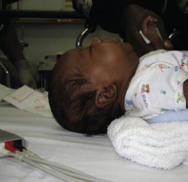
FIGURE 245.2 Pediatric airway. Placing a towel under the shoulders helps better align the airway by raising the torso, optimizing breathing and visualization of the airway.
In pediatric patients with compromised respiratory effort, there is a natural tendency to rush toward endotracheal (ET) intubation, even in the face of suboptimal intubating conditions. This is a common but potentially fatal mistake.
Almost all children can be effectively ventilated by bag-mask ventilation (BMV). As noted above, proper airway positioning is the key to effective BMV. BMV in children is much easier than in adults because of the child’s smaller size and more compliant physiology; it is also much easier to generate appropriate tidal volumes and airway pressures. However, it is also easier to produce compromising gastric distention, ventilatory compromise, and unwanted aspiration.
The ubiquitous tendency to overventilate the young child in an emergency situation, both in terms of volume and rate, should be resisted. In the absence of direct pulmonary injury, slow, steady breaths (saying “squeeze . . . release, release”) are all that is required. Furthermore, hyperventilation may be detrimental to the child with traumatic brain injury (TBI), inducing cerebral vasoconstriction and worsening focal or global ischemia.
Indications for emergent intubation of pediatric patients are the same as those for adults: airway obstruction, ineffective respiratory effort or hypoventilation, respiratory distress or hypoxemia, profound neurologic injury (Glasgow Coma Scale [GCS] <8), or altered mental status with combativeness (GCS >8).
A length-based system should be used to select the appropriately sized ETT and intubation equipment. An age-based formula is also commonly used. Choices of induction and paralytic agents are addressed more fully in Chapter 218.
Intubating children in the prehospital environment has been shown to be largely unnecessary and may produce worse outcomes than appropriately performed BMV. The unique anatomy of young children, combined with the rarity of pediatric procedures, results in a situation in which skill development and retention is suboptimal. The rate of unsuccessful intubation and complications is significantly higher than intubation in the emergency department (ED), whereas the benefits remain unproven. The vast majority of pediatric patients can be transported to the ED safely, using BMV or supraglottic devices where BMV is problematic.
In the exceedingly rare instance in which severe orofacial or direct laryngeal injury precludes performing ET intubation, needle cricothyrotomy can be considered. Cricothyrotomy is nearly impossible in infants and young children because the cricothyroid membrane is virtually nonexistent prior to age 4. Because the child can almost always be ventilated by bag-mask techniques, even with severe laryngeal injury, needle cricothyrotomy should be a procedure of last resort. Use of supraglottic airway devices, such as the laryngeal mask airway, may be a viable temporizing measure prior to attempting a surgical airway.
Circulation
Pediatric patients demonstrate more physiologic reserve than adults, and manifestations of hemorrhagic shock may not be obvious for the same degree of blood loss. In addition, persistent hypotension is as likely to be from head injury as uncontrolled hemorrhage, and this should be kept in mind before proceeding with aggressive fluid resuscitation.
As in adults, tachycardia is an early and important response to inadequate tissue perfusion. Infants in particular have a limited ability to increase contractility and must therefore maintain perfusion by increasing heart rate. Unfortunately, as reflected by the wide variation in normal heart rates in Table 245.1, tachycardia is not as specific a sign of blood loss as in adults. Children can greatly accelerate their heart rate from recognized or unrecognized pain, fear, and anxiety, as well as volume loss.
TABLE 245.1
Pediatric Vital Signs

Shock in children is more likely to present in a compensated fashion, with a normal blood pressure until profound blood loss occurs. A drop in blood pressure is considered more ominous and more likely to represent an advanced state of shock. Once blood pressure has been compromised, meticulous attention and urgent intervention must occur to prevent circulatory collapse.
Clinicians may be at risk of underestimating the presence of shock or the risk of imminent cardiovascular collapse because of a poor sense of what constitutes normal pediatric vital signs. Age-appropriate ranges for pediatric vitals sign should be posted in clear view for easy reference in critical circumstances. The formula [70 + (2 times age in years) = systolic blood pressure] for children over the age of 1 is a good marker to gauge the lower limit of acceptable blood pressure in the trauma setting, where pain and anxiety may increase baseline blood pressures. Any child below this level should be considered hypotensive and in decompensated shock until proven otherwise.
A rapid bolus infusion of 20 cc/kg of crystalloid (normal saline or lactated Ringer solution) should be administered in any child in whom significant blood loss and hemorrhagic shock, compensated or decompensated, are suspected.
The principles of blood replacement in children are similar to those in adults. Once two crystalloid boluses have been administered, blood products should be considered for patients still in hemorrhagic shock or actively bleeding. A single “unit” equivalent of packed red blood cells in the pediatric patient is generally considered to be 10 cc/kg and is expected to raise the hemoglobin 3 gm/dL in the absence of ongoing hemorrhage. In the hemodynamically stable pediatric patient who has sustained significant blood loss, no clear consensus exists on what level of blood loss or hemoglobin level mandates transfusion.
There are limits to the reserves of pediatric victims of blunt trauma. Those presenting with severe hypotension (SBP <50) or cardiac arrest have an almost universally dismal outcome, rarely surviving without devastating neurologic disability. The vast majority of these patients die from associated head or SCI, not exsanguinating hemorrhage. The only survivors are likely to be patients without head injury who have a witnessed ED arrest and resuscitation.
Emergency venous access in the young pediatric patient outside specialized pediatric centers is one of the most challenging issues facing the emergency physician. The introduction of intraosseous needles with mechanical insertion devices (e.g., EZ IO™) has dramatically altered the landscape of pediatric venous access. Traditional teaching has been that advanced access methods should be instituted if peripheral access cannot be obtained in three attempts or 90 seconds. However, practically speaking, in the unstable, critically injured child, the patient should be simultaneously prepped for intraosseous insertion as peripheral lines are attempted. Cannulation of the proximal tibia, distal femur, or humeral head with an intraosseous needle can often be accomplished in seconds. Essentially any pharmacologic agent can be injected through an intraosseous line, including epinephrine, bicarbonate, antibiotics, blood products, phenytoin, and even iodinated computerized tomography (CT) contrast agent (4). Aggressive fluid resuscitation can be optimized by using higher-pressure infusion devices such as pressure bags to maximize flow rates if necessary. In awake and semiconscious patients, pain with infusion can be an issue; after line placement, 0.5 mg/kg of preservative-free lidocaine can be infused to anesthetize the marrow cavity. Pediatric venous access is discussed in detail in Chapter 219.
Neurologic Injury
Providing the “ABCs” is critical not only to the survival but to the functional outcome of the head-injured child. The presence of a significant head injury does not alter the focus on the airway and circulation, but early awareness should heighten the clinician’s concern for even brief drops in blood pressure and oxygenation. Hypotension and hypoxia are both critical determinants of neurologic outcome in children with severe TBI.
During the primary survey, the goal is simply to provide an initial, general assessment, and the best way to rapidly assess overall CNS injury is the patient’s level of consciousness. At this stage, the AVPU (Alert, Voice, Pain, Unresponsive) mnemonic is preferred to the Pediatric GCS until there is time for a more in-depth evaluation. By using this brief, simple assessment, the emergency staff can gain a sense of whether significant CNS injury is present.
SECONDARY SURVEY
After the initial survey of the patient’s respiratory and circulatory status, the child must be fully undressed for an adequate secondary survey. Although it is important to ensure that the child is undressed rapidly and completely, infants and small children are especially vulnerable to hypothermia, an issue that is easily overlooked in a busy resuscitation. Temperature is the forgotten vital sign in a critical resuscitation and it should be monitored closely.
Although the purpose is to identify additional injuries that are not immediately life-threatening, one must not neglect closer reassessment of the cardiorespiratory system during the secondary survey. The systematic examination is essentially the same as one would perform in an adult trauma patient, except that some findings may be more subtle in young or frightened children. Two factors may result in deviation from a systematic approach to the secondary survey: (1) a dramatic injury or (2) a frightened, difficult to calm child. Awareness of these issues will prevent injuries from being missed in an otherwise disciplined, systematic, head-to-toe examination.
Head and Brain
The relatively low mortality rate for pediatric blunt trauma rises exponentially in the presence of TBI. TBI is one of the leading causes of traumatic deaths in children, with more than 5,500 deaths per year in the United States. Almost 500,000 ED pediatric visits for head trauma take place every year.
The principles of diagnosis and management of severe TBI (defined as GCS ≤8) are similar in children and adults. Diagnosis can usually be made on clinical grounds and management dictated by findings on diagnostic imaging (i.e., CT). The primary injury occurs at the time of impact and is unalterable. However, secondary injury takes place over the ensuing hours to days and is amenable to therapy. Prehospital, ED, and intensive care management of secondary injury may have a profound effect on outcome.
Initially, the major focus should center on prevention of secondary injury from concurrent hypoxia, hypercarbia, and hypotension. Even a relatively brief decrease in cerebral perfusion pressure results in cerebral ischemia and worsened outcomes (5). Ischemic injury is the primary insult in the first 6 to 12 hours, after which a second phase of hyperemia and increase in intracranial pressure become increasingly critical. Meticulous attention to blood pressure and oxygenation status is critical in optimizing the outcome in patients with serious neurologic injury. Hypoxia (oxygen saturation <90%), either from hypoventilation or concomitant pulmonary injury, is associated with increased mortality and must be corrected immediately. Blood pressure should be maintained with normal saline if possible and with vasopressors if necessary. Both the number and duration of hypotensive episodes prior to stabilization have been linked to significant increases in mortality.
Hyperosmolar agents and hyperventilation are the two mainstays of the management of acute brain herniation. Mannitol (0.25 to 1 mg/kg) has been the traditional hyperosmolar agent of choice and has long been used for the management of severe increased intracranial pressure with surprisingly little evidentiary support (5). More recent pediatric studies and the current recommendations of the Brain Trauma Foundation suggest that the use of hypertonic 3% saline may be superior to mannitol (5). Acute dosing recommendations for hypertonic 3% saline range from 6.5 to10 cc/kg.
Hyperventilation, which is very common albeit inadvertent in the pediatric resuscitation, reduces PaCO2, inducing vasoconstriction, decreased cerebral blood flow, and decreased intracranial pressure, at the price of decreased brain oxygenation and induced ischemia. Hyperventilation in the setting of severe TBI continues to be used despite a lack of evidence supporting this practice (6). If used acutely as a temporizing measure to control severe increases in ICP and to prevent or limit acute herniation, the PaCO2 should not be driven down below 30 mm Hg and should ideally be kept in the range of 32 to 35 mm Hg. Even a single episode of severe hypocarbia has been shown to produce an increase in mortality (6).
Cervical Spine and Cerebrovascular Injury
A bimodal distribution of cervical spine injury has been noted, with peaks between the ages of 2 to 4 years and 13 to 15 years. Motor vehicle–related injuries (automobile crashes and pedestrian-struck) are responsible for about half of pediatric cervical spine injuries (2). Athletic misadventures cause between one-quarter and one-half of cervical spine injuries and predominantly affect adolescent boys. Not surprisingly, American football followed by diving are the highest risk activities.
The majority of cervical spine injuries in children under age 8 occur between the occiput and C3 (2). Injury patterns in older children tend to resemble those of adults, with a more equal distribution across all levels. However, even in younger children, fractures of the lower cervical spine (C4 to C7) are possible, and care must be taken to visualize the entire cervical spine when imaging is done. As many as 7% of children with spine injuries are found to have noncontiguous lesions (i.e., at more than one level), reinforcing the recommendation to image the entire spine when a fracture is identified. In addition to fractures, other injuries relatively unique to young children include atlanto-occipital dissociation, atlanto-axial rotary subluxation, and vertebral endplate fractures.
Even in young children with injury to the osseous structures of the cervical spine, traumatic myelopathy (spinal cord injury), is uncommon. Beyond early childhood, thoracic and lumbar SCI is nearly as frequent as traumatic cervical myelopathy. When SCI is diagnosed, the administration of methylprednisolone is expressly discouraged in the most recent guidelines of the American Association of Neurological Surgeons (7). A solid scientific foundation for this practice has never been established in children and recent data strongly suggests that harm outweighs any theoretical benefits of this practice.
SCIWORA, originally described by Pang in 1982, refers to the presence of SCI in the absence of fracture noted on conventional radiography (plain radiography and CT imaging). Ligamentous laxity of the pediatric spine allows for excessive movement and recoil of the spinal column without fracture. SCIWORA can occur at any level but is more common in the thoracic and cervical spine. Most studies report that younger children are more likely to manifest SCIWORA than older children, and when they do, it is likely to be more rostral and more severe. SCIWORA is a well-recognized phenomenon, but it is quite rare, found in less than 15% of pediatric patients with traumatic myelopathy. Neurologic signs or symptoms without a clear explanation on conventional imaging should be further investigated with magnetic resonance neuroimaging (MRI).
Blunt carotid and vertebral cerebrovascular injuries do occur in children, and risk factors mimic those in adults (8). Vertebral artery injuries occur almost exclusively with cervical spine injuries, particularly C1 through C3. Carotid injuries, resulting from severe cervical hyperextension or rotation, are more strongly associated with major thoracic trauma. Other, lesser risk factors include basilar skull fracture and unexplained neurologic deficit or unexplained GCS <8 (i.e., normal head CT) (8).
When cerebrovascular injury is present, subsequent stroke risk is high and the window of opportunity that exists before neurologic signs and symptoms ensue should not be ignored. While the incidence in pediatric blunt trauma may be under 1%, the use of recognized risk factors may produce a yield above 20% (8). Any patient undergoing a thoracic CT scan or found to have a cervical spine fracture should be strongly considered for cerebrovascular CT angiography.
Thoracic Trauma
Severe chest trauma is second only to TBI as a cause of death in pediatric trauma patients. About 10% of children with blunt torso trauma are ultimately diagnosed with a thoracic injury, about half of whom have more than one finding (9). Major chest trauma is a marker for severe blunt force impact, and the presence of chest trauma in the setting of polytrauma is associated with a 20-fold increase in mortality. Severe thoracic trauma is also associated with other major injuries such as cerebrovascular, head, and cervical spine injury.
Abnormalities on the chest examination are reasonably sensitive for thoracic injury. However, increased chest wall compliance means that significant internal injury may exist in the absence of visible external injury (9). The majority of internal thoracic injuries in children are not accompanied by rib fracture. When present, fractures of more than one rib is associated with a significantly greater mortality rate (42% vs. 18%).
Predictors that have been associated with intrathoracic injury include (1) hypotension, (2) abnormal chest examination or chest auscultation, (3) altered mental status, and (4) femur fracture (9).
Hemothorax, pneumothorax, pulmonary contusion, and rib fractures are the most commonly encountered pediatric chest injuries (9). Fortunately, other more life-threatening tracheobronchial, esophageal, cardiac, and thoracic vascular injuries are extremely uncommon (10).
Pneumothorax is more common with major chest trauma in children than in adults and is associated with a significant increase in mortality. Other concurrent intra- or extrathoracic injuries are frequently present and should be sought after. While the presence of pneumomediastinum heightens concern for other major intrathoracic injuries, it generally has little clinical significance. Both simple pneumothorax and hemothorax can generally be managed with an appropriately sized tube thoracostomy.
Vascular injuries are the most lethal thoracic injuries but fortunately are exceedingly rare in children (10). Traumatic aortic injury is less common in children than adults and is more often discovered in ambulatory or bicycling children who are struck by a moving vehicle.
Cardiac contusion in blunt pediatric trauma is diagnosed by a combination of elevated cardiac enzymes, the presence of arrhythmias (usually noted in the ED) and depressed cardiac output on echocardiogram. As in adults, blunt cardiac injury occurs in the context of multisystem trauma, and isolated cardiac injury is rare.
Abdomen
After the head, the abdomen is the most commonly injured body area from MVCs. Of those who are restrained by seatbelts, children in the age range of 4 to 8 years who transition too early from child to adult belts are the most likely to be injured. The proper restraint is a booster seat, which provides optimal placement of the seat belt and allows the belt to sit on the pelvis. The pelvis is able to withstand greater force without injury than the viscera of the mid-abdomen.
The most valuable part of the pediatric abdominal examination is palpation. Verbal children should be asked if and where they hurt and the examination is then begun away from that site. Younger children are frequently frightened, and verbal responses to leading questions regarding either the presence or absence of pain should be interpreted cautiously; unsolicited complaints of pain should be taken very seriously. Serial examination of the abdomen may be necessary for accurate assessment and may reveal significant findings that were missed on the initial examination.
The significance of a seat belt sign or other visible evidence of abdominal trauma has been questioned. A recent study of 1,963 children with blunt torso trauma and visible evidence of abdominal trauma highlights the importance of this finding: 112 (5.7%) required active medical or surgical intervention (11). In the context of a seat belt sign, the presence of free fluid on abdominal CT predicts operative intervention (12). Chance fractures of the lumbar spine, less commonly seen with the near-universal use of three-point seat belts, can still occasionally be seen in inappropriately restrained children and may be associated with hollow viscous injury.
Each intra-abdominal organ has a unique anatomy and physiology that necessitates a particular approach. Solid organ injuries of the liver, spleen, or kidney in children have been managed nonoperatively since the 1980s with a success rate of over 90%, regardless of age (see Chapter 251).
Pelvic Fractures
Pediatric pelvic fractures are uncommon and are the result of high impact injuries, most notably pedestrians struck by vehicle and MVCs. Skeletal immaturity (prior to age 14 in boys and age 12 in girls) is protective and leads to less severe fracture patterns that are amenable to more conservative management. Severe pelvic hemorrhage and the need for blood transfusion is less common in children than adults, and any increase in mortality is almost exclusively related to concomitant injuries.
Torode and Zieg grade 4 injuries (unstable ring fractures usually involving the posterior elements) are very rare, but recent work suggests that more aggressive surgical management results in improved outcomes. While bony injuries are often very forgiving in children, the pelvis does not undergo significant remodeling once union occurs, emphasizing the importance of establishing anatomic reduction and joint congruity. Pelvic asymmetry and leg-length discrepancy (from premature sacroiliac epiphyseal closure), scoliosis, chronic low back and sacroiliac pain, and hip dysplasia are all long-term complications of suboptimal management.
Injuries with Delayed Diagnosis
The rate of missed injuries in pediatric trauma patients has been reported to range from 1% to 19% but probably lies at the lower end of this range. Children with a delayed diagnosis are more likely to initially present with altered mental status, require emergent intubation, and have higher severity scores and more identified injuries. However, as many as 50% of delayed diagnoses do occur in patients with a low ISS score (<4) and an initial diagnosis of a single injury.
Fractures comprise the majority of delayed diagnoses, and most missed injuries are of relatively low consequence. However, delayed diagnosis of intra-abdominal injuries can result in significant morbidity. Gastrointestinal injuries are, not surprisingly, the most commonly missed intra-abdominal injury; thus, vigilance for these injuries must be maintained (11,13).
Pain Management
Effective pain management is a particular problem in young patients. This is probably due to a combination of inadequate experience in managing significant pain in pediatric patients, fear of overdosing young infants and children, and failure to fully appreciate the extent of anxiety and discomfort in younger patients. Nonverbal children are especially difficult to assess, both in terms of their degree of pain and in their response to analgesia. The inability to fully recognize pain in young children offers a subconscious excuse to undertreat these patients.
One additional barrier to effective pain management is the difficulty or discomfort of placing an intravenous line, particularly if it is solely for narcotic administration. A recent and effective solution to this problem is the administration of intranasal fentanyl. Typical doses are 1.5 to 2 μg/kg, with a maximum of 100 μg due to volume constraints. Nitrous oxide can also provide relief and can be used early on, prior to placement of an intravenous line.
Parental Presence and Family-Centered Care
Many medical providers, particularly those who provide pediatric care on a less regular basis, would prefer that parents not be present during a major resuscitation, either because of concerns regarding family interference, perceived psychological distress to the family, or increased stress on providers. Parents, however, would clearly like the option to choose whether to be present or not, and this choice is supported by American Academy of Pediatrics policy. Recent evidence supporting this policy demonstrates that family presence does not negatively affect the efficiency of pediatric trauma or medical resuscitation (see Chapter 293).
ED EVALUATION
Laboratory Studies
Laboratory studies should be ordered on patients in shock, with an altered level of consciousness, or with evidence of multisystem injury. At a minimum, these should include immediate bedside glucose testing, hemoglobin, electrolyte panel, and type and screen or crossmatch. This can be achieved with a single iSTAT analysis and type and hold.
Additional studies may be indicated depending on the clinical scenario and local institutional approach. Aspartate transaminase (AST) and/or alanine transaminase (ALT) are incorporated into diagnostic imaging protocols at some institutions (13,14). Although liver enzymes may not be a routine part of the trauma evaluation in all centers, they are felt to be useful in identifying the presence of occult intra-abdominal injury when the abdominal examination is equivocal, for example, in patients with altered mental status or head injury, in toddlers and young children, and in infants in whom nonaccidental trauma is suspected (11). Coagulation studies should be ordered in patients with severe TBI, where the incidence of coagulopathy may be as high as 40%. Urine pregnancy testing should be routinely performed in adolescent girls over the age of 12.
Urinalysis should be examined for gross hematuria when available (13). Many pediatric studies suggest using a low threshold of microscopic hematuria as a driver for further imaging. However, isolated microscopic hematuria in the absence of another indication for abdominal imaging is not strongly associated with abnormal findings on CT (13). Interestingly, the presence of hematuria is less suggestive of genitourinary injury than to other intra-abdominal organs.
DIAGNOSTIC IMAGING
CT and Radiation Risk
The value of CT to diagnose traumatic injury in adults is well established. However, growing children are more susceptible to the effects of ionizing radiation than adults and have a longer period of time to develop a malignancy after exposure. It is estimated that the lifetime risk of developing cancer from an abdominal or head CT performed before the age of 10 is between 1 in 1,000 and 1 in 1,500. Therefore, thoughtful risk assessment should be employed to decide which children, if any, need CT imaging.
A cranial CT results in an average exposure of about 2 mSv, more than 100 times that of a plain chest x-ray (0.02 mSv) (Fig. 245.3). For comparison, a neck CT exposes the patient to 2 to 3 mSv, a chest CT 3 to 7 mSv, a CT of the abdomen and pelvis up to 14 mSv, and a “pan-scan” 21 mSv. In one study, the mean radiation exposure for admitted pediatric trauma patients was 17.9 mSv and 18.4 mSv for those who were discharged (15). These doses fall well within the range associated with an increased risk for leukemia, thyroid, and solid cancers.

FIGURE 245.3 Ionizing radiation exposure by diagnostic study (mSv).
In adults, CT is routinely obtained to look for blunt aortic injury and cervical spine fracture, and its utility is well documented. The diagnostic yield for CT scanning is lower in children and its utility relative to plain radiography has not been convincingly demonstrated (15–18). Yet radiation exposure in pediatric trauma has become more indiscriminate, with a high percentage of scans being done in patients with relatively low acuity (ISS 6) and few positive findings (19%) (11,16). A composite of pediatric decision rules to determine the need for imaging is shown in Table 245.2.
TABLE 245.2
Risk Factors for Identifying Injury

Head Injury
That children with obvious neurologic findings or unequivocally altered mental status after head trauma need an immediate head CT is not controversial. A major dilemma in pediatric emergency medicine, however, is the approach to diagnostic imaging of the child with minor head trauma and normal or near-normal mental status (i.e., GCS 13–15) (Table 245.2). This issue is particularly challenging in preverbal children (see Chapter 246, Major Head Injury; and Chapter 247, Minor Head Injury).
It should be remembered that head injury decision rules generally do not identify which children require CT imaging. Rather, they identify a low-risk population that does not require neuroimaging. The PECARN rule splits the non-low-risk population into those who require a CT and those who can be managed alternatively with clinical observation. Clinical judgment is still necessary to prevent unnecessary imaging.
Cervical Spine Injury
Cervical spine injury in children is distinctly uncommon and is noted in less than 1% of blunt trauma patients, compared with 2.4% of adult blunt trauma patients. Because the numbers of young children in major validation studies are low, guidelines such as NEXUS, and the Canadian Cervical Spine rules cannot be comfortably applied to young children. Most pediatric decision rules are derived from case series.
There are no clear pediatric guidelines for clinical clearance of the pediatric cervical spine. For this reason, imaging of pediatric trauma patients tends to be quite liberal, particularly in nonpediatric centers (16). In a study of radiation exposure in pediatric trauma, of patients getting at least one CT scan, 97% of patients had a head CT (of which 24% were positive) and 62% had a neck CT (of which only 3% were positive) (16). The extent of imaging found in many studies probably reflects the widespread belief, especially outside of pediatric trauma centers, that young children are difficult if not impossible to clear clinically (17). Recent work has begun to firmly establish that cervical spine imaging is not necessary in all patients, and that physicians can identify low-risk pediatric populations that can be cleared clinically (19).
Findings associated with a risk of intracranial injury include (1) altered mental status, (2) focal neurologic findings, (3) neck pain, (4) restricted range of motion, (5) thoracic trauma, and (6) mechanism predisposing to cervical spine injury such as diving or high-risk MVC (19).
The results of these studies do not imply that all children who fail to fall into the “low-risk” group require diagnostic imaging. Patients who do not fall into these low-risk parameters require heightened diagnostic judgment, not an automatic default to the radiology department.
Even though adult practice has steadily moved toward CT as the imaging study of choice, such a consensus does not exist in the pediatric literature. Evidence of equal benefit is lacking, and plain radiography appears to be more sensitive in children. The Canadian Consensus and other guidelines suggest that there is still a primary role for plain radiography as the initial assessment tool in children (3,17). CT should be reserved for situations in which there is diagnostic uncertainty for clinical or radiographic reasons. Pediatric patients with an “unreliable” clinical examination should be treated cautiously and conservatively, but the simple fact that young children are preverbal does not automatically categorize their examination as “unreliable” (17) (see Chapter 249).
Thoracic Trauma
Chest radiography is a sensitive screening test for significant intrathoracic injury in children, particularly for those that require immediate surgical intervention (20). Other studies have noted that as many as one-third of thoracic injuries identified on CT are missed on plain chest film, the vast majority being pulmonary contusions, small pneumothoraces, and nondisplaced rib fractures (9,10,18). With the increase in “pan-scanning,” the use of routine chest CT in children, with or without preceding chest x-ray, has greatly increased, without an apparent increase in detection of significant pathology. One study of 353 pediatric patients who had chest CT as part of their evaluation found no life-threatening injuries that were not suggested by plain chest x-ray, even as the use of chest CT doubled during the study period (20).
While CT has clearly been shown to be more sensitive than supine chest x-ray in identifying thoracic injuries, the clinical significance of those additional discoveries is questionable and management is rarely altered (10,18,20). Furthermore, evidence suggests that the great majority of significant intrathoracic injuries are located in the lower thoracic cavity and can actually be identified on abdominopelvic CT (9,10).
Plain chest films therefore remain the imaging study of first choice in pediatric chest trauma. CT should be reserved for patients with an abnormal chest x-ray or in whom major or potential life-threatening injuries (e.g., vascular, esophageal, or bronchopleural) are suspected, or for further assessment of major bony injury such as vertebral or sternal fractures (10).
Abdominal Trauma
Abdominal CT has become the near-gold standard for the identification of intra-abdominal injury. However, identifying which child needs intra-abdominal imaging remains a challenge. As few as 10% of CT scans performed for blunt abdominal trauma are diagnostic of injury (13). In an attempt to limit the number of unnecessary CT scans of the abdomen and pelvis, a number of decision rules (as opposed to guidelines) have been created over the past decade (13,14). These rules rely on combinations of physical examination findings and laboratory studies. While published pediatric algorithms provide some guidance as to what constitutes a low-risk group, the specificity and positive predictive value of these prediction rules is poor and if strictly applied, they would result in overimaging. In contrast to the application of cervical spine guidelines, there is a much greater need to combine clinical judgment with currently available evidence (13).
A recursive partitioning analysis of 1,095 pediatric trauma patients with blunt torso trauma, with a 10% rate of intra-abdominal injury, found three physical findings that were associated with intra-abdominal injury—hypotension, abdominal tenderness, and femur fracture. There were three laboratory findings as well—
elevated liver transaminases, microscopic hematuria (>5 RBC/hpf), and low hematocrit. More recently, a larger multi-center study of more than 12,000 pediatric victims of blunt torso trauma which looked only at historical and physical examination variables found that the most important predictors of intra-abdominal injury were, in decreasing order of importance, physical evidence of trauma to the abdomen, altered mental status (GCS ≤13), abdominal tenderness, physical evidence of trauma to the thoracic wall, complaint of abdominal pain, decreased breath sounds, and vomiting (11).
For clinicians relying primarily on clinical findings for decision making, the presence of visible abdominal trauma, abdominal tenderness, the presence of unexplained hypotension, and a low GCS (i.e., unreliable examination) are most highly associated with the need for intervention (11,13,14). Thoracic trauma and femur fracture both serve as markers for major impact force and should prompt strong consideration of abdominal imaging.
The abdominal CT occasionally demonstrates the presence of free fluid in the absence of identifiable organ injury. Any free fluid is suspicious for injury, but its mere presence does not mandate laparotomy and should be addressed with serial abdominal examinations (12). The amount of free fluid is clearly associated with the risk of intestinal injury; a significant probability of intestinal injury is found with large amounts of free fluid, whereas smaller amounts are more likely secondary to mesenteric and other injuries.
The decision to take a child to the operating room is largely a clinical one, based on physiologic parameters, either at presentation or over the ensuing period of observation. Patients taken to the operating room based solely on CT findings have a significantly lower rate of therapeutic laparotomy than patients taken for clinical reasons.
Pelvic and Extremity Trauma
The need for routine conventional pelvic radiographs is limited. Pediatric patients experience less than half the proportion of pelvic fractures of adults with similar mechanisms of injury. However, children with pelvic fractures have the same incidence of intra-abdominal injury as adults (around 15%), and thus serious consideration of abdominal CT imaging is warranted. Fortunately, the incidence of life-threatening bleeding from these fractures appears to be low in the pediatric population. It should be noted that, compared with CT imaging, conventional radiography is less sensitive than in adults, only 54% sensitive in one study.
Ultrasound FAST Examination
In experienced hands, the Focused Assessment of Sonography in Trauma (FAST) examination can be very helpful in identifying unstable trauma patients who need immediate operative intervention. It can be performed rapidly and without delay in the resuscitation bay while other life-saving interventions are simultaneously taking place.
An important limitation of the FAST examination in pediatric patients with blunt abdominal trauma is that, compared with CT scanning, it may not be sensitive enough to identify all clinically significant hemoperitoneum (21). FAST examination is even less sensitive for identifying intra-abdominal organ injury when there is little or no associated free fluid (21). Therefore, unless unstable, patients in whom there is clinical concern for intra-abdominal injury should advance to CT imaging regardless of FAST scan results. Furthermore, while the presence of free fluid on FAST examination is very suspicious for injury, it is not an unequivocal indication for operative intervention; most pediatric patients with intra-abdominal injury will be managed medically (21).
In summary, the FAST examination is most useful in identifying which unstable pediatric trauma patient should receive immediate operative intervention. A negative FAST examination should not be relied upon for decision making in stable patients and CT remains the imaging modality of choice.
Disposition
In many areas of the country, pediatric victims of serious traumatic injury are preferentially directed toward established trauma centers with expertise in pediatric patients or to pediatric trauma centers. These centers provide evidenced-based care based on the latest protocols and guidelines. Trauma care is a team effort and in most centers it is a collaborative effort between emergency medicine (or pediatric emergency medicine) and trauma surgery.
Activation of the trauma team is dependent upon triage protocols that are designed to provide timely, optimal care to seriously injured patients without delivering expensive, inefficient care to those who do not need it.
The use of anatomic and physiologic criteria seems to be significantly more effective than traditional consideration of mechanism of injury for identifying the need for high resource utilization (22). Such an approach maintains similar sensitivity for identifying traumatic injuries while reducing overactivation of strained resources.
The American College of Surgeons has mandated six triage criteria (ACS-6) for the highest level of activation for both adult and pediatric trauma centers:
• age-specific hypotension
• respiratory compromise
• provision of blood prior to transfer from another hospital
• gunshot wound to chest, abdomen, or neck
• GCS <8 or reduction of GCS by two or more from initial assessment
While these are adult criteria, they have been shown to be reasonably effective in triaging pediatric trauma patients. The addition of age-defined tachycardia and findings that indicate poor perfusion, and the need for prehospital fluid resuscitation with greater than 40 cc/kg crystalloid, improves the sensitivity of these criteria in children, with only a modest decrease in specificity.
Dedicated pediatric trauma centers, and adult trauma centers with added qualifications in pediatric trauma care, have increasingly been recognized to provide care that is superior to that available at adult level 1 or level 2 trauma centers or at general EDs. Mortality differences are difficult to demonstrate because of the low rate of traumatic death in children, but a focus on morbidity as revealed through “processes of care” have revealed stark differences (23). Adult surgeons managing trauma in the community setting are significantly more likely to intervene operatively than pediatric surgeons at a pediatric specialty center. These concerns go beyond just the potentially limited pediatric skills of the immediate providers but address the breadth of the support services for the critically injured pediatric patient: surgery, anesthesia, orthopedics, and especially pediatric intensive care services (23).
Ideally, this would mean that pediatric trauma patients are guided toward centers with the greatest pediatric expertise, balanced by the extent and threat of the injury, risk, inconvenience, distance from the specialized center, and resource expenditure. That having been said, access to trauma centers is not universal, particularly in the first hours. The great majority of pediatric patients continue to be evaluated and treated, at least in the initial phase of their illness, at nonspecialized, general EDs. Children with major traumatic injuries should be transferred to a designated pediatric trauma center, or adult trauma center with pediatric expertise, where such designated regional referral centers exist. Ideally, established written protocols for interfacility should exist prior to transfer. Such an agreement would define reciprocal responsibilities, process for selecting the most appropriate transport service, and clinical indications for transfer.
Common Pitfalls
• Failure to appreciate age-related norms and the recognition of compensated shock in children.
• Failure to avoid even brief periods of hypotension or hypoxia in children with severe TBI.
• Failure to provide appropriate analgesia to the distressed child.
• Lack of familiarity with pediatric-specific criteria for the management of pediatric trauma patients.
• Overreliance on CT imaging in the assessment of pediatric trauma patients.
REFERENCES
1. http://www.cdc.gov/injury/wisqars/index.html. Accessed February 4, 2013.
2. Garton HJL, Hammer MR. Detection of pediatric cervical spine injury. Neurosurgery. 2008;62:700–708.
3. Nigrovic LE, Rogers AJ, Adelgais KM, et al. Utility of plain radiographs in detecting traumatic injuries of the cervical spine in children. Pediatr Emerg Care. 2012;28:426–432.
4. Guy J, Haley K, Zuspan SJ. Use of intraosseous infusion in the pediatric trauma patient. J Pediatr Surg. 1993;28:158–161.
5. Kochanek PM, Carney, N, Adelson PD, et al. Guidelines for the acute medical management of severe traumatic brain injury in infants, children, and adolescents - second edition: Hyperosmolar therapy. Pediatr Crit Care Med.2012;13(suppl):S37–S41.
6. Kochanek PM, Carney, N, Adelson PD, et al. Guidelines for the acute medical management of severe traumatic brain injury in infants, children, and adolescents - second edition: Hyperventilation. Pediatr Crit Care Med.2012;13(suppl):S58–S60.
7. Hurlbert RJ, Hadley MN, Walters BC, et al. Pharmacological therapy for acute spinal cord injury. Neurosurgery. 2013;72:93–105.
8. Kopelman TR, Berardoni NE, O’Neill PJ, et al. Risk factors for blunt cerebrovascular injury in children: Do they mimic those seen in adults? J Trauma. 2011;71:559–564.
9. Holmes JF, Sokolove PE, Brant WE, Kuppermann N. A clinical decision rule for identifying children with thoracic injuries after blunt torso trauma. Ann Emerg Med. 2002;39:492–499.
10. Patel RP, Hernanz-Schulman M, Hilmes MA, et al. Pediatric chest CT after trauma: Impact on surgical and clinical management. Pediatr Radiol. 2010;40:1246–1253.
11. Holmes JF, Lillis K, Monroe D. Identifying children at very low risk of clinically important blunt abdominal injuries. Ann Emerg Med. 2013;62(2):107–116.
12. Venkatesh KR, McQuay N Jr. Outcomes of management in stable children with intra-abdominal free fluid without solid organ injury after blunt abdominal injury. J Trauma. 2007;62:216–220.
13. Holmes JF, Sokolove PE, Brant WE, et al. Identification of children with intra-abdominal injuries after blunt trauma. Ann Emerg Med. 2002;39:500–509.
14. Streck CJ Jr, Jewett BM, Wahlquist AH, et al. Evaluation for intra-abdominal injury in children after blunt torso trauma: Can we reduce unnecessary abdominal computed tomography by utilizing a clinical prediction model? J Trauma Acute Care Surg. 2012;73:371–376.
15. Brunetti AM, Mahesh M, Nabaweesi R, et al. Diagnostic radiation exposure in pediatric trauma patients. J Trauma. 2011;70:E24–E28.
16. Mueller DL, Hatab M, Al-Senan R, et al. Pediatric radiation exposure during the initial evaluation for blunt trauma. J Trauma. 2011;70:724–731.
17. Chung S, Mikrogianakis A, Wales PW, et al. Trauma Association of Canada Pediatric Subcommittee National Pediatric cervical spine evaluation pathway: Consensus guidelines. J Trauma. 2011;70:873–884.
18. Renton J, Kincaid S, Ehrlich PE. Should helical CT scanning of the thoracic cavity replace the conventional chest X-ray as a primary assessment tool in pediatric trauma? An efficacy and cost analysis. J Pediatr Surg. 2003;38:793–797.
19. Leonard JC Kuppermann N, Olsen C. et al. Factors associated with cervical spine injury in children after blunt trauma. Ann Emerg Med. 2011;58:145–155.
20. Markel TA, Kumar R, Koontz NA, et al. The utility of computed tomography as a screening tool for the evaluation of pediatric blunt chest trauma. J Trauma. 2009;67: 23–28.
21. Fox JC, Boysen M, Gharahbagian L, et al. Test characteristics of focused assessment of sonography for trauma for clinically significant abdominal free fluid in pediatric blunt abdominal trauma. Acad Emerg Med. 2011;18:477–482.
22. Krieger AR, Wills HE, Green MC, et al. Efficacy of anatomic and physiologic indicators versus mechanism of injury criteria for trauma activation in pediatric emergencies. J Trauma Acute Care Surg.2012;73:1471–1477.
23. Stylianos S, Nathens AB. Comparing processes of pediatric trauma care at children’s hospitals versus adult hospitals. J Trauma. 2007;63:S96–S100.