
Josh Easter and Rick Place
Traumatic brain injury (TBI) is a leading cause of death and disability in the pediatric population. Roughly 60,000 children aged 0 to 19 years are hospitalized every year for TBI. Of these, about 10%, or 6,000, die of their injuries; well over half of them are older teenagers (11).
In very young children, injury is primarily due to falls. In adolescents TBI is the result of falls, motor vehicle accidents, and impact by an object such as a motor vehicle (11). Such age-related differences have significant implications in terms of specific injury patterns and prognosis.
Rapid identification and aggressive management of children with severe TBI (sTBI) improves outcomes (3). A retrospective review of pediatric deaths following TBI classified nearly one-third as potentially preventable with early intervention (32).
CLINICAL PRESENTATION
The infant’s brain differs from its mature counterpart in that the higher water content and incomplete myelinization make the young child more vulnerable to diffuse brain injury. The child’s head is relatively larger and heavier in proportion to the body, creating a higher fulcrum of rotation in the neck, which explains the greater propensity for high cervical spine injuries. The calvarium is more fragile in very young infants and more likely to fracture with minor impacts. While sutures may remain open until about 18 months of age, it is unclear whether they are effective in dissipating the force of an impact or in providing room for expansion in case of increased intracranial pressure.
The possibility of intracranial bleeding should be considered in any child with a significant head injury. The skull in young children is relatively flexible, and intracranial hemorrhage may be present in the absence of skull fracture (18,20).
Epidural Hemorrhage
Epidural hematomas are uncommon in infancy, as the dura is still firmly attached to the skull. In toddlers and older children, skull fractures arising from direct blows may lacerate meningeal arteries, the great majority of which are temporal/temporoparietal in location (20). Epidural hematoma may be venous in origin, however, arising from rotational injuries that damage sinus emissary veins. The mechanism may frequently be relatively minor; falls from less than 5 ft are the most common cause of epidural bleeds in young children and infants (34).
The classic presentation is a brief loss of consciousness followed by a lucid interval and then rapid deterioration as arterial blood accumulates to create a mass effect. The presentation varies and some children may have a relatively normal neurologic examination at the time of presentation (20). However, most exhibit headache, vomiting, or lethargy.
Epidural hematomas appear as lentiform (biconvex) hyperdensities on CT imaging, displacing the brain away from the skull (Fig. 246.1). As the dura usually remains firmly attached to the sutures, the hematoma generally does not cross suture lines (18). Areas of mixed hyperdensity and hypodensity, known as the “swirl sign,” suggest active bleeding at the time of the scan and typically requires immediate neurosurgical evacuation.

FIGURE 246.1 Epidural hematoma. This 8-year-old boy presented after a sledding accident. He had no loss of consciousness but complained of headache and vomiting. A head computed tomography scan shows the classic biconvex hyperdensity of an epidural hematoma. He proceeded to the operating room, where a large mass of clotted blood was removed.
The vast majority of children with epidural hematomas require operative intervention; neurosurgical consultation should be obtained emergently, regardless of the current mental status (20,34). Although conservative management has been advocated for patients with a small epidural hematoma (e.g., <1 cm) and a GCS of 15, the majority of these patients ultimately proceed to surgery (20). Delay of neurosurgical intervention is the only factor that has been associated with increased mortality; even patients with a dilated (“blown”) pupil are likely to survive and do well if operated on within 90 minutes. Outcomes are generally good and are largely dependent upon condition at presentation; children who have an initial normal neurologic examination uniformly do well (20).
Subdural Hemorrhage
Subdural hemorrhage is generally caused by an impact that produces severe acceleration/deceleration or rotational forces. Blood accumulates because of tearing of the bridging veins between the dura and the arachnoid membrane (Table 246.1). Injuries to cerebral cortical arterioles can also lead to arterial hemorrhage into the subdural space. Acute subdural hemorrhages (less than 72 hours old) commonly appear as homogenous, hyperdense, concave or crescent-shaped lesions on CT imaging. Unlike epidural bleeds, subdural hematomas are not limited by dural suture attachments and may cross sutures lines.
TABLE 246.1
Intracranial Lesions in Children

Subdural hemorrhages are most common in infants and are usually the result of nonaccidental trauma (31). Bleeding that appears chronic (hypodense on CT imaging), is seen in the interhemispheric space or posterior fossa, or is noted at multiple sites, is highly suggestive of nonaccidental trauma. Beyond infancy, subdural hematomas are usually frontal and parietotemporal and are associated with a significant direct blow to the head (18). Association with other brain injuries is significantly more common than with epidural hematomas and the prognosis tends to be worse.
Children with a midline shift or mass effect require urgent operative management. Those with severe alteration in neurologic status often have concomitant brain injuries (e.g., diffuse axonal injury, cerebral contusions) and are sometimes managed nonoperatively.
Severe TBI results in both primary and secondary injuries. Primary brain injury, such as hematoma and shear injury, is a direct result of the precipitating event. Secondary injuries arise minutes to hours to days following the initial insult and are modifiable by therapeutic intervention. Secondary brain injury is exacerbated by hypoxemia, hypotension, elevated intracranial pressure (ICP), and seizure activity. Even brief episodes of hypotension or hypoxia after TBI are associated with worse outcomes (33). The primary goal of emergency management is to minimize secondary injury with timely intervention to optimize blood pressure, oxygenation, ICP, and cerebral blood flow.
Acutely elevated ICP, resulting from focal mass effect (e.g., epidural hematoma) or more global injury (e.g., diffuse axonal injury) may have deleterious effects on cerebral perfusion and the delivery of critical metabolic substrates, including oxygen and glucose, to the CNS (12). Cerebral blood flow is directly related to cerebral perfusion pressure (CPP), which is calculated as the mean arterial pressure minus the intracranial pressure (CPP = MAP − ICP). The ICP is measured most accurately by an intracranial pressure monitor.
In adults, the critical threshold for CPP lies in the range of 50 to 70 mm Hg, but in young children, it may be as low as 40 mm Hg, with a 100% mortality rate below this threshold reported in one retrospective study (2,10,12,27). Above this level, however, a threshold beyond which active maintenance of CPP contributes to improved outcomes has not been well defined (2,3). It is likely that there are different CPP thresholds at different ages (25,27).
Hypotension (variously defined as a MAP or systolic pressure below the fifth percentile) can reduce CPP, leading to cerebral hypoxia and exacerbating secondary injury (1,33). Even a single episode of hypotension harbors a risk for poor functional outcome after sTBI in children, particularly in the first 6 hours post injury. Attention to this critical parameter has been suboptimal. In a recent study of children with sTBI presenting to a level 1 trauma center, 39% of children had documented hypotension, but it was addressed in less than half and the outcome was worse in those patients.
Increases in ICP can also be associated with cerebral hypoperfusion, regardless of the blood pressure. Extra-axial blood collections, vasogenic edema secondary to neuronal cell death, and loss of vascular autoregulation can lead to increased ICP. Pediatric studies have consistently demonstrated that elevations of ICP >20 mm Hg are associated with significantly increased adverse neurologic outcomes.
Severe TBI is defined as a Glasgow Coma Scale (GCS) of ≤8, moderate TBI as a GCS of 9 to 13, and mild TBI as a GCS of 14 to 15. The Pediatric GCS should be used for young children. Providers must reassess children frequently to promptly detect increased ICP; a child who arrives with a GCS suggestive of mild TBI may rapidly deteriorate and require emergent intervention.
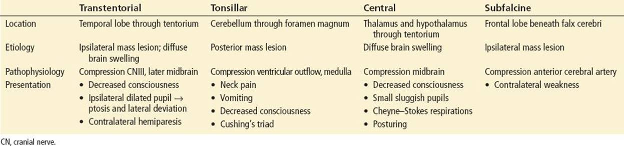
Herniation occurs when brain parenchyma is displaced due to intracranial mass effect from intracranial hematoma or malignant cerebral edema. The four common cerebral herniation syndromes transtentorial, tonsillar, central, and subfalcine, are described in Table 246.2. Acute signs of herniation include anisocoria (pupillary asymmetry greater than 1 mm), pupillary dilatation, nonreactive pupils (less than 1 mm reactivity), unilateral weakness, extensor posturing, Cheyne–Stokes breathing (rapid, deep breathing alternating with apnea), and Cushing’s triad (hypertension, bradycardia, and irregular respirations). Since few children manifest the stereotyped sequence of events as classically described, any signs of herniation are ominous and must be addressed emergently. Progressive herniation results in neurologic devastation or ultimately death.
TABLE 246.2
Common Cerebral Herniation Syndromes
ED EVALUATION
Cranial computed tomography (CT) is the cornerstone of the initial diagnostic evaluation of the severely head injured child. Its purpose is to identify lesions amenable to surgical intervention or identify severe cerebral edema. Important findings include evidence of brain contusion, subdural or epidural hematoma, intracerebral hematoma, diffuse axonal injury, acute brain swelling, pneumocephalus, subarachnoid hemorrhage, midline shift, or cerebral herniation.
Standard Advanced Trauma Life Support (ATLS) protocols apply to all patients with TBI, and attention should be paid to possible associated injuries, given that these patients are unable to communicate effectively (32,22). As many as 60% of patients with sTBI present with serious concomitant injuries (23). The potential for associated cervical spine injury is of particular concern, and patients with major TBI should be presumed to have cervical spine injury. Cervical spine immobilization should be maintained throughout the ED stay, regardless of initial results of cervical spine imaging.
As noted, scrupulous attention must be paid to the presence of hypotension and hypoxia (33). This means identifying visible or occult hemorrhage and aggressively managing hemorrhagic shock. Ongoing hemorrhage clearly complicates attempts at maintaining CPP in patients with marginal blood pressure. Similarly, respiratory compromise, either from TBI-related hypoventilation or from pulmonary injury must be rapidly addressed. Such concerns are not restricted to the initial resuscitation; secondary insults from hypotension, hyperglycemia, hypocarbia, and hypoxia must be prevented if at all possible (22).
A standard laboratory panel including complete blood count and electrolyte panel is often obtained in multiply injured patients, regardless of the presence of associated injuries. Elevated liver transaminases may alert the clinician to an unrecognized intra-abdominal injury. A full coagulation panel also should be obtained, given that coagulation abnormalities are common and of prognostic importance in patients with sTBI (21).
KEY TESTING
• CT of head
• CBC, electrolytes, coagulation studies
• Imaging for associated injuries (e.g., cervical spine, abdominal trauma)
ED MANAGEMENT
Airway and Breathing: Patients with severe TBI typically require intubation for of loss of airway protective reflexes or due to hypoventilation (Fig. 246.2). A GCS of less than 8 has traditionally been used as an indication for intubation; this is typically performed as rapid sequence orotracheal intubation with in-line cervical spine immobilization.

FIGURE 246.2 Emergency management of children with severe traumatic brain injury.
Several adjuncts have traditionally been used to blunt procedurally induced elevations in intracranial pressure in patients with head injury. More recent literature, however, has concluded that their effects in children are not clinically significant. Thus, defasciculation prior to the use of succinylcholine is no longer considered to be necessary (5,28). Similarly, pretreatment with intravenous lidocaine is no longer recommended (5,28).
Etomidate is an ideal sedative agent, as it has minimal effects on blood pressure and lowers ICP following sTBI (6,28). However, because of concern about adrenal suppression, its use should be limited to a single dose in the ED (6). Thiopental and midazolam have potential neuroprotective effects but can lead to hemodynamic instability. Recent practice guidelines have removed head injury as a relative contraindication to the use of ketamine, which may actually be beneficial in terms of intracranial pressure and cerebral perfusion (8). Recent data suggests the use of ketamine for refractory elevations of ICP or prior to procedural interventions actually improves ICP in sedated ICU patients (17).
Ventilator settings should be adjusted to maintain oxygen saturations between 93% and 99%, while minimizing application of positive pressure, which impairs cerebral venous drainage (24). End-tidal carbon dioxide (EtCO2)should be monitored closely. Unless needed for the emergent treatment of acute herniation, hyperventilation should be avoided and PaCO2 maintained between 35 and 40 mm Hg (7,24).
Circulation: Isotonic fluid (normal saline or lactated Ringer’s solution) should be administered as necessary to achieve euvolemia. If hypotension persists despite aggressive resuscitation with crystalloid or blood products, vasopressors may be considered.
Positioning: The effect of position on intracranial pressure appears to exhibit interpatient variability and the optimal position may be patient-specific (19). Placement of the head in the midline and elevation of the head of the bed to approximately 30 degrees appear to promote venous drainage and reduce ICP, though with an equivocal effect on CPP (19). Elevation beyond 30 degrees is likely to reduce CPP and should be avoided.
Sedation: Analgesia and sedation can improve ICP by reducing pain and stress. Administration of analgesics and sedatives in small, frequent doses can help to reduce ICP while minimizing hypotension. While there are theoretical benefits to particular agents, none have been compared empirically. However, reports of increased mortality from metabolic acidosis associated with propofol infusions in critically ill children have led to recommendations against its use in children with TBI (6). Similarly, multiple doses of etomidate are not recommended due to the possibility of adrenal suppression (6). Midazolam (0.1 mg/kg/hr) may be a more appropriate choice in the absence of overt hypotension.
Neuromuscular Blockade: Neuromuscular agents reduce ICP by decreasing airway and intrathoracic pressure, thereby promoting increased cerebral drainage. They also reduce metabolic demand and spikes in ICP by preventing muscle contraction. However, they mask neurologic findings and seizure activity and (except for use in RSI) should only be employed when sedatives do not correct elevations in ICP.
Anticonvulsants: The incidence of early posttraumatic seizures, defined as within 7 days of injury, is approximately 10% in children. Recommendations for prophylactic anticonvulsants are weaker than those in adults, but they may be considered when significant risk factors for posttraumatic seizures, such as depressed skull fracture, penetrating injury, cerebral contusion, or retained fragments, are present.
Hypothermia: Prophylactic hypothermia in the setting of severe pediatric TBI has not been shown to improve outcomes (14,15).
Management of herniation and severely elevated ICP: Aggressive intervention to address severely elevated ICP have been demonstrated to improve outcomes (9).
Interventions such as sedation, paralysis, judicious hyperventilation, hyperosmolar therapy, cerebrospinal fluid drainage, and surgical decompression improve mortality (7). Hypocapnia causes cerebral vasoconstriction, reducing cerebral blood flow, volume, and ICP. This may effectively reverse cerebral herniation as part of an emergent life-saving salvage strategy. However, children with sTBI should not be hyperventilated to a PaCO2 below 30 mm Hg (7).
In children with sTBI, a single episode of PaCO2 <30 mm Hg in the first 48 hours has been associated with increased mortality (odds ratio ∼ 1.5). Two or more episodes of severe hypocarbia increase the odds ratio to 4 (16). Emergency providers should utilize EtCO2 monitors to ensure that PaCO2 is maintained in an appropriate range of 35 to 40 mm Hg, or 30 to 35 mm Hg during hyperventilation, for impending herniation.
Patients with increased ICP or herniation should receive intravenous mannitol (usual dose 1 gm/kg; recommended administration time 30 minutes). Mannitol, an osmotic diuretic, reduces ICP partly through the immediate reduction of cerebral blood volume (with maintained cerebral blood flow) and intracranial pressure, an effect that lasts less than 75 minutes (30). Mannitol also osmotically draws water from the brain parenchyma into the systemic circulation, reducing intraparenchymal volume and ICP; an effect which lasts up to 6 hours. Mannitol should not be used prophylactically but rather should be reserved for patients manifesting signs of imminent herniation.
Hypertonic saline is an alternative hyperosmolar agent. It appears to reduce ICP through mechanisms similar to those of mannitol, with production of an osmotic gradient across an intact blood–brain barrier (30). It causes less osmotic diuresis and hypotension than mannitol and is preferable for patients with multisystem trauma. While severe hypernatremia (>160 mEq/L) has been associated with poor outcomes in pediatric sTBI, this is unlikely to occur with therapeutic hyperosmolar treatment (26). There is currently insufficient evidence to support the use of saline concentrations greater than 3%, although this issue remains unsettled (26,27). While mannitol has long been the traditional hyperosmolar agent of choice used in the treatment of increased ICP, current recommendations favor the use of hypertonic (3%) saline (30).
CRITICAL INTERVENTIONS
• Early aggressive treatment of hypotension or hypoxia
• Recognition of concomitant traumatic injuries
• Adequate sedation during and after intubation
• Hyperosmolar therapy and mild hyperventilation (PCO2 30 to 35 mm Hg) for herniation
• Early consultation with neurosurgery or transfer to pediatric trauma center
DISPOSITION
Management of pediatric patients with severe multi-system trauma is clearly associated with improved outcome; this is no different for children who have suffered severe head trauma (4). Admission to a specialized pediatric intensive care unit allows for the early aggressive management that has been demonstrated to improve outcome of sTBI in children.
Common Pitfalls
• Failure to recognize and promptly treat even brief episodes of hypoxemia or hypotension.
• Failure to reassess the neurologic examination frequently to identify signs of increased ICP.
• Failure to consider nonaccidental trauma in infants with TBI, particularly those with subdural hematoma.
• Excessive hyperventilation to PaCO2 <30 mm Hg.
• Failure to consider concomitant injuries, including cervical spine injuries.
REFERENCES
1. Samant UB 4th, Mack CD, Koepsell T, et al. Time of hypotension and discharge outcome in children with severe traumatic brain injury. J Neurotrauma. 2008;25:495–502.
2. Downard C, Hulka F, Mullins RJ, et al. Relationship of cerebral perfusion pressure and survival in pediatric brain-injured patients. J Trauma. 2000;49:654–658.
3. Stein SC, Georgoff P, Meghan S, et al. Relationship of aggressive monitoring and treatment to improved outcomes in severe traumatic brain injury. J Neurosurg. 2010;112(5):1105–1112.
4. Oyetunji TA, Haider AH, Downing SR, et al. Treatment outcomes of injured children at adult level 1 trauma centers: Are there benefits from added specialized care. Am J Surg. 2011;201:445–449.
5. Luten RC, Mick NW. Differentiating aspects of the pediatric airway. In: Walls RM, Murphy MF, eds. Manual of Emergency Airway Management. 4th ed. Philadelphia, PA: Lippincott, Williams & Wilkins; 2012.
6. Kochanek PM, Carney, N, Adelson PD, et al. Guidelines for the acute medical management of severe traumatic brain injury in infants, children, and adolescents -second edition: Analgesics, sedatives, and neuromuscular blockade. Pediatr Crit Care Med. 2012;13:S64–S67.
7. Kochanek PM, Carney N, Adelson PD, et al. Guidelines for the acute medical management of severe traumatic brain injury in infants, children, and adolescents -second edition: Hyperventilation. Pediatr Crit Care Med.2012;13:S58–S60.
8. Green SM, Roback MG, Kennedy RM, et al. Clinical practice guideline for emergency department ketamine dissociative sedation: 2011 update. Ann Emerg Med. 2011;57:449–461.
9. Kochanek PM, Carney N, Adelson PD, et al. Guidelines for the acute medical management of severe traumatic brain injury in infants, children, and adolescents -second edition: Indications for intracranial monitoring. Pediatr Crit Care Med. 2012;13:S11–S17.
10. Kochanek PM, Carney N, Adelson PD, et al. Guidelines for the acute medical management of severe traumatic brain injury in infants, children, and adolescents -second edition: Cerebral perfusion pressure thresholds. Pediatr Crit Care Med. 2012;13:S24–S29.
11. Faul M, Xu L, Wald MM, et al. Traumatic Brain Injury in the United States: Emergency Department Visits, Hospitalizations, and Deaths. Atlanta, GA: Centers for Disease Control and Prevention, National Center for Injury Prevention and Control; 2010.
12. Stein DM, Hu PF, Brenner M, et al. Brief episodes of intracranial hypertension and cerebral hypoperfusion are associated with poor functional outcome after severe traumatic brain injury. J Trauma.2011;71:364–374.
13. Wakai A, Roberts I, Schierhout G, et al. Mannitol for acute traumatic brain injury. Cochrane Database Syst Rev. 2013;8:CD001049. doi:10.1002/14651858.CD001049.pub5
14. Hutchison JS, Ward RE, Lacroix J, et al. Hypothermia therapy after traumatic brain injury in children. N Engl J Med. 2008;358:2447–2456.
15. Adelson PD, Wisniewski SR, Beca J, et al. Comparison of hypothermia and normothermia after severe traumatic brain injury in children (Cool Kids): A phase 3, randomized controlled trial. Lancet Neurol. 2013;12:546–553.
16. Curry R, Hollingworth W, Ellenbogen RG, et al. Incidence of hypo- and hypercarbia in severe traumatic brain injury before and after 2003 pediatric guidelines. Pediatr Crit Care Med. 2008;9:141–146.
17. Bar-Joseph G, Guilburd Y, Tamir A, et al. Effectiveness of ketamine in decreasing intracranial pressure in children with intracranial hypertension. J Neurosurg Pediatr. 2009;4:40–46.
18. Pediatric neurosurgery: Surgery of the developing nervous system. David G McLone editor. Saunders 2001. Fourth edition.
19. Tasker RC. Intracranial pressure: Influence of head-of-bed elevation, and beyond. Pediatr Crit Care Med. 2012;13:116–117.
20. Gerlach R, Dittrich S, Wilfried Schneider W, et al. Traumatic epidural hematomas in children and adolescents: Outcome analysis in 39 consecutive unselected cases. Pediatr Emerg Care. 2009;25:164–169.
21. Talving P, Lustenberger T, Lam L, et al. Coagulopathy after isolated severe traumatic brain injury in children. J Trauma. 2011;71:1205–1210.
22. Fujita Y, Algarra NN, Vavilala MS, et al. Intraoperative secondary insults during extracranial surgery in children with traumatic brain injury. Childs Nerv Syst. 2014; [epub ahead of print]
23. Stewart TC, Alharfi IM, Fraser DD. The role of serious concomitant injuries in the treatment and outcome of pediatric severe traumatic brain injury. J Trauma Acute Care Surg. 2013;75:836–842.
24. Ramaiah VK, Sharma D, Ma L, et al. Admission oxygenation and ventilation parameters associated with discharge survival in severe pediatric brain injury. Childs Nerv Syst. 2013;29:629–634.
25. Bennett TD, Riva-Cambrin J, Keenan HT, et al. Variation in intracranial pressure monitoring and outcomes in pediatric traumatic brain injury. Arch Pediatr Adolesc Med. 2012;166:641–647.
26. Nakagawa K, Chang CW, Koenig MA, et al. Treatment of refractory intracranial hypertension with 23.4% saline in children with severe traumatic brain injury. J Clin Anesth. 2012;24:318–323.
27. Bell MJ, Adelson PD, Hutchison JS, et al. Differences in medical therapy goals for children with severe traumatic brain injury – an international study. Pediatr Crit Care Med. 2013;14:811–818.
28. Martinon C, Duracher C, Blanot S, et al. Emergency tracheal intubation of severely head-injured children: Changing daily practice after implementation of national guidelines. Pediatr Crit Care Med.2011;12:65–70.
29. Coles JP, Minhas PS, Fryer TD, et al. Effect of hyperventilation on cerebral blood flow in traumatic head injury: Clinical relevance and monitoring correlates. Crit Care Med. 2002;30:1950–1959.
30. Kochanek PM, Carney, N, Adelson PD, et al. Guidelines for the acute medical management of severe traumatic brain injury in infants, children, and adolescents - second edition: Hyperosmolar therapy. Pediatr Crit Care Med.2012;13:S36–S41.
31. Matschke J, Voss J, Obi N, et al. Nonaccidental head injury is the most common cause of subdural bleeding in infants <1 year of age. Pediatrics. 2009;124:1587–1594.
32. Sharples PM, Storey A, Aynsley-Green A, et al. Avoidable factors contributing to death of children with head injury. BMJ. 1990;300:87–91.
33. Zebrack M, Dandoy C, Hansen K, et al. Early resuscitation of children with moderate-to-severe traumatic brain injury. Pediatrics. 2009;124:56–64.
34. Ciurea AV, Kapsalaki EZ, Coman TC, et al. Supratentorial epidural hematoma of traumatic etiology in infants. Childs Nerv Syst. 2007;23:335–341.