
Janet G. Alteveer and Dunisha G. Ranasuriya
Ophthalmologic disorders in children represent a wide variety of diseases, both congenital and acquired, the diagnosis of which hinges on a careful history and a thorough examination of the eye. The elements of the examination change with patient age and developmental stage. Most children with ophthalmologic emergencies present to the ED with eye redness, eye swelling, or abnormal vision.
THE PEDIATRIC EYE EXAMINATION
A complete pediatric eye examination includes inspection of the eyelids, conjunctivae, pupil, and iris; evaluation of the extraocular muscles; fundoscopy; and testing of visual acuity. This examination is an essential part of the evaluation in any child presenting primarily with ophthalmologic conditions such as conjunctivitis, periorbital edema, visual change or loss, and trauma or neurologic complaints.
Inspection of the eyelids and conjunctivae should document the presence of erythema, ecchymosis, edema, proptosis, or masses. The cornea should be evaluated for normal contour and clarity. A bulging or hazy cornea in a neonate may indicate congenital glaucoma. The cornea should be stained using topical anesthetic drops and fluorescein test strips after rest of the examination is completed so as not to interfere with visual acuity testing. Shining a Wood light on the cornea in a darkened room will reveal stain adherent to corneal defects caused by an abrasion, herpetic infection, or ulcer. The pupil should be assessed for contour, bilateral symmetry, and reactivity to light. An abnormally shaped or reactive pupil may indicate defects in the iris or disruption of the afferent or efferent nerve pathways to the eye.
Extraocular muscle testing can be accomplished by 4 months of age. By this age, a normal, full-term infant should be able to fixate on and follow a small, brightly colored toy held 6 to 12 in from the baby’s face. By 5 to 6 months, the infant should fixate and follow an object through the full range of motion. By 2 to 3 years of age, most children, with gentle encouragement, will follow a light or finger.
Evaluation of the posterior chamber via direct ophthalmoscopy is dependent on the child’s age and cognitive development, becoming easier as cooperation increases. The use of a short-acting topical parasympatholytic such as tropicamide (Mydriacyl 0.5%) will facilitate the examination of the retina. To encourage the child to look beyond the examiner and away from the direct light, use a brightly colored toy or have the parent wave at the child from directly behind the examiner. The minimum examination in an infant is the documentation of a normal red reflex. The presence of an abnormal red reflex (leukocoria or white reflex), which could indicate a cataract or intraocular tumor, mandates formal consultation with a pediatric ophthalmologist.
The corneal light reflex, or Hirschberg test, is useful in detecting strabismus or an ocular muscle palsy. It is conducted by shining a penlight, held 3 ft (1 m) away, centrally onto the child’s face. In a normal child, the light will reflect centrally on both pupils when the child is fixated on the light or a small toy held in front of the face. If the light reflex is eccentric on one eye, a strabismus or palsy is present.
Visual acuity testing is an extremely important aspect of the complete eye examination. The manner of testing is dependent on the age and cognitive ability of the child. Visual acuity also varies by age (Table 255.1). To assess an infant, have the baby fixate (on a bright toy or light) and cover one eye gently with your thumb. If the uncovered eye has normal vision, the child will continue to fixate on the object. If the uncovered eye has poor vision, the child may lose interest, push the covering thumb away or protest.
TABLE 255.1
Visual Acuity by Age

By 3 years of age, depending on developmental and cognitive ability, more objective visual testing can be achieved. The Allen chart, HOTV matching test, or “Tumbling E” game can be used. By 5 years of age, most cognitively normal children can use the Snellen chart. Testing with the Snellen, Tumbling E, or HOTV chart should be performed at a distance of 20 ft (6 m). The Allen test can be performed at 10 ft (3 m).
In all of these charts, letters or objects are presented as a group. Children with poor vision have increased difficulty identifying objects when presented as a group (the crowding phenomenon) and will perform better when objects are presented singly.
CONJUNCTIVITIS AND THE RED EYE
CLINICAL PRESENTATION
Conjunctivitis results in 5 million outpatient visits per year (1) or 1% to 4% of all pediatric ambulatory complaints (2). The causative agent may be either bacterial, viral, allergic or chemical. Bacterial conjunctivitis predominates in infants and young children: 55% to 78% in recent studies (3). Common pathogens in young children are nontypable Haemophilus influenzae, Streptococcus pneumoniae, and Moraxella catarrhalis. Staphylococcus Aureus may be the causative agent in any age group and the incidence of methicillin-resistant S. aureus (MRSA) approaches 30% in some urban areas (4).
Neisseria meningitides and Neisseria gonorrhoeae are uncommon pathogens. Gonococcal conjunctivitis may occur occasionally outside of the neonatal period and in the prepubescent child who is not sexually active. Sexual abuse must be ruled out in these cases; however, transmission from infected caregivers not abusing the child has been reported (5).
Infectious neonatal conjunctivitis is most commonly caused by Chlamydia trachomatis, followed by N. gonorrhoeae (ophthalmia neonatorum), and herpes simplex virus (HSV). The infant is infected at birth during passage through the vaginal canal. Women are routinely screened during pregnancy and prior to delivery so that risk factors would be absence of prenatal care, substance use, or history of a sexually transmitted infection. Neonatal prophylaxis for ophthalmia neonatorum is recommended universally in the United States. Currently only 0.5% erythromycin ophthalmic ointment is available in the United States, however, only silver nitrate solution is effective in prevention of infection by a penicillinase-producing gonococcus. The incubation period for gonococcal conjunctivitis is 2 to 7 days. Neonatal chlamydial conjunctivitis develops a few days to several weeks after birth. Fifty percent of infants born to infected women acquire C. trachomatis, and, of those, 25% to 50% develop conjunctivitis. Neonatal ophthalmic prophylaxis is not effective in the prevention of chlamydial conjunctivitis. Neonatal HSV infection occurs in 1 per 3,000 to 1 per 20,000 live births. The infant is mostly at risk if the mother has a primary infection at the time of delivery. The risk to an infant born to a mother with a recurrent infection is much lower. Neonatal HSV infections are of three types: generalized infection (25%), localized central nervous system (CNS) infection (30%), or disease of the skin, eye, and mouth (45%) (5). In addition, the above neonates may develop conjunctivitis due to gram-negative bacteria such as Klebsiella or Pseudomonas while in the NICU.
Bacterial conjunctivitis is most often bilateral and occurs predominantly in infants and preschool-aged children. It tends to come on abruptly and not be associated with fever or sore throat. In a recent study the history of “gluey” or “sticky” eyelids in the morning and physical findings of mucoid or purulent discharge demonstrated the highest likelihood for bacterial disease, whereas watery discharge and burning were more likely indicative of viral etiologies. Other low-risk predictors for bacterial disease include age greater than 6 years, onset between April and November, watery discharge, and the absence of adherent eyelids upon awakening. The presence of three factors predicted a negative conjunctival culture 76% of the time, while the presence of all four factors was predictive of a negative culture 92% of the time (1). N. gonorrhoeae typically causes copious purulent exudates in all age groups referred to as a waterfall discharge. A neonate with ophthalmia neonatorum may appear toxic, with a copious purulent discharge. C. trachomatis infection causes a beefy red conjunctiva (5).
Viral conjunctivitis is often bilateral, occurs in school-aged children, often has a copious and clear discharge (however, a mucopurulent discharge may be present) and the presence of a conjunctival inflammatory membrane is diagnostic. Marked periorbital edema can be present, and should not be mistaken for periorbital cellulitis (POC). Significant eye pain suggests the likelihood of an etiology other than simple conjunctivitis, such as corneal abrasion, herpetic infection, or anterior uveitis.
In the summer, adenoviruses may predominate, particularly among children older than 6 years. Adenoviral conjunctivitis may be associated with two distinct clinical syndromes: pharyngoconjunctival fever and epidemic keratoconjunctivitis. Pharyngoconjunctival fever is caused by adenovirus types 3 and 7. It is most commonly spread by person-to-person contact but has also been reported to be spread through swimming; thus, the infection may be referred to as “swimming pool” conjunctivitis. Adenovirus types 8, 19, and 37 cause epidemic keratoconjunctivitis. This entity is transmitted by direct contact with infected people or contaminated instruments and is often iatrogenic. Attention to frequent hand washing prevents transmission (6).
Contact lens use is increasing in children and adolescents who may not always adhere to proper hygiene and lens disinfection procedures, put themselves at increased risk of pseudomonas and acanthamoeba infection (7).
Nontypable H. influenzae commonly causes concomitant otitis and conjunctivitis, which may play a role in the etiologies of infection in children less than 6 years old, where 25% with bacterial conjunctivitis also have otitis media.
HSV infection of the eye may be bilateral and is frequently accompanied by involvement of the skin and mouth. Skin and mouth lesions may not be present at the time of presentation, however; furthermore, the characteristic dendrites (seen on fluorescein staining) may be observed in only 30% of patients (8). This may lead to significant delay in diagnosis in cases where HSV presents only as a nonspecific appearing recurrent blepharoconjunctivitis with no fluorescein findings. Failure to diagnose HSV stromal disease in a timely manner can lead to significant scarring and potential vision loss (8). Herpes zoster virus (HZV) infection of the first division of the trigeminal nerve may affect the eye. There may be characteristic vesicular lesions on the forehead and eyelid. The conjunctivae are usually injected and fluorescein staining may show pseudodendrites. HZV is a unilateral infection and often, periorbital pain precedes the appearance of the vesicles (5).
Rarely, the red eye may be due to keratitis from HSV or HZV infection. Herpesviruses can cause ocular involvement during primary or recurrent infections. HSV is ubiquitous and is transmitted from person to person during primary and recurrent infections that may or may not be symptomatic. Periocular inoculation can occur from the HSV-1 perioral infections. Herpes zoster represents a reactivation of latent varicella-zoster virus. When it occurs in the dermatomal distribution of the trigeminal nerve, it can cause keratitis with conjunctivitis (5).
The most common cause of allergic conjunctivitis in children is seasonal rhinoconjunctivitis. The symptoms are caused by a localized IgE-mediated hypersensitivity reaction to airborne allergens, with resultant release of histamine and other products from mast cells. Pollens and grasses are the most common allergens. In up to 25% of children, ocular findings may be the sole manifestation (6). The findings are caused by the release of products from mast cells, predominantly histamine. Allergic conjunctivitis is bilateral with variable conjunctival injection and watery eye discharge. Other allergic symptoms such as boggy nasal turbinates, watery nasal discharge, and allergic shiners (dark circles around the eye), may or may not be present.
Chemical conjunctivitis is most often caused by ocular medications. Neomycin has the greatest incidence of contact sensitivity. Silver nitrate was a common cause in the neonate. Accidental environmental exposure to a toxin, such as cleaning agents, is also common. Chemical conjunctivitis presents with conjunctival injection and often, a burning sensation.
A host of other entities may present as a red eye. Corneal abrasion may present with a unilateral red eye. In the preverbal child it may be difficult to obtain the history of trauma to the eye. There is generally tearing, pain, and photophobia. Anterior uveitis classically presents with eye pain, a constricted pupil, potentially decreased visual acuity, and perilimbic injection of the conjunctiva (injection that is limited to the area of the conjunctiva surrounding the cornea) (9). Five percent of uveitis in the United States presents in children younger than 16 years and 41% of cases are associated with juvenile rheumatoid arthritis. Toxoplasmosis accounts for approximately 10% of cases. HSV and HVZ may also cause uveitis. Uveitis in children is a chronic disease with a high incidence (up to 20%) of developing blindness in one eye (9).
DIFFERENTIAL DIAGNOSIS
The differential diagnosis of the red eye is extensive and includes bacterial, viral, allergic, and chemical causes. More specific considerations should include Kawasaki disease, blepharitis, dacryocystitis, corneal abrasions, dry eye syndromes, and Parinaud oculoglandular syndrome (catscratch disease) involving the conjunctiva and an ipsilateral preauricular lymph node.
ED EVALUATION
The ED evaluation of the red eye begins with a detailed history and physical, including a complete age-appropriate eye examination. A slit-lamp examination in an older child who is cooperative is helpful. Outside of the neonatal period, conjunctival cultures are usually obtained only in recurrent cases or those unresponsive to initial treatment. An office-based, rapid adenovirus detector test is available with a sensitivity of 89% and a specificity of 94%; it may be useful in the clinically unclear case (10). Fluorescein staining should be considered, especially with severe photophobia or corneal changes. Keratitis may appear as punctuate spots on the corneal surface. A Gram stain should be obtained if the discharge is copious, suggesting N. gonorrhoeae infection.
All young infants in whom the diagnosis of ophthalmia neonatorum or skin, eye, and mouth disease secondary to HSV is being considered must undergo a full evaluation for disseminated disease. This evaluation must include blood and cerebrospinal fluid (CSF) studies (complete blood count, chemistries, blood culture, CSF culture for bacteria and viruses, glucose, protein, Gram stain), conjunctival cultures for bacteria and viruses should also be obtained. In suspected gonococcal infection, a Gram stain of the eye drainage may be diagnostic and aid in early confirmation. A Tzanck preparation may be useful in herpetic infections. A conjunctival scraping for epithelial cells is more sensitive than swab cultures. When a chlamydial infection is suspected, cultures or specimens for rapid antigen testing of the conjunctiva and nasopharynx should be obtained using a Dacron swab (5). A chemical conjunctivitis is suggested when a history of ocular exposure to a toxin or ophthalmic medication is obtained in the presence of conjunctival injection.
Anterior uveitis is diagnosed with the slit lamp, showing cells (leucocytes) and flare (protein) in the anterior chamber. A corneal abrasion will also be evident on fluorescein staining with or without slit-lamp evaluation.
Fluorescein staining should identify the abrasion. Eversion and examination of the palpebral conjunctiva is necessary to ensure that there is no retained foreign body.
KEY TESTING
• Fluorescein examination
• Gram stain in suspected gonoccocal conjunctivitis
• Slit-lamp evaluation in the cooperative child
ED MANAGEMENT
The management of conjunctivitis is dependent on the suspected etiology and the age of the child. Treatment ranges from supportive care to hospital admission for parenteral antibiotics. Bacterial conjunctivitis, although a self-limited disease, is most frequently treated with topical ophthalmic antibiotics because this results in a bacterial cure and faster clinical improvement (11). This in turn allows children to return to day care or school and their parents to return to work. The ideal topical antibiotic should be broad spectrum, rapidly bactericidal, nontoxic to the eye, easy to use, inexpensive, and have low-resistance rates to the most common pathogens. Unfortunately bacterial resistance continues to rise: 29% of H. influenzae and 60% of S. pneumoniae exhibit resistance to penicillins and only 44% of the S. pneumoniae sp are included in the pneumococcal vaccine; S. aureus resistance to the older quinolones is increasing and while the newer quinolones (moxifloxacin and besifloxacin) have improved profiles against gram-positive organisms; they are expensive. Polymyxin B/trimethoprim has decreased efficacy against H Influenzae and S pneumoniae however trimethoprim is still effective against MRSA (4). Sulfonamides are no longer recommended because of increasing bacterial resistance and significant discomfort on instillation. Gentamicin and tobramycin are toxic to the corneal epithelium and should be reserved for gram-negative organisms (4). Choice of topical antibiotic should reflect the antibiogram of the community and also be somewhat case specific: is this a young child with presumed H. influenzaesp. or other common organism? Is there a risk of MRSA? Or is this a contact lens wearer with a high risk of pseudomonas sp?
In the instance of conjunctivitis–otitis syndrome, oral therapy with an appropriate antibiotic to treat H. influenzae, such as amoxicillin–clavulanate, will cure both the otitis and the conjunctivitis. Oral therapy for conjunctivitis without otitis has been shown to decrease the occurrence of otitis in the ensuing 2 weeks. Further data are needed to substantiate the efficacy of oral treatment of conjunctivitis. Meningococcal and gonococcal conjunctivitis is treated with systemic antibiotics.
Table 255.2 lists the etiologies of neonatal conjunctivitis with the indicated treatment.
TABLE 255.2
Etiology of Neonatal Conjunctivitis and Therapy
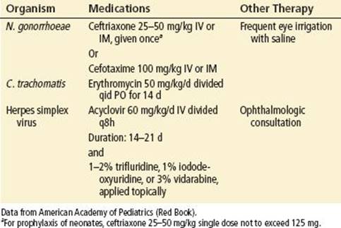
Localized HSV infections involving the eye should be treated with acyclovir 60 mg/kg/d in three divided doses for children younger than 1 year of age, and for older children, 1,500 mg/m2/d in three divided doses for 7 to 10 days. Higher dosing and intravenous (IV) dosing (maximum 80 mg/kg/d) are used for disseminated or CNS disease (5,8). Some sources also advocate initiating topical antivirals (5). Acyclovir-resistant herpesvirus (an issue usually only with HIV-infected children previously exposed to acyclovir) is treated with foscarnet 80 to 120 mg/kg/d in two or three divided doses, although there is insufficient data for the appropriate dosage in children (5).
Adenoviral conjunctivitis is treated with supportive therapy. To prevent the spread of infection to other family members, the family should be counseled to wash hands frequently and to avoid sharing washcloths. The infected child should be kept away from immunocompromised persons and neonates, because this group is at increased risk for severe disease. The child should also be kept home from school or day care for 5 to 7 days.
The treatment of allergic conjunctivitis is avoidance of the allergen and topical therapy with vasoconstrictors and cool compresses. There are a number of topical H1 blockers on the market as well as mast cell stabilizers such as lodoxamide or nedocromil. These latter may be more effective than topical nonsteroidal anti-inflammatory drugs (NSAIDs); however, they may require several days to a few weeks to effect symptom improvement (6).
Corticosteroid-containing eye drops have no place in the treatment of acute conjunctivitis. They may be necessary in the treatment of uveitis or severe keratitis but should be used only after consultation with an ophthalmologist. Long-term use of steroid drops can endanger vision by causing cataracts or glaucoma. Short-term use may inhibit the hosts’ ability to limit infection and prolong viral shedding in contagious conjunctivitis. Corticosteroid/antibiotic combinations may also promote or increase the severity of fungal keratitis.
The cornerstone of treatment of chemical exposure is decontamination: copious flushing of eye exposure with water or normal saline. This is followed by visual acuity testing, fluorescein staining, and slit-lamp evaluation.
Corneal abrasions are treated symptomatically and often with antibiotic drops as well. Contact lens wearers should be prescribed a fluoroquinolone preparation because of the concern for pseudomonas. Eye patching has not been shown to be of benefit and may retard healing in abrasions <10 mm in length (12). Cycloplegics, thought to relieve pain from ciliary muscle spasm, also may not be effective. Ketorolac drops, although safe, are expensive (6).
DISPOSITION
Bacterial conjunctivitis, with the exception of gonococcal disease, outside of the neonatal period is an outpatient disease. Follow-up with a primary care provider should be recommended. School districts have policies with regard to when children with presumed bacterial conjunctivitis may return to school. Many will allow children who have been on 24 hours of antibiotic drops to return to class. Viral conjunctivitis is much more infectious; if there is keratitis they should be referred to an ophthalmologist for follow-up.
If large corneal defects, especially those involving the visual axis are detected, emergent ophthalmologic evaluation is necessary. All cases of suspected ophthalmia neonatorum, HSV, and HZV ophthalmic infection as well as anterior uveitis should be referred to a pediatric ophthalmologist emergently. The risk for ocular damage and permanent visual loss is significant.
Neonatal, herpetic, meningococcal, and gonococcal conjunctivitis requires inpatient therapy at a facility well versed in pediatric care with pediatric ophthalmologic resources. When chlamydial infection is suspected, consultation with a pediatrician is helpful.
Significant chemical exposures should be referred to an ophthalmologist.
Common Pitfalls
• Failure to recognize herpetic infection of the eye. Delay in treatment can often lead to visual loss, CNS disease, or systemic disease.
• Inappropriate use of corticosteroid-containing eye drops, unless discussed with an ophthalmologist.
• Ignoring the presence of eye pain which may indicate corneal abrasion, herpetic infection or anterior uveitis
PERIORBITAL&NDASH;ORBITAL CELLULITIS AND THE SWOLLEN EYE
CLINICAL PRESENTATION
Children with a swollen, red eye are a relatively common problem in the ED. POC, also called preseptal, and orbital cellulitis (OC) are distinct clinical entities with different pathogenesis and treatment (13). POC is defined as an inflammatory or infectious process superficial to the orbital septum while OC is an inflammation or infection deep to the orbital septum. OC may involve the ocular structures, causing abscess formation, most commonly subperiosteal, and vision loss. The veins draining the periorbital skin and soft tissues, orbital structures and ethmoid and maxillary sinuses are valveless and anastomosing, allowing easy spread of infection to the cavernous sinus and CNS (14).
POC may occur via two different routes. The skin barrier near the eye may be broken by local trauma, an insect sting, or an HZV lesion with local spread of infected material to the periorbital tissues. Alternatively, there may be seeding of periorbital tissues from bacteremia, often several days after onset of an upper respiratory infection (URI). OC develops from sinusitis, most often beginning with formation of a subperiosteal abscess that then spreads into the orbital tissues. The ethmoid sinus develops earliest, so younger children present with ethmoiditis and ethmoid sinusitis. In older children, the frontal sinuses can be the origin of infection. Uncommonly, OC results from extension of infection through natural bony dehiscences of the ethmoid bones, which form the medial wall of the orbit. In one recent study, OC occurred with a similar frequency in older and younger children and ethmoid sinusitis was present in 98% of the study population. Figure 255.1 shows the close proximity of the sinuses to the orbital cavity (13).

FIGURE 255.1 General anatomy imaging and technology CT and MRI.
The thin skin and loose connective tissue of the eyelid allow for easy and rapid accumulation of fluid. A child with POC/OC may often present within hours of onset with a red and swollen eyelid. Ninety-five percent of cases are unilateral, with the actual color of the lid described as violaceous or purple. Seventy-five percent of children are febrile. Two-thirds will have an upper respiratory infection; one-third, a history of minor trauma; one-fourth, an acute otitis media; and one-fifth, an associated conjunctivitis (15). In bacteremic or posttraumatic POC, palpation of the periorbital tissues will elicit tenderness, but the eye itself should move freely and painlessly. In inflammatory POC due to an underlying sinusitis, the periorbital tenderness is variable. The edema may initially present in the early morning and be intermittent with resolution of edema as the day progresses. In advanced OC, the eye may be proptotic and extremely painful to voluntary movement of the extraocular muscles and visual acuity may be reduced. There may be diplopia on upward gaze. The child may appear “toxic” or irritable, with early meningeal signs.
A bacterial pathogen in POC/OC is identified, mostly, by blood culture in only 30% of cases. Prior to the introduction of the Haemophilus influenzae type b (HiB) and S. pnemoniae vaccines, 80% of the cultures in bacteremic POC, were positive for H. influenzae; with the remaining cases due to S. pneumoniae. Widespread usage of vaccines has resulted in a significant decrease in the incidence of bacteremic POC (16). The agents causing OC or inflammatory POC are the same that cause acute sinusitis: S. pneumoniae, nontypable H. influenzae, and M. catarrhalis (14). Since 2000, however there has been a significant decrease in invasive pneumococcal disease, as well as a decrease in drug-resistant S. pneumoniae. Consequently there has been a concomitant increase in both disease and colonization by nonvaccine serotypes; fortunately these organisms are less likely to cause invasive disease.
DIFFERENTIAL DIAGNOSIS
The differential diagnosis of the swollen eye beyond orbital and periorbital cellulitis includes local allergic reaction, trauma, sinusitis, bulbar or retrobulbar neoplasms, and dacryoadenitis (inflammation or infection of the lacrimal gland).
ED EVALUATION
The examination of the eye must include not only gentle palpation of the periorbital area but also an assessment of whether the globe is proptotic, the function of the extraocular muscles, and whether there is any pain with movement of the eye. Visual acuity, pupillary function, and confrontation visual fields must be assessed, if necessary, with the use of lid retractors. Specialists currently use a staging process of stage I to IV with stage I being POC and stage II through IV reflecting increasingly severe levels of OC (15).
Laboratory studies are of limited value, although a white blood cell count >15,000 may suggest bacteremia. Cultures of any wounds near the eye should be obtained; keeping in mind, conjunctival or nasopharyngeal swabs are generally not helpful. Blood cultures have been reported to be positive in up to 30% of children; however 70% to 80% of these were H. influenzae in the prevaccine era (13). It seems like blood cultures will identify fewer and fewer organisms. In the pre-HiB era, lumbar puncture (LP) was considered almost mandatory in the young child because of the invasive and virulent character of H. influenzae bacteremia. Currently, most authors reserve LP for children younger than 1 year of age with increased risk of S. pneumoniae bacteremia (13), those who are “toxic” or irritable, or those who have not yet received or completed the Hib and pneumococcal vaccination series.
Computed tomography (CT) of the orbit is useful in delineating sinusitis, subperiosteal and orbital abscesses, orbital involvement, or intracranial extension. It should be reserved for the child who presents with proptosis, pain on extraocular movement, limitation of eye movements, double vision, vision loss, ANC >10, 000 cell/μL or inability to conduct a full eye examination (12,13).
KEY TESTING
• Culture of any open wound(s) near the eye
• Blood cultures
• CT with IV contrast
ED MANAGEMENT
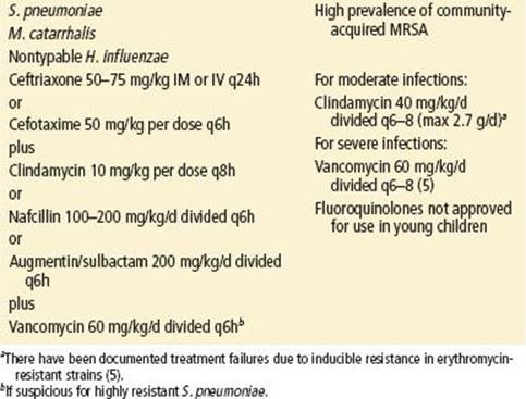
For OC: After appropriate cultures have been obtained, an IV line should be started and antimicrobial therapy initiated with an eye to covering the most likely organisms (Table 255.3). Subspecialty consultation with an ophthalmologist is mandatory.
TABLE 255.3
Antibiotic Treatment of Orbital Cellulitis
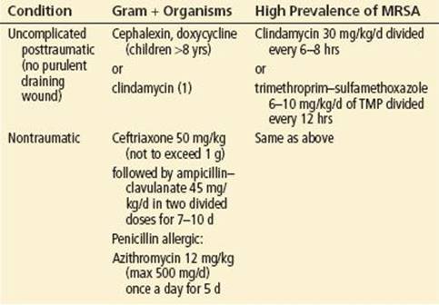
For POC: The choice of outpatient antibiotics should reflect the level of antibiotic resistance within the community to S. pneumoniae, group A Streptococcus, and S. aureus. Blood cultures should be drawn prior to antibiotics. Treatment recommendations are presented in Table 255.4.
TABLE 255.4
Outpatient Treatment of POC
DISPOSITION
All children with OC should be admitted to the hospital. Immediate consultation with an ophthalmologist and/or otolaryngologist should be undertaken if there are any physical findings suggestive of orbital involvement (proptosis, pain on eye movement, or decreased visual acuity), if there is difficulty obtaining an adequate eye examination, or if there has been failure to respond to parenteral antimicrobial therapy. Surgical evacuation is currently recommended for those patients who develop optic nerve findings such as decreasing visual acuity, afferent pupillary defects, visual field deficits, or who do not respond to 24 to 36 hours of medical management (13). The presence of sinusitis should prompt an otolaryngologic evaluation for possible endoscopic sinus surgery.
Recently, selected children with POC have been managed on an outpatient basis. There are several criteria that must be met before the child can be discharged: (1) There must be no orbital involvement (proptosis, ophthalmoplegia, or decreased visual acuity), (2) the child should be well-appearing, and (3) caregivers must demonstrate willingness to take the child home and to return the child immediately to the ED should the child’s condition deteriorate. The child will need to have daily examinations to document resolution until blood cultures are negative (usually 48 hours) (15,17).
Common Pitfalls
• Failure to conduct a thorough eye examination, thus missing signs of orbital involvement.
• Failure to consult an ophthalmologist and or an otolaryngologist early in cases of orbital involvement, which could result in a poor outcome, possible visual impairment, or loss of the eye.
VISUAL DISTURBANCE
CLINICAL PRESENTATION
The clinical presentation of visual loss varies with age and developmental stage. An infant may be brought to the ED because parents have noted unilateral swelling of the globe or haziness of the cornea. Parents may complain that the 2- to 3-month-old infant does not seem able to fixate on and follow objects. There may be no complaints at all, and the physician may discover one of these findings incidentally or note an abnormal red reflex or the presence of nystagmus.
The parents of a preverbal child may bring them to the ED because of perceived strabismus or a persistent head tilt. Intraocular tumors may present with decreasing visual acuity, leukocoria (absent red reflex), strabismus, or gradually increasing proptosis and corneal edema. Rhabdomyosarcoma may present explosively, with rapid development of proptosis and decreased visual acuity (18). Craniopharyngiomas may present with advanced visual field defects, with or without headache, and sometimes with an unusual vertical “see-saw” nystagmus. By contrast, optic neuritis (ON) presents with sudden, severe unilateral or bilateral visual loss. There may be a history of an antecedent viral infection. On examination, there is reduced vision, nonreactive pupils, and swollen optic nerves. Hemorrhages and exudates may be seen on the optic disc.
Table 255.5 lists the differential diagnoses of abnormal vision.
TABLE 255.5
Differential Diagnosis of Abnormal Visiona

Children with complaints related to vision rarely present to the ED. When they do, the visit is prompted by their own complaints of abnormal vision or complaints from their parents, teachers, or school nurses. Occasionally, the ED physician, during the course of an examination, discovers a particular finding that necessitates urgent ophthalmologic referral. The complaints and etiologies for visual loss can vary greatly with the age of the child.
Nystagmus in childhood may be congenital or acquired. The etiologies of congenital nystagmus are essentially the same as those causing visual impairment in infancy. Nystagmus that develops in the older child is usually either drug-induced or associated with severe visual loss or intracranial neoplasm.
The most common complaint in the toddler and preschool-aged child seen in the primary care office (and, therefore, the pediatric ED as well) is strabismus or ocular malalignment. At birth, most babies display mild exotropia (outward deviation). This neonatal misalignment typically resolves by 3 months of age, and any strabismus after this age is abnormal. Large-angle esotropia (inward deviation) is also abnormal in infants (19).
Cataracts, glaucoma, intraocular tumors, optic nerve problems, and nystagmus may all present in infancy. The incidence of congenital cataracts is 1 to 4 of 10,000 live births in industrialized countries and 5 to 15 of 10,000 births in the developing world. Cataracts usually present in the newborn nursery (20). However, with home births and the easy movement of populations between developing countries and the industrialized world, it is conceivable that the first presentation may be in the ED. Cataracts are strongly associated with several chromosomal abnormalities (trisomy 13, 18, 21), perinatal infections (rubella, toxoplasmosis, or HSV), and inborn errors of metabolism (20).
Primary glaucoma has an incidence of <1 in 10,000 live births in Western countries; certain south Asian populations have up to a ninefold increased incidence related to increased rates of consanguinity in marriage. Most cases present before 6 months of age and 80% within the first year of life. Secondary glaucoma may occur as a result of prior eye surgery, uveitis, or in association with syndromes such as Sturge–Weber. Children with secondary glaucoma can present later than those with primary glaucoma.
Cortical visual impairment, defined as bilateral visual impairment in the presence of normal ocular structures and pupillary light reflex and absence of nystagmus, has increased in the United States in the past decade to become one of the most common causes of childhood blindness. The most common cause is perinatal hypoxia; however, many other entities such as CNS infections, CNS malformations, epilepsy, malignancy, drugs, and head trauma may result in cortical visual impairment (21).
Retinoblastoma is the most common intraocular malignancy. The average age of presentation is 18 months, with 90% occurring prior to 5 years of age (22). One-third of tumors is bilateral and present earlier than unilateral tumors. The 10% of cases that present in older children are unilateral and not of the familial type (22).
Visual loss in childhood may result from compression of the optic nerve by tumor, inflammation of the optic nerve, and heredodegenerative conditions. Ten percent of rhabdomyosarcomas occur in the eye, arising from the intraocular muscles. The peak incidence is at age 8, with 75% occurring before age 10. Optic Neuritis (ON) in the child may be a postviral autoimmune phenomenon. The most common preceding infections are measles, mumps, varicella, pertussis, mononucleosis. Optic neuropathy has been reported in a few children with Lyme disease. Until recently, it was felt there was a weak association in children between ON and multiple sclerosis (MS). However, a recent small study showed that children with unilateral ON had a 36% progression rate within 2 years to MS. The presence of extraocular white matter lesions on MRI, bilateral ON, and extraocular clinical findings led to higher rates of MS diagnosis (11).
Optic gliomas and craniopharyngiomas are the two most common tumors that compress the optic nerve. Optic gliomas are intrinsic tumors of the optic nerve. They usually occur between 4 and 8 years of age. There is a strong association with neurofibromatosis. Craniopharyngiomas are the third most common brain tumor of childhood, representing 10% of all tumors in childhood. They arise from the embryonic rests of Rathke pouch near the sella turcica. Craniopharyngiomas affect visual acuity by compressing the optic chiasm or the optic nerve (22).
DIFFERENTIAL DIAGNOSIS
The differential diagnosis for visual disturbance includes, cataracts, glaucoma (either primary or secondary), intraocular tumors, nystagmus, or ON (Table 255.5).
ED EVALUATION
When a tumor is suspected, neuroimaging, beginning with a CT scan, is imperative. Particularly, any finding of proptosis, with or without decreased vision, necessitates an urgent CT scan or MRI. The evaluation of ON includes magnetic resonance imaging and an LP. The LP usually reveals a monoclonal pleocytosis and mild protein elevation.
KEY TESTING
• CT scan if tumor is suspected
• MRI and LP if ON is suspected
ED MANAGEMENT
The emphasis in management, in the majority of these conditions, should be on recognition of the potential severity of the problem; timely neuroimaging, when applicable; and prompt, sometimes emergent, referral. Surgery is the mainstay of treatment for glaucoma, but medications such as pilocarpine or a β-blocker are often used initially to control intraocular pressure while awaiting surgery.
Therapy for ON is based on randomized clinical trials conducted in adults. The optic neuritis treatment trial (ONTT) demonstrated that IV steroids resulted in a more rapid improvement in vision, a benefit that waned at 12 months. There was, however, a significant reduction in progression in adults to multiple sclerosis at 2 years, but this effect was lost at 5 years. Oral steroids (prednisone) alone were conclusively shown to be detrimental. The decision to treat with IV steroids remains somewhat controversial but is frequently offered by ophthalmologists to children with bilateral disease because of a worse prognosis for visual recovery. The steroid therapy of choice is methylprednisolone 15 to 30 mg/kg/d for 10 to 14 days. This is followed by a slow prednisone taper over 1 to 2 months. Too rapid a taper has been associated with a recurrence of ON and compounded visual loss. In spite of severe initial visual deficits, 80% of children had excellent visual recovery.
The treatment of cataracts, tumors, or glaucoma is largely surgical. Because infancy, particularly the first 2 months of life, represents a critical period in normal visual maturation, failure to remove the obstruction to clear vision could result in irreversible amblyopia, a functionally blind eye or difficulties with stereoscopic vision (19).
DISPOSITION
Tumors, glaucoma, cataracts, and ON often involve urgent admission to the hospital. The facility, as well as the consultant, should be experienced in pediatric eye disorders. Because the growth of some tumors, such as rhabdomyosarcoma, can be explosive, consultation with an experienced ophthalmologist is emergent.
The presence, in an infant, of leukocoria or a swollen anterior chamber requires urgent referral to an ophthalmologist comfortable with the treatment of pediatric cataracts, tumors, and glaucoma.
Because nystagmus can represent a structural lesion as well as amblyopia, there should be referral to an ophthalmologist on a semiurgent basis.
The finding of strabismus in the preverbal or preschool child mandates outpatient referral to an ophthalmologist.
Children with known risk factors for secondary glaucoma should undergo periodic routine ophthalmic evaluation.
Common Pitfalls
• Failure to perform an age-appropriate, complete eye examination.
• Failure to refer a time-dependent process urgently to an appropriate ophthalmologist.
NASOLACRIMAL DUCT OBSTRUCTION AND DACRYOCYSTITIS
CLINICAL PRESENTATION
Congenital nasolacrimal duct obstruction (CNLDO) is a common problem of infancy, with 20% of children exhibiting some symptoms within the first year of life. Canalization of the nasolacrimal duct may not be complete at birth. In most of these children, a membranous obstruction is the etiologic factor; however, a nasolacrimal duct cyst may also result in obstruction (23). Ninety-six percent of infants with CNLDO show spontaneous resolution of the obstruction by the first birthday. Refer to Figure 255.2 for anatomy of the nasolacrimal duct.
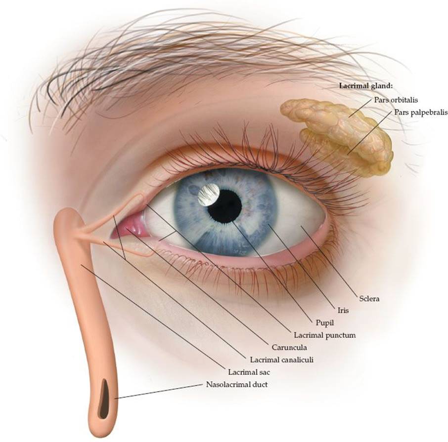
FIGURE 255.2 Eye anatomy; left eye outer labeled. (Asset courtesy of Anatomical Chart Company, ©2014.)
Dacryocystitis, or infection of the nasolacrimal duct, may be acute or chronic. It may occur as a complication of CNLDO or of acquired nasolacrimal duct obstruction (ANLDO). In the neonatal period, dacryocystitis is rare, occurring in <2% of infants with CNLDO. However, if a duct cyst is the etiology of the obstruction, the risk of dacryocystitis is greater, with earlier presentation, usually by 2 weeks of age (23). The causative organisms are most often S. aureus, Staphylococcus epidermidis, and α-hemolytic streptococci. Corynebacterium diphtheriae and Epstein–Barr virus have also been reported (24). The risk of bacteremia is higher in the neonatal period than in the older infant or child. ANLDO may occur as the result of ethmoidal sinusitis or maxillary fracture through the wall of the nasolacrimal duct (24).
Symptoms of CNLDO may be present at birth or may be delayed for several weeks, until normal tear production develops. Signs include excessive tear lake, tear overflow, or a mucoid discharge that is produced by the lacrimal sac. The tearing is often distressing to the parent. There may be crusting or stickiness of the eye; however, the conjunctivae are clear, the eye is neither red nor swollen and the patient should not be febrile.
Dacryocystitis is characterized by erythematous and swollen skin over the lacrimal sac and purulent drainage from the punctum. Fever may be present and POC or OC may develop as a complication of dacryocystitis (24).
DIFFERENTIAL DIAGNOSIS
The differential diagnosis for dacryocystitis includes corneal abrasion, local trauma with infection (i.e., insect bite), conjunctivitis, and in advanced cases OC or POC.
ED EVALUATION
A careful history and physical examination should differentiate CNLDO from dacryocystitis. In the latter case, cultures of any purulent drainage, as well as blood cultures, need to be obtained. A complete blood count may be helpful, with a high white blood cell count indicating a higher risk of bacteremia. In the infant who is 1-month old or younger, a full sepsis workup, including lumbar puncture, should be done.
KEY TESTING
• Culture of drainage and blood cultures
• Full sepsis workup in the infant less than 1 month of age
ED MANAGEMENT
In dacryocystitis, antibiotics to cover Staphylococcus and Streptococcus species should be started immediately after cultures are obtained. The choice of particular antibiotics should reflect Staphylococcus and Streptococcusresistance and the increase in MRSA within each community
Recommended agents include:
• Clindamycin 40 mg/kg/d IV in three or four divided doses
• Nafcillin 150 mg/kg/d IV doses
• Cefuroxime 150 mg/kg/d IV in three divided doses
In the penicillin-allergic infant or child, vancomycin can be used (5).
DISPOSITION
All patients with acute dacryocystitis should be admitted to the hospital due to the potential complications of sepsis and/or meningitis. Immediate consultation with an ophthalmologist is required, because early nasolacrimal duct probing is important for successful treatment of acute dacryocystitis. Because neonatal dacryocystitis is often associated with a nasolacrimal duct cyst, a complete intranasal examination at the time of surgery and marsupialization of any cyst is imperative to prevent recurrence (23).
The treatment of chronic or “low-grade” dacryocystitis is primarily surgical, with duct probing within several weeks of presentation. Systemic antibiotics are not necessary, although some consultants recommend a topical antibiotic ointment such as polymyxin B (23).
Dacryocystitis after facial fracture or sinusitis requires referral to the appropriate specialist: ophthalmology, otolaryngology, or plastic surgery (23).
Conservative management of CNLDO includes warm water compresses (to remove the mucoid discharge) and nasolacrimal duct massage; performed by occluding the common canaliculus with the index finger and then stroking firmly downward. This maneuver increases the hydrostatic pressure in the nasolacrimal sac, with the ultimate goal of overcoming the membranous obstruction. Massage the duct in repetitions of 5 to 10 passes, 4 to 6 times a day is recommended. Erythromycin ophthalmic ointment applied 4 times a day is also recommended; however, its efficacy in speeding recovery is not proven. Referral to an ophthalmologist is recommended for cases that do not resolve with conservative management. Probing will cure 90% of these infants (23). Newer, minimally invasive techniques such as balloon dacryocystoplasty, lacrimal stents, conjunctivoplasty, and endoscopic dacryocystorhinostomy are continually being refined and may be offered in individual cases.
Common Pitfalls
• Failure to consider bacteremia, sepsis, or meningitis in the neonate or young infant.
• Failure to consult an ophthalmologist early in cases of acute dacryocystitis.
REFERENCES
1. Pichichero ME. Bacterial Conjunctivitis in Children: Antibacterial treatment options in an era of increasing drug resistance. Clin Pediatr (Phila). 2011; 50(1):7–13.
2. Sheik A, Hurwitz B, van Schayck CP, et al. Antibiotics versus placebo for acute bacterial conjunctivitis. Cochrane Database Syst Rev. 2012;(9):CD004875.
3. Campolattaro BN, Lueder GT, Tychsen L. Spectrum of pediatric dacryocystitis: Medical and surgical management of 54 cases. J Pediatr Ophthalmol Strabismus. 1997;34:143–153.
4. Olitsky SE, Nelson LB. Common ophthalmologic concerns in infants and children. Pediatr Clin North Am. 1998;45:993–1012.
5. Bacal DA, Rousta ST, Hertle RW. Why early vision screening matters? Contemp Pediatr. 1999;16:155–163.
6. Teoh DL, Reynolds S. Diagnosis and management of pediatric conjunctivitis. Pediatr Emerg Care. 2003;19:48–55.
7. Wong VW, Lai TY, Chi SC, et al. Pediatric ocular surface infections: A 5-year review of demographics, clinical features, risk factors, microbiological results and treatment. Cornea. 2011;30:995–1002.
8. Patel PB, Diaz MC, Bennett JE, et al. Clinical features of bacterial conjunctivitis in children. Acad Emerg Med. 2007;14(1):1–5.
9. Golde KT, Gardiner MF. Bacterial Conjunctivitis is Children: A current review of pathogens and treatment. Int Ophthalmol Clin. 2011;51(4):85–92.
10. Wald ER. Conjunctivitis in infants and children. Pediatr Infect Dis J. 1997;16(2):
S17–S20.
11. Wilejto M, Shroff M, Buncie JR, et al. The clinical features, MRI findings, and outcome of optic neuritis in children. Neurology. 2006;67:258–262.
12. Turner A, Rabiu M. Patching for corneal abrasion. The Cochrane Collaboration. John Wiley & Sons. 2007;3.
13. Nageswaran S, Woods CR, Benjamin DK Jr, et al. Orbital cellulitis in children. Pediatr Inf Dis J. 2006;25:695–699.
14. Wald ER. Periorbital and orbital infections. Ped Rev. 2004;25(9):312–320.
15. Zetterstrom C, Lundvall A, Kugekberg M. Cataracts in children. J Cataract Refract Surg. 2005;31:824–839.
16. Whitney CG, Farley MM, Hadler J, et al. Decline in invasive pneumococcal disease after introduction of protein-polysaccharide conjugate vaccine. N Eng J Med. 2003;348:1737–1746.
17. Starkey CR, Steele RW. Medical management of orbital cellulitis. Pediatr Infect Dis J. 2001;20:1002–1025.
18. Givner LB. Periorbital versus orbital cellulitis. Pediatr Infect Dis J. 2002;21:
1157–1158.
19. Liu S, Pavan-Langston D, Colby K. Pediatric herpes simplex of the anterior segment. Ophthalmology. 2012;119:2003–2008.
20. Meltzer JA, Kunkov S, Crain EF. Identifying children at low risk for bacterial conjunctivitis. Arch Pediatr Adolesc Med. 2010;164(3):263–267.
21. American Academy of Pediatrics. Chlamydial infections, gonococcal infections, herpes simplex, and varicella zoster. In: Pickering LK, Baker CJ, Kimberlin DW, eds. Red Book: Report of the Committee on Infectious Diseases. 29th ed. Elk Grove Village, IL: 2012:272–281,336–344,398–408,724–789,841.
22. Castillo BV Jr, Kaufman L. Pediatric tumors of the eye and orbit. Pediatr Clin North Am. 2003;50:149–172.
23. Papadopoulos M, Cable N, Rahi J, et al. The British infantile and childhood glaucoma (BIG) eye study. Invest Ophthalmol Vis Sci. 2007;48(9):4100–4106.
24. Donohue SP. Pediatric strabismus. N Engl J Med. 2007;356(10):1040–1047.