
Elizabeth L. Thorpe and Melissa A. Vitale
Earache can be caused by infections or can result from referred pain from pharyngitis, odontogenic pain, external otitis, parotitis, or a foreign body in the ear canal. Infections of the ear can involve the middle ear (otitis media) and mastoid air cell system (mastoiditis), as well as the outer ear (otitis externa [OE]).
ACUTE OTITIS MEDIA
Acute otitis media (AOM) is the most common reason for antibiotic prescriptions in children, and a leading cause of acute care visits (1). Young children are prone to develop AOM secondary to their eustachian tube (ET) anatomy (short, narrow, horizontal, and floppy) and their frequent viral upper respiratory infections (URIs). URIs cause ET dysfunction via inflammation and consequent edema as well as decreased ciliary activity. This prevents physiologic drainage of middle ear secretions into the nasopharynx, and creates a negative pressure that draws bacteria from the nasopharynx into the middle ear. Accordingly, AOM is most commonly diagnosed in children less than 2 years of age, especially in those who attend daycare and experience frequent URIs. Other risk factors include smoke exposure, lack of breast-feeding in the first 6 months of life, pacifier use beyond the first 6 months of age, and craniofacial anomalies (e.g., Down syndrome, cleft palate).
The most common pathogens are Streptococcus pneumoniae and nontypeable Haemophilus influenzae followed by Moraxella catarrhalis. Since the introduction of the heptavalent pneumococcal conjugate vaccine (PCV-7), the microbiology of AOM has shifted toward a predominance of H. influenzae and non-PCV-7 strains of S. pneumoniae (2,3). While the proportion of penicillin (PCN)-resistant S. pneumoniae and beta-lactamase–positive H. influenzaehas remained stable in children with first-time AOM, there has been a notable increase in resistant strains in children with treatment failures or recurrent AOM (2). Several highly resistant serotypes of S. pneumoniae (19 A, 7 F, 6 C) that are not included in PCV-7 have emerged as major pathogens causing the incidence of S. pneumoniae to be comparable to pre-PCV-7 rates (2). In 2010, the 13-valent pneumococcal conjugate vaccine (PCV-13) was introduced. Preliminary studies of nasopharyngeal carriage rates in children less than 2 years of age with AOM who received PCV-13 showed a marked decrease in the prevalence of serotypes 19 A, 7 F, and 6 C (4). Viruses also cause AOM and may be clinically indistinguishable from bacterial AOM; common viral etiologies include picornavirus, respiratory syncytial virus, and enterovirus. Of note, infants under 8 weeks of age with AOM most often have the typical pathogens, but can also have Group B streptococcusand gram-negative bacilli (5). In children with tympanostomy tubes, Staphylococcus aureus and Pseudomonas aeruginosa are also common, with methicillin-resistant S. aureusbecoming more prevalent (6).
CLINICAL PRESENTATION
The symptoms of AOM have an acute onset, usually within 48 hours of clinical presentation, and include otalgia, fever, excessive crying or fussiness, restless sleep, and decreased appetite. Otalgia has the highest likelihood ratio in predicting the presence of AOM (3). In nonverbal children, otalgia may be indicated by tugging or rubbing the ear. The absence of fever does not rule out AOM and in fact, studies have demonstrated that presence of fever cannot predict the diagnosis of AOM (7).
DIFFERENTIAL DIAGNOSIS
When AOM is not present, the most likely cause of otalgia is referred pain from infection or trauma in regions with shared innervation, namely the pharynx, nares, and teeth. ET dysfunction causing negative pressure and temporal–mandibular joint pathology can also cause otalgia.
ED EVALUATION
In an attempt to increase the diagnostic accuracy of AOM as well as decrease the unnecessary use of antibiotics, guidelines have been published and most recently revised in 2013 (8). According to these guidelines, AOM is diagnosed if the tympanic membrane (TM) is bulging severely or moderately, indicating a middle ear effusion (MEE), or there is new onset of otorrhea not due to OE (8). AOM can also be diagnosed if there is mild bulging of the TM and onset of ear pain within the last 48 hours or intense erythema of the TM (8). In the absence of a MEE, AOM cannot be diagnosed.
Examination of the TM is facilitated by using the largest speculum that fits comfortably in the canal to allow proper visualization. Pneumatic otoscopy involves assessing TM mobility by insufflating air into the external ear canal. This technique can aid in confirming the presence of a MEE, in which case the TM is not mobile; however, this will not be successful as a diagnostic test unless the speculum forms a tight seal on the ear canal. If the TM cannot be visualized because of cerumen, the cerumen can be removed with a curette or by gentle irrigation with warm water. Cerumenolytics can be instilled before irrigation. MEE can signify either AOM or otitis media with effusion (OME); it is the presence of inflammatory changes that distinguishes AOM from OME. A red TM and the acute onset of otalgia that interferes with a child’s normal activities are consistent with inflammation of the middle ear. It is important to note that although redness of the TM can represent inflammation, it can also be secondary to vascular engorgement caused by fever or crying. Relying on TM redness alone will result in the overdiagnosis of AOM. This finding, combined with the acute onset of symptoms and evidence of MEE on examination is more specific for AOM.
It is important to assess for complications of AOM on physical examination by including the mastoid process and cranial nerves (refer to Chapter 68). A cholesteatoma (a white cystic mass behind or involving the TM) can develop within the middle ear, leading to a facial nerve palsy secondary to inflammation of the facial nerve as it traverses the inner and middle ear.
Adjunctive Diagnostics
Tympanocentesis, needle aspiration of MEE, is useful to establish a bacterial diagnosis. It should be considered in neonates and patients with immunosuppression, persistent treatment failure and/or suppurative complications (1). This is most commonly performed by an otolaryngologist.
KEY TESTING
• No specific diagnostic tests are generally indicated.
ED MANAGEMENT
The primary goals of treatment are to relieve symptoms and prevent complications. Appropriate management decisions are based upon the child’s age, allergies, recent antibiotic exposure, and the severity of the infection. Refer to Table 256.1 for drug dosages. Febrile or irritable neonates require hospitalization for parenteral antibiotics. In the rare circumstance that a cranial nerve palsy is present, an emergent myringotomy should be performed by an otolaryngologist.
TABLE 256.1
Oral Antibiotic Treatment Options for AOM

Pain Management
Pain from AOM may be significant in the first few days of infection, and therefore it is important to manage in the emergency department (ED). Studies have shown that children less than 2 may have pain for longer periods than older children with AOM, even extending up to 3 to 7 days after initiating antibiotics (9). Oral analgesics such as ibuprofen and acetaminophen are effective in controlling pain from AOM and should be initiated even if antibiotics are not started (8). In rare cases, opioids may be needed. There is not sufficient evidence to support the routine use of topical analgesics (8). If used, they should only be utilized in the presence of an intact TM. Other adjunctive medications, namely decongestants, antihistamines, and systemic steroids, are not beneficial and are associated with increased side effects.
Decision for Antibiotics
The decision to treat a child with antibiotics is based on the child’s age and severity of symptoms (Fig. 256.1). For children age 6 to 24 months, antibiotics should be initiated for AOM with otorrhea, bilateral AOM, or AOM with severe symptoms (defined as toxic appearance, otalgia for more than 48 hours, temperature of 39°C or above in the past 48 hours, or uncertain access to a follow-up visit) (8). For a certain diagnosis of unilateral AOM without severe symptoms, antibiotics may be started, or watchful waiting may be used (see below). For children older than 24 months, antibiotics are recommended for AOM with otorrhea or AOM with severe symptoms (8). For unilateral or bilateral AOM without severe symptoms, either antibiotics or watchful waiting may be utilized. For children less than 6 months,antibiotics should be initiated for AOM. However, it is important to also evaluate for alternative sources of infection in the urine, blood, and potentially the CSF, if fever is present in this age group.
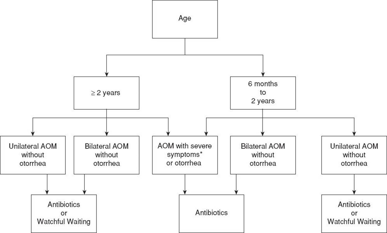
FIGURE 256.1 Algorithm for Management of Acute Otitis Media. *Severe symptoms are defined as toxic appearance, otalgia for more than 48 hours, temperature of 39°C or above in the past 48 hours, or uncertain access to a follow-up visit.
A management strategy of “watchful waiting” with the avoidance of antibiotics for certain patient populations initially gained support in the United States in the early 2000s. When compared to those who successfully avoided antibiotics with watchful waiting, patients treated with antibiotics had higher rates of multidrug-resistant pathogens, and suffered more antibiotic sides effects, most notably diarrhea (10). In addition to decreasing bacterial resistance, proponents also sited the high rate of spontaneous resolution of bacterial AOM. If the “watchful waiting” strategy is chosen, follow-up must be assured and antibiotics must be able to be initiated if symptoms worsen or fail to improve within 48 to 72 hours. In the ED setting, this may be facilitated by giving the parents a prescription to hold for 2 to 3 days that is filled if symptoms are not improved over this time period. If follow-up cannot be assured, the “watchful waiting” strategy should not be utilized (8).
Antimicrobial Therapy
Because S. pneumoniae and H. influenzae remain the most common pathogens in first-time AOM, initial therapy is directed at these organisms. High-dose (HD) amoxicillin (90 mg/kg/d divided bid; Table 256.1) is the first-line agent in treating AOM (8). In addition, amoxicillin is relatively inexpensive, and tolerated well by children in regards to taste and side effects. For children that have had amoxicillin within the past 30 days, have purulent conjunctivitis on examination, or have a history of recurrent AOM that did not respond to amoxicillin, additional beta-lactamase coverage is needed and HD amoxicillin–clavulanate is the recommended initial agent (8). HD amoxicillin–clavulanate is an ES (extra strength) preparation which allows HD amoxicillin dosing (90 mg/kg/d based on the amoxicillin component, divided bid) while keeping the clavulanate portion in a range less likely to cause diarrhea and other side effects. HD amoxicillin–clavulanate is preferred for patients with concomitant conjunctivitis because of the association of nontypeable H. influenzae with conjunctivitis–otitis (11). For patients with a penicillin allergy, reasonable first-line choices include cephalosporins (Table 256.1).
Most children treated for AOM will not feel significantly better within the first 24 hours of treatment. However, if signs and symptoms fail to improve after 48 to 72 hours, second-line therapy should be started (8). Second-line therapy should take into consideration the spectrum of the “failed” antibiotic in order to provide coverage against the most likely organism(s) untreated by this regimen. Treatment failure with HD amoxicillin suggests beta-lactamase producing organisms; thus, options include HD amoxicillin–clavulanate, select second-generation cephalosporins and ceftriaxone (Table 256.1) (8). Of these oral agents, HD amoxicillin–clavulanate is preferred because it is more biologically potent than the second-generation cephalosporins and proven highly effective against both beta-lactamase producing pathogens as well as penicillin nonsusceptible S. pneumoniae (12). Although a single injection of ceftriaxone (50 mg/kg IM) can be used for first-line therapy in patients who are vomiting or who cannot take oral antibiotics, a three-dose (3-day) regimen is recommended if the AOM was unresponsive to initial antibiotic therapy (8). For resistant AOM, some experts recommend a dose of 75 mg/kg of ceftriaxone followed in 48 hours by a second dose of 75 mg/kg. While no specific clinical trials have been conducted using tympanocentesis to document eradication, this regimen is supported by the pharmacokinetics and pharmacodynamics of ceftriaxone (13).
Children with AOM and otorrhea secondary to acute TM perforation are managed similarly to patients without a perforation. However, some clinicians advocate the concomitant use of ototopical agents to hasten the resolution of AOM and prevent the development of OE (14). Because the perforation may seal before sterilization of the middle ear, topical agents cannot be used as monotherapy.
There is no standard therapy for children with tympanostomy tubes and otorrhea from AOM. Monotherapy with topical agents, specifically ciprofloxacin–dexamethasone, is as effective as systemic agents for the typical pathogens and is also effective for P. aeruginosa, a common pathogen in this setting for which no oral systemic antibiotic is approved for children (15). Studies have shown that topical ciprofloxacin–dexamethasone compared to oral antibiotics results in more rapid resolution of symptoms (15). Moreover, topical agents do not lead to bacterial resistance, have fewer side effects, and are more convenient for the patient compared with systemic therapy. Ciprofloxacin–dexamethasone dosing for AOM with tympanostomy tubes is the same as for OE (Table 256.2), four drops twice daily for 7 days. However, ofloxacin dosing for AOM with tympanostomy tubes differs and should be dosed as five drops instilled twice a day for 10 days. Pumping the tragus after the installation of ear drops can improve delivery through the tube. Concomitant treatment with systemic antibiotics is advocated by some clinicians for those children with systemic signs and symptoms or with copious exudate that may interfere with the administration of topical agents. In these cases, use of an agent with additional staphylococcal coverage, such as HD amoxicillin–clavulanate, is useful as S. aureus is common in those with chronic drainage. Although routine culture of otorrhea is unnecessary, careful culturing with appropriately sized swabs after cleaning the ear canal can be helpful in those with persistent infection.
TABLE 256.2
Ototopical Antibiotics for Treatment of Otitis Externa

DISPOSITION
If no evidence of serious systemic infection is present, children may be managed as outpatients. A follow-up appointment with the primary care physician should occur in 2 to 3 weeks to monitor recovery of the MEE. Instruction should be given for patients to seek care sooner if symptoms, including fever, do not resolve in 48 to 72 hours or signs of worsening infection develop. Children with toxicity, other serious systemic infections, intracranial complications, or clinically significant immunodeficiencies and febrile neonates should be hospitalized and placed on parenteral antibiotics.
Common Pitfalls
• Overdiagnosis of AOM secondary to failure to remove cerumen from ear canal.
• Diagnosis based solely upon redness of the TM.
• Failure to distinguish OME from AOM.
• Failure to consider other more serious coexisting diagnosis, such as sepsis or meningitis.
MASTOIDITIS
Mastoiditis is a suppurative complication of AOM. It most commonly affects children less than 2 years of age, but can be seen in all age groups. The overall incidence of mastoiditis has decreased since the introduction of antibiotics for the treatment of AOM. Mastoiditis is a spectrum of disease ranging from infectious inflammation of mastoid air cells to abscess development and subsequent spread to contiguous areas. The most common bacterial pathogens are S. pneumoniae and Streptococcus pyogenes, followed by S. aureus, P. aeruginosa, and nontypeable H. influenzae. In the post-PCV-7 era, S. pneumoniae remains the predominant pathogen with serotype 19 A and other drug-resistant serotypes becoming increasingly common (16).
CLINICAL PRESENTATION
Acute mastoiditis (AM) represents a spectrum of disease. AM without periostitis or osteitis is the initial subclinical stage involving inflammation of the lining of the mastoid air cells. Because the mastoid is directly connected to the middle ear, almost all episodes of AOM are thought to be associated with some inflammation of the mastoid. Diagnosis of this stage is often made incidentally when cranial imaging, obtained for other reasons, reveals cloudy mastoids without evidence of bony erosion. There are no specific clinical signs or symptoms of mastoiditis in this stage, and no special treatment is required.
AM with periosteitis occurs when the infection spreads into the periosteum. Patients present clinically with postauricular edema, erythema, and tenderness. The postauricular crease may be absent. The auricle may be displaced downward and outward. Fever, ear pain, and irritability are common, especially in younger children. Up to half of children presenting with mastoiditis may not have had AOM diagnosed within the preceding weeks (17). This may occur when a middle ear infection resolves but granulation tissue or edema obstruct the narrow tube connecting the middle ear to the mastoid thus preventing drainage and resolution of the mastoiditis. In addition, studies have demonstrated that up to half of patients with mastoiditis were treated prior with antibiotics for AOM (17,18), suggesting that appropriate treatment of AOM may not prevent the development of mastoiditis. Most importantly, since the introduction of AOM “watchful waiting” guidelines in 2004, no significant increase in the incidence of AM has been noted (18).
AM with osteitis occurs when the infection results in erosion of the bony septae that separate the mastoid air cells, resulting in an empyema; making some children appear toxic. Infection can spread laterally creating a postauricular subperiosteal abscess, anteriorly creating a preauricular abscess, inferiorly toward the sternocleidomastoid muscle, medially into the petrous air cells and inner ear labyrinth, or posteriorly into the calvarium.
Chronic mastoiditis is most commonly a manifestation of chronic suppurative otitis media (otitis with perforation and persistent otorrhea) that has failed outpatient therapy. However, it can occur in a child with ineffectively treated AM.
Differential Diagnosis
The differential diagnosis of AM includes OE with postauricular cellulitis, perichondritis of the pinna, and postauricular lymphadenitis. In each of these, middle ear disease is usually absent, and with perichondritis of the pinna and postauricular lymphadenitis, the posterior auricular crease usually remains intact. Postauricular lymphadenitis is usually seen in the presence of scalp infection or inflammation.
ED EVALUATION
If symptoms and signs of mastoiditis are present, a CT scan of the temporal bones should be performed. Key findings of mastoiditis are destruction and coalescence of the mastoid air cells. Historically, a head CT was not recommended unless signs and symptoms of intracranial complications were present. However more recently, a case series of 70 patients with AM showed that of 10 patients with intracranial complications (parasinus empyema, sigmoid sinus thrombosis, epidural abscess, and subdural abscess), none had neurologic signs or symptoms on presentation. This suggests that a head CT with contrast should strongly be considered in all patients presenting with suspected AM (19).
KEY TESTING
• CT scan of the mastoid bone
• CBC
• Palpation of the mastoids to check for tenderness.
ED MANAGEMENT
It is recommended that all patients with mastoiditis be admitted for treatment (20). However, some clinicians will advocate an attempt at outpatient treatment in mild cases when close follow-up can be assured. HD amoxicillin–clavulanate will treat the most common pathogens, with the exception of P. aeruginosa. Fluoroquinolones are relatively contraindicated in young children because of theoretic, but unproven, concerns about cartilage toxicity. When P. aeruginosa is suspected, tympanocentesis can be helpful in directing therapy. Any patient with AM presenting with fever, ill appearance or osteitis should be admitted for parenteral antibiotics and otolaryngology consultation (20). Traditional treatment involved intravenous antibiotics and mastoidectomy with the removal of necrotic bone. More recently, the use of myringotomy with or without the placement of tympanostomy tubes has obviated the need for mastoidectomy in many cases. Serial CT scans are used to document resolution of infection. Ampicillin–sulbactam or third-generation cephalosporins are acceptable for initial intravenous antibiotic therapy. In the post-PCV7 era, the resistance of S. pneumoniae to both PCN and ceftriaxone is increasing and therefore some have suggested empiric treatment with both ceftriaxone and clindamycin (16). For patients who have proven ceftriaxone-resistant S. pneumoniae or in those for which there is a high index of suspicion, vancomycin can be added. If P. aeruginosa is suspected, initial, empiric antipseudomonal coverage with ceftazidime or piperacillin–tazobactam can be used. Subsequent therapy can be modified based upon culture results from tympanocentesis.
Chronic mastoiditis may be treated with a parenteral antipseudomonal antibiotic. Otolaryngology consultation is advised as mastoidectomy is necessary for patients that do not improve with medical management.
DISPOSITION
Patients with early, mild cases of AM with periosteitis may be managed as outpatients using antibiotic therapy combined with close follow-up. All other cases require hospital admission, parenteral antibiotics, and otolaryngology consultation.
Common Pitfalls
• Failure to recognize AM or distinguish AM from OE with cellulitis, perichondritis of the pinna or postauricular lymphadenitis.
• Failure to consider concurrent intracranial complications of AM such as meningitis and brain abscess.
OTITIS EXTERNA
OE is a diffuse inflammation or infection of the external ear canal which is most often acute in nature (refer to Chapter 68). Although swimming is the most common precipitant, a break in the epithelial lining from any cause (e.g., trauma, foreign body, and eczematous skin disorder) can result in OE. The lifetime incidence of OE is 10% and peaks between the ages of 7 and 12 years; it is rare in those less than 2 years (21). The most common bacterial pathogen is P. aeruginosa followed by S. aureus. Fungal OE (Candida and Aspergillus species) is rare in the United States and occurs more often in cases of chronic OE and refractory infection (22).
CLINICAL PRESENTATION
The diagnosis of OE is consistent with a history of rapid onset of symptoms over 48 hours, including otalgia, otorrhea, pruritus, and sense of ear fullness. Additional but less common symptoms include hearing loss or jaw pain. The infection may spread to the surrounding soft tissue resulting in a periauricular cellulitis and perichondritis. Bone infections can occur with spread to the mastoid or cranium. Necrotizing (malignant) OE is a rare, but aggressive infection (usually caused by Pseudomonas species) may be potentiate systemic spread (refer to Chapter 68).
DIFFERENTIAL DIAGNOSIS
The differential diagnosis includes middle ear infections, other external ear canal disorders (furunculosis, herpes zoster oticus) and mastoiditis. OE and AOM can often be distinguished clinically by the presence or absence of pain with manipulation of the outer ear. However, if an episode of AOM is complicated by TM perforation, the purulent drainage can cause a secondary OE. Furunculosis is caused by an infected hair follicle in the distal portion of the external canal causing a localized OE. This condition responds well to drainage and antistaphylococcal antibiotics. Ramsay Hunt syndrome is a Herpes zosterinfection in the distribution of the facial nerve presents with vesicles in the external auditory canal or auricle, facial nerve paralysis, hearing loss, and vertigo. Finally, mastoiditis may be difficult to distinguish from OE with postauricular cellulitis, and may require diagnostic imaging to clarify the diagnosis. Loss of the postauricular fold and tenderness over the mastoid are more common in mastoiditis.
ED EVALUATION
The diagnosis of OE is based on history and physical examination. Key examination findings are tenderness elicited by manipulation of the tragus or pinna and diffuse ear canal edema or erythema (21). Canal wall edema may progress to obstruction, trapping debris and secretions in the proximal canal and preventing visualization of the TM (refer to Chapter 68).
KEY TESTING
• Careful physical examination, looking for purulent discharge and manipulation of tragus or pinna to elicit pain
ED MANAGEMENT
Management of OE consists of controlling inflammation and infection; thus, it is important for the appropriate topical otic agents to reach the inflamed, edematous canal. In mild OE, secretions can be dry mopped with rolled tissue or swabs prior to antibiotic administration. In severe OE with near-obstruction of the canal, a wick made of compressed cellulose or gauze can be inserted. The topical antibiotic is then dripped onto the absorbent wick allowing delivery of the medication to the obstructed portion of the canal. If the wick does not fall out on its own as the canal edema decreases, it should be removed in 2 to 3 days. In the absence of an ear wick, the topical antibiotic should be instilled in the outer ear canal while the patient is lying with the affected side up. Medication delivery is enhanced by gently moving the pinna to and fro. The patient should remain on their side for 3 to 5 minutes. The canal may be then left open (21).
Topical antibiotics are recommended as first-line treatment for uncomplicated OE (21). Oral antibiotics are reserved for immunocompromised hosts and cases where infection extends beyond the external ear canal, including concomitant AOM. Classes of ototopicals currently used to treat OE include acidifying agents, antiseptics, antibiotics, and steroids. Topical acidic solutions inhibit bacterial growth, but are not commonly used because they cause local burning and irritation. Topical antimicrobials aimed at treating the leading causative organisms (P. aeruginosa and Staphylococcus spp) are the mainstay of therapy (Table 256.2). Polymyxin B–neomycin–hydrocortisone agents have been used extensively for years with good results; however, they have been associated with hypersensitivity and ototoxicity and should be avoided if the TM is perforated. Topical fluoroquinolones (ofloxacin and ciprofloxacin) are now used more commonly due to a decreased risk of ototoxicity and hypersensitivity. Newer research also shows that quinolones may be superior to traditional agents regarding both bacterial eradication and cure of OE (23). The two fluoroquinolones differ in that the ciprofloxacin preparation contains dexamethasone whereas the ofloxacin preparation does not. Fluoroquinolones are the only topical agents approved in OE with TM perforation. Studies comparing topical antibiotics with and without a steroid have shown comparable clinical cure rates (24). Fungal OE treatment involves cleansing with acidifying agents (2% acetic acid or 90% alcohol solution); antifungal drops are reserved for severe infections (25). Eczematous OE treatment is the same as for the primary disorder.
DISPOSITION
In the absence of an accompanying cellulitis with systemic symptoms, children can be managed at home. Reevaluation should occur if symptoms do not improve in 48 to 72 hours. Follow-up is also recommended in severe cases when complete examination of the external canal and TM is not possible or when an ear wick is placed. Patients should be advised to abstain from swimming for 7 to 10 days during treatment; hearing aids should not be inserted until pain and otorrhea has resolved (21). Prophylaxis against recurrent swimmer’s ear includes application of drops containing equal parts vinegar and alcohol (to alter pH and dry the canal, respectively) immediately after swimming. A hairdryer set on low and held one foot from the ear can also be used to dry the ear canal after swimming. The use of earplugs for prevention of OE is controversial. Earplugs will keep the canal dry only when properly fitted and may cause local irritation and impaction of debris/cerumen.
CRITICAL INTERVENTIONS
• Examination of the TM with pneumatic otoscopy, paying careful attention to patient positioning, comfort, and speculum size to optimize visualization and seal.
• Careful follow-up for patients when the “watchful waiting” strategy is adopted.
• Otitis media is rare in patients less than 6 months old. Pay careful attention to alternative sources of infection such as meningitis in neonates and young infants.
• CT scanning of the temporal bone and mastoid air cells in cases of suspected mastoiditis.
Common Pitfalls
• Failure to diagnose concurrent AOM.
• Failure to use a wick in severe cases.
• Failure to recognize complicating conditions (periauricular cellulitis and necrotizing OE; immunocompromised status) that require systemic antibiotics.
ACKNOWLEDGMENTS
The authors gratefully acknowledge the contributions of Noel S. Zuckerbraun, Katalin I. Koranyi, and Robert W. Hickey, who wrote the previous versions of this chapter.
REFERENCES
1. Media SoMoAO. Diagnosis and management of acute otitis media. Pediatrics. 2004;113:1451–1465.
2. Casey JR, Adlowitz DG, Pichichero ME. New patterns in the otopathogens causing acute otitis media six to eight years after introduction of pneumococcal conjugate vaccine. Pediatr Infect Dis J.2010;29(4):304–309.
3. Coker TR, Chan LS, Newberry SJ, et al. Diagnosis, microbial epidemiology, and antibiotic treatment of acute otitis media in children: A systematic review. JAMA. 2010;304(19):2161–2169.
4. Cohen R, Levy C, Bingen E, et al. Impact of 13-valent pneumococcal conjugate vaccine on pneumococcal nasopharyngeal carriage in children with acute otitis media. Pediatr Infect Dis J. 2012;31(3):297–301.
5. Turner D, Leibovitz E, Aran A, et al. Acute otitis media in infants younger than two months of age: Microbiology, clinical presentation and therapeutic approach. Pediatr Infect Dis J. 2002;21(7):669–674.
6. Fishman I, Sykes KJ, Horvat R, et al. Demographics and microbiology of otorrhea through patent tubes failing ototopical and/or oral antibiotic therapy. Otolaryngology-head and neck surgery. Otolaryngol Head Neck Surg.2011;145(6):1025–1029.
7. Laine MK, Tahtinen PA, Ruuskanen O, et al. Symptoms or symptom-based scores cannot predict acute otitis media at otitis-prone age. Pediatrics. 2010;125(5):e1154–e1161.
8. Lieberthal AS, Carroll AE, Chonmaitree T, et al. The diagnosis and management of acute otitis media. Pediatrics. 2013;131(3):e964–e999.
9. Rovers MM, Glasziou P, Appelman CL, et al. Antibiotics for acute otitis media: A meta-analysis with individual patient data. Lancet. 2006;368(9545):1429–1435.
10. Tahtinen PA, Laine MK, Huovinen P, et al. A placebo-controlled trial of antimicrobial treatment for acute otitis media. N Engl J Med. 2011;364(2):116–126.
11. Corbeel L. What is new in otitis media? Eur J Pediatr. 2007;166(6):511–519.
12. Dagan R, Hoberman A, Johnson C, et al. Bacteriologic and clinical efficacy of high dose amoxicillin/clavulanate in children with acute otitis media. Pediatr Infect Dis J. 2001;20(9):829–837.
13. Gudnason T, Gudbrandsson F, Barsanti F, et al. Penetration of ceftriaxone into the middle ear fluid of children. Pediatr Infect Dis J. 1998;17(3):258–260.
14. Bluestone CD, Klein J. Otitis Media in Infants and Children. 3rd ed. Philadelphia, PA: WB Saunders Company; 2001.
15. Dohar J, Giles W, Roland P, et al. Topical ciprofloxacin/dexamethasone superior to oral amoxicillin/clavulanic acid in acute otitis media with otorrhea through tympanostomy tubes. Pediatrics.2006;118(3):e561–e569.
16. Choi SS, Lander L. Pediatric acute mastoiditis in the post-pneumococcal conjugate vaccine era. Laryngoscope. 2011;121(5):1072–1080.
17. Thompson PL, Gilbert RE, Long PF, et al. Effect of antibiotics for otitis media on mastoiditis in children: A retrospective cohort study using the United kingdom general practice research database. Pediatrics. 2009;123(2):424–430.
18. Ho D, Rotenberg BW, Berkowitz RG. The relationship between acute mastoiditis and antibiotic use for acute otitis media in children. Arch otolaryngol Head Neck Surg. 2008;134(1):45–48.
19. Luntz M, Bartal K, Brodsky A, et al. Acute mastoiditis: The role of imaging for identifying intracranial complications. Laryngoscope. 2012;122(12):2813–2817.
20. Smith JA, Danner CJ. Complications of chronic otitis media and cholesteatoma. Otolaryngol Clin North Am. 2006;39(6):1237–1255.
21. Rosenfeld RM, Brown L, Cannon CR, et al. Clinical practice guideline: Acute otitis externa. Otolaryngol Head Neck Surg. 2006;134(4):S4–S23.
22. Martin TJ, Kerschner JE, Flanary VA. Fungal causes of otitis externa and tympanostomy tube otorrhea. Int J Pediatr Otorhinolaryngol. 2005;69(11):1503–1508.
23. Dohar JE, Roland P, Wall GM, et al. Differences in bacteriologic treatment failures in acute otitis externa between ciprofloxacin/dexamethasone and neomycin/polymyxin B/hydrocortisone: Results of a combined analysis. Curr Med Res Opin. 2009;25(2):287–291.
24. Rosenfeld RM, Singer M, Wasserman JM, et al. Systematic review of topical antimicrobial therapy for acute otitis externa. Otolaryngol Head Neck Surg. 2006;134(4Suppl):S24–S48.
25. Osguthorpe JD, Nielsen DR. Otitis externa: Review and clinical update. Am Fam Physician. 2006;74(9):1510–1516.