
Cristina M. Estrada and Timothy G. Givens
Infections of the upper airway are potentially life-threatening in that they may rapidly progress to obstruction, respiratory failure, and arrest. Inflammation of the supraglottic, glottic, or subglottic regions may produce laryngeal obstruction. Likewise, abscess formation may occur in the peritonsillar, retropharyngeal, or parapharyngeal spaces, leading to pharyngeal obstruction. Because of the confined spaces in which they occur, these entities share many clinical traits regardless of the offending organism.
Croup is a clinical syndrome characterized by a bark-like cough, hoarseness, inspiratory stridor, and respiratory distress of variable severity. The two most common forms are viral laryngotracheobronchitis (LTB) and spasmodic croup. The former is due to infection with parainfluenza viruses types 1, 2, and 3; respiratory syncytial virus; influenza virus type A; human coronavirus NL63 (HCoV-NL63); adenovirus; or rhinoviruses (1,2). The virus causes inflammation of the subglottic tissues and sometimes of the tracheal mucosa. The supraglottic structures are normal. Spasmodic croup is of uncertain etiology, is seen more commonly in patients with recurrent episodes of croup unaccompanied by infectious symptoms, and a family history of asthma, or allergies. Treatment of spasmodic croup ultimately does not differ from that of LTB.
The peak incidence of LTB is during late fall and early winter. It can occur in all age groups but is most prevalent during the second year of life. There is a 2:1 male-to-female ratio, with a recurrence rate of 5%. Most cases of croup are mild to moderate and self-limited.
Epiglottitis, in contrast, is an acute life-threatening bacterial process. The epiglottis and the aryepiglottic folds become critically inflamed, while the glottis and subglottis remain unaffected. Epiglottitis is typically caused by Haemophilus influenzae type B (HIB); however, since the introduction of HIB vaccination the incidence of epiglottitis has declined precipitously among children (3). Epiglottitis occurs year round and can present in any age group, but it most often afflicts preschool children aged 2 to 6 years. Due to widespread HIB vaccination the burden of disease has shifted to older children, adolescents, and even adults. The onset is usually rapid, with a fulminant course leading to respiratory arrest from either airway obstruction or respiratory fatigue associated with a partial airway obstruction. When properly managed, the signs and symptoms of acute infection resolve within 24 to 72 hours. Epiglottitis does not recur following the first infection.
Bacterial tracheitis is a croup variant also known as pseudomembranous croup. This relatively uncommon illness occurs in children between 6 months and 8 years of age. Its distinguishing features are the presence of thick mucopurulent secretions. Although most commonly caused by Staphylococcus aureus or methicillin-resistant Staphylococcus aureus (MRSA), other pathogens such as Streptococcus pneumoniae, Haemophilus influenzae, α-hemolytic streptococcus, pneumococcus, and Moraxella catarrhalis have also been implicated (4). Owing to the evolution in treatment options including widespread immunization against H. influenzae type b and the use of corticosteroids for the treatment of viral croup, bacterial tracheitis has eclipsed croup and epiglottitis in terms of overall morbidity.
Infections of the retropharyngeal and peritonsillar spaces may range from cellulitis to frank abscess formation. Retropharyngeal abscess (RPA) typically afflicts patients younger than 6 years, probably because of the prominence of retropharyngeal lymph nodes in young children, which involute over time. RPA usually develops by lymphatic spread, though extension from contiguous tissues, local trauma, or presence of a foreign body may be the inciting event. Peritonsillar abscesses represent extension of tonsillar infections and are more common in adolescents and adults rather than younger children. Both varieties of abscesses are polymicrobial in origin, with S. aureus, streptococcal species, and anaerobes the usual offenders (5).
CLINICAL PRESENTATION
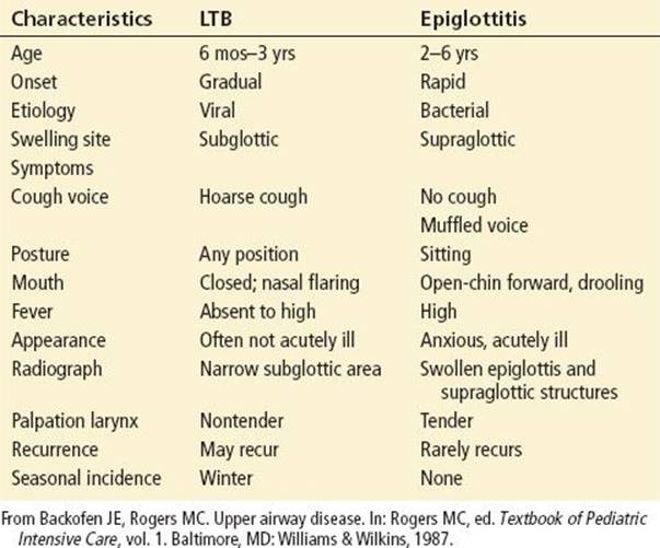
LTB is characterized by a 1- to 2-day prodrome of coryza with a low-grade fever, followed by inspiratory stridor and the characteristic barking cough, with mild to moderate respiratory distress. Symptoms peak by 2 to 3 days, with respiratory distress worsening when the patient becomes agitated or cries; improvement and resolution is noted by 5 to 7 days (6). Patients presenting with wheezing, tachypnea, and stridor have a combined illness of bronchiolitis with overlying croup. Clinical scoring systems may assist physicians in assessing the degree and progression of respiratory distress (Table 261.1). In spasmodic croup, the same barking cough, stridor, and labored breathing are observed, but the patient is afebrile and does not have the upper respiratory symptoms preceding LTB. Patients with spasmodic croup are usually responsive to cool, humidified air and often improve by the time they reach the emergency department (ED).
TABLE 261.1
Comparison Between Epiglottitis and Laryngotracheobronchitis
On the clinical disease spectrum, bacterial tracheitis lies between croup and epiglottitis. Similar to patients with LTB, bacterial tracheitis patients may have a viral prodrome; however, they may rapidly develop septic appearance with high fever and signs of upper airway obstruction. As opposed to epiglottitis, bacterial tracheitis patients often cough, are able to lie flat, and have no problems managing their secretions. Lastly, they often fail to respond to standard treatment for croup, including racemic epinephrine and systemic steroids (4).
Patients with epiglottitis typically have an abrupt onset of sore throat, high fever, and one or more of the “four D’s”: dysphagia, dysphonia, drooling, and distress. Inspiratory stridor may be audible, but there is usually no cough. Secretions, laryngospasm, and fatigue can further compromise breathing. Classically, patients assume a tripod sitting position, with the mandible extended forward in an effort to maintain airway patency. Patients appear very apprehensive and are often septic (3).
Both retropharyngeal and peritonsillar abscesses present with vague symptoms of fever and odynophagia which may evolve into a septic appearance. Patients with RPA may feed poorly, have stridor, exhibit difficulty handling secretions, and develop neck stiffness, pain or torticollis, all of which may combine to create a confounding picture consistent with meningitis. Those afflicted with peritonsillar abscesses may complain of unilateral ear or neck pain and speak with a “hot potato voice.” Development of trismus is a clear clue to the presence of a peritonsillar abscess, and a unilateral erythematous bulge of the soft palate around the tonsil with uvular deviation away from the affected side is visible on inspection of the oropharynx (5). The absence of uvular deviation is an important pertinent negative in the evaluation of the adolescent patient with throat pain.
DIFFERENTIAL DIAGNOSIS
Atypical clinical features should prompt other considerations. Occasionally, a child with LTB caused by parainfluenza or influenza virus will present with stridor and wheezing and be diagnosed with bronchiolitis. Epiglottitis relative to croup is differentiated by its more acute and fulminant presentation (Table 261.1); in addition, a history of HIB vaccination does not entirely rule out epiglottitis. Ludwig angina, a rapidly spreading inflammation of the sublingual, submandibular, and submaxillary spaces (more common in adults) and retropharyngeal and peritonsillar abscesses are suspected based on careful inspection of the sublingual space and posterior pharynx. If membranes are noted in the oropharynx, diphtheria and infectious mononucleosis should be considered. Bacterial tracheitis is a primary consideration when a patient continues to deteriorate in spite of treatment or fails to respond to the standard management for LTB. Afebrile stridulous patients with a history of coughing or choking should be evaluated for possible foreign-body aspiration (see Foreign Body Chapter 273) (7). Patients with recurrent or persistent stridor or LTB during an atypical time of year should be considered at risk for the following diagnoses: congenital airway anomalies, acquired tracheal stenosis secondary to previous intubations, congenital heart disease with associated vascular anomalies, tracheal hemangiomas, or recurrent angioneurotic edema.
ED EVALUATION
Croup is typically a clinical diagnosis; laboratory and radiographic studies are usually unnecessary (6). If the history and physical examination suggest LTB, the physician may apply a clinical scoring system to assess the patient’s degree of compromise (Table 261.2). Sequential scoring is done after each intervention (8). An inspiratory soft tissue lateral neck radiograph and chest radiograph may be helpful in distinguishing LTB from other more ominous clinical entities (Fig. 261.1–Fig. 261.3). Classic radiographic signs of epiglottitis include the thumb sign (enlarged epiglottis), swelling of the aryepiglottic folds, blunting of the vallecula, and obliteration of the piriform sinuses. In LTB, the “steeple sign” (a gradual narrowing at the subglottic area) may be observed, however, all supraglottic structures should appear normal. The pseudomembranes of bacterial tracheitis, radiographically, may make the tracheal air column appear hazy. Radiographic widening of the soft tissue between the air column and cervical vertebrae is indicative of RPA or cellulitis. Since retropharyngeal cellulitis without abscess formation can be managed with antibiotics alone, a CT scan may prevent a surgical exploration for a stable patient in whom RPA was suspected.
TABLE 261.2
Clinical Croup Score

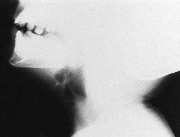
FIGURE 261.1 Normal inspiratory soft tissue lateral neck radiograph.

FIGURE 261.2 Epiglottitis on inspiratory soft tissue lateral neck radiograph. Supraglottitis.

FIGURE 261.3 Croup. Steeple sign on a soft tissue neck radiograph.
KEY TESTING
• Inspiratory soft tissue lateral neck radiographs may be performed when the diagnosis is in question.
• Chest radiographs can demonstrate the presence of a steeple sign in croup.
• CT scan with intravenous contrast may be helpful in delineating the extent of abscesses, in diagnosing complications, and in planning surgical intervention.
• Laboratory studies are of limited diagnostic utility in children with croup.
ED MANAGEMENT
Maintenance or provision of an adequate airway is the first priority in any illness in which airway obstruction is possible. A child in respiratory distress should not be separated from parental comfort unless respiratory arrest is imminent (6). Minimizing manipulation serves to reduce agitation and avoids increasing the child’s work of breathing or worsening respiratory distress; for this reason venipuncture should be deferred if possible. The physical examination consists of inspection and observation at a comfortable distance from the patient. Oxygen should be delivered in a manner tolerable to the child. Strong subjective airway assessment skills are vital under these circumstances. When epiglottitis or critical tracheitis is established as the diagnosis, immediate arrangements should be made to transport the patient to an operating room for secure placement of an artificial airway. A physician experienced in airway management must accompany the child during transport to the operating room.
Each institution must establish an epiglottitis and critical tracheitis protocol that can be rapidly activated to mobilize personnel once these diagnoses are established (9). Patients with epiglottitis or critical tracheitis should be taken to the operating room immediately, where inhalational anesthesia is given with the patient in a sitting position (10). The hypopharynx is then visualized, and rapid intubation is performed with an endotracheal tube 0.5 to 1 mm smaller in diameter than predicted for the patient’s age and weight. A surgeon skilled in pediatric tracheostomy should be prepared to perform an emergency tracheostomy in the event attempts at intubation fail. Intravenous access and laboratory studies are usually obtained after induction of anesthesia.
For patients who present to the ED with impending or complete respiratory arrest, the airway must be addressed immediately in the ED. These patients may include those with more advanced cases of epiglottitis or tracheitis or LTB patients with croup scores >7. Bag-valve-mask (BVM) ventilation with 100% oxygen should be initiated. Higher positive pressures are sometimes necessary, requiring a two-handed technique to obtain an adequate chest rise. If these measures are unsuccessful within 15 to 30 seconds, a rapid oral intubation should be attempted. Ventilatory failure should be suspected in critical upper airway obstruction in patients who do not respond quickly to initial therapeutic maneuvers, who lose head control or become lethargic. Because of airway narrowing, an endotracheal tube size smaller in diameter than that predicted for the child’s age and weight should be used if intubation becomes a necessity. Tips for intubating children with epiglottitis (or other types of critical upper airway narrowing) include using a smaller size endotracheal tube, using a stylet, and looking for an air bubble at the glottic opening while an assistant compresses the chest. If intubation is impossible, emergency cricothyrotomy or tracheostomy may need to be performed (see Chapter 218).
In milder cases of LTB, the treatment provided depends on the patient’s level of respiratory distress. In mild cases (croup score <4), a trial of humidification therapy may be offered. However, no significant benefit of cool mist therapy has ever been proven, and a recent randomized, controlled trial of children with moderate croup failed to demonstrate the effectiveness of mist therapy in improving the clinical symptoms of croup (11).
There is ample evidence to suggest that corticosteroids are of benefit to patients with croup (12). Steroids promote resolution of laryngeal edema through their anti-inflammatory activity. Dexamethasone (Decadron), 0.15 to 0.6 mg/kg, administered either orally or as a single intramuscular injection, has been shown to reduce the severity of symptoms, rate of hospital admissions, need for subsequent nebulized epinephrine treatments, and duration of hospital stay or observation in the ED (13,14). It reduces the number of return visits to the ED and speeds the resolution of symptoms in children with mild croup (13,14). Dexamethasone has a half-life of 54 hours, so one dose is usually sufficient. Inhaled budesonide has a rapid onset (within 2 to 4 hours) and has also been shown to be effective in mild to moderate croup (15).
Both oral and intramuscular routes of administration have been shown to be effective (12), and a randomized clinical trial comparing equivalent doses of intramuscular and oral dexamethasone showed no difference between the two in terms of clinical efficacy (16). The ease of administration of oral dexamethasone over nebulized or intramuscular agents makes the oral route preferable in most cases. The other routes become options in complicated croup requiring admission or associated with vomiting. As the effects of corticosteroid treatment may take up to 6 hours to appear, early administration is important. All patients who require epinephrine therapy should also receive steroids.
Patients with LTB and a clinical score ≤4 or inspiratory stridor at rest should receive oxygen and nebulized racemic epinephrine (0.05 mL/kg per dose [maximum dose, 0.5 mL] of a 2.25% solution diluted in 3 mL of saline and delivered with humidified oxygen, repeated as necessary) (8,17). Equivalent doses of either racemic epinephrine or (Levo) L-epinephrine have been shown to be equally effective (18). The L-epinephrine dose is 0.5 mL/kg of a 1:1,000 concentration to a maximum of 5 mL per dose. There should be almost immediate clinical improvement owing to reduction of edema and vasoconstriction of the inflamed mucosa through stimulation of the α-adrenergic receptors. Parents should be encouraged to deliver the nebulized treatment in a nonthreatening manner. Heart rate should be monitored during treatment with nebulized sympathomimetics. The treatment should be stopped if the heart rate exceeds 200 beats/min or if dysrhythmias occur.
In the past, patients who received nebulized epinephrine were admitted to the hospital because of concern about a rebound effect, a clinical worsening thought to be due to vasodilatation and increased mucosal edema as the adrenergic effects abate. This phenomenon usually occurs within 2 to 4 hours after epinephrine treatment. Recently, the need for hospitalization has been reassessed; several studies suggest discharge from the ED is safe after 2 to 4 hours of observation, if the patient has no stridor at rest, normal air entry, normal color, and a normal level of consciousness (1).
Heliox, a combination of helium and oxygen gas, has a low viscosity and a low specific gravity and allows for greater laminar air flow through the respiratory tract (16). In clinical trials, heliox has not been shown to be more effective than humidified oxygen or racemic epinephrine in reducing croup scores (7,19).
Antibiotics are unnecessary for patients with LTB. Patients with epiglottitis or bacterial tracheitis should receive intravenous antibiotics as soon as IV access can be safely established. Epiglottitis can be treated with ceftriaxone (100 mg/kg/d at 12-hour intervals), cefuroxime (150 mg/kg/d at 8-hour intervals), cefotaxime (150 mg/kg/d at 8-hour intervals), or chloramphenicol (100 mg/kg/d at 6-hour intervals). Adequate coverage for S. aureus should be a consideration in cases of bacterial tracheitis. Bronchospasm and excessive secretions can be treated with β 2-agonist nebulization (e.g., albuterol) and suctioning. Additional supportive treatment includes antipyretics and intravenous fluids for patients with compromised oral intake.
Both retropharyngeal and peritonsillar abscesses are treated with intravenous antibiotics, with or without incision and drainage. Gram-positive and anaerobic coverage are standard. Antibiotic choices include clindamycin (30 mg/kg/d) or ampicillin–sulbactam (50 mg/kg per dose every 6 hours intravenously) (20).
CRITICAL INTERVENTIONS
• Have appropriate airway equipment available for the rare child with critical airway obstruction and respiratory distress.
• Administer oral or IM dexamethasone in a timely manner to all patients with LTB or spasmodic croup.
• Administer racemic epinepherine via nebulization to patients with stridor at rest, increased work of breathing, with a croup score >4.
DISPOSITION
LTB patients with a clinical score ≤4 (significant respiratory distress), those with persistent stridor at rest, and those with minimal or no response to treatment with nebulized adrenergic agents should be admitted, as should those with bacterial tracheitis and infections of the retropharyngeal space.
Instructions to parents whose children are sent home include monitoring the child’s work of breathing (respiratory rate, retra-ctions, and agitation), implementing a method of rapid and easy communication access between physician and family, and ensuring minimal physical exertion and stimulation during the convalescent phase of the illness.
Common Pitfalls
• Separating children from parents during initial assessment or during administration of nebulized medications or oxygen.
• Performing medical procedures (venipuncture, vital signs, laboratory, and x-ray studies) while patient is acutely symptomatic in lieu of administering steroids or nebulized epinephrine.
• Delaying the establishment of an artificial airway or not having the appropriate equipment readily available to manage the rare critical airway.
• Failure to consider bacterial tracheitis or RPA in a patient not responding appropriately to treatment.
REFERENCES
1. Sung JY, Lee HJ, Eun BW, et al. Role of human coronavirus NL63 in hospitalized children with croup. Pediatr Infect Dis J. 2010;29(9):822–826.
2. Rihkanen H, Rönkkö E, Nieminen T, et al. Respiratory viruses in laryngeal croup of young children. J Pediatr. 2008;152(5):661–665.
3. Guldfred LA, Lyhne D, Becker BC. Acute epiglottitis: Epidemiology, clinical presentation, management and outcome. J Laryngol Otol. 2008;122(8):818–823.
4. Hopkins A, Lahiri T, Salerno R, et al. Changing epidemiology of life-threatening upper airway infections: The reemergence of bacterial tracheitis. Pediatrics. 2006;118(4):1418–1421.
5. Goldstein NA, Hammersclag MR. Peritonsillar, retropharyngeal, and parapharyngeal abscesses. In: Feigin RD, Cherry JD, Demmler-Harrison GJ, et al., eds. Textbook of Pediatric Infectious Diseases. 6th ed. Philadelphia, PA: Saunders; 2009:177.
6. Cherry JD. Clinical practice. Croup. N Engl J Med. 2008;358(4):384–391.
7. Weber JE, Chudnofsky CR, Younger JG, et al. A randomized comparison of helium-oxygen mixture (Heliox) and racemic epinephrine for the treatment of moderate to severe croup. Pediatrics.2001;107(6):E96.
8. Westley CR, Cotton EK, Brooks JG. Nebulized racemic epinephrine by IPPB for the treatment of croup: A double-blind study. Am J Dis Child. 1978;132(5):484–487.
9. Shah RK, Roberson DW, Jones DT. Epiglottitis in the Hemophilus influenzae type B vaccine era: Changing trends. Laryngoscope. 2004;114(3):557–560.
10. Rogers MC. Textbook of Pediatric Intensive Care, vol 3. Baltimore, MD: Williams & Wilkins; 1996:69–70.
11. Neto GM, Kentab O, Klassen TP, et al. A randomized controlled trial of mist in the acute treatment of moderate croup. Acad Emerg Med. 2002;9:873–879.
12. Russell KF, Liang Y, O’Gorman K, et al. Glucocorticoids for croup. Cochrane Database Syst Rev. 2011;(1):CD001955.
13. Super DM, Cartelli NA, Brooks LJ, et al. A prospective randomized double-blind study to evaluate the effect of dexamethasone in acute laryngotracheitis. J Pediatr. 1989;115:323–329.
14. Geelhoed GC, Macdonald WB. Oral dexamethasone in the treatment of croup: 0.15 mg/kg vs. 0.3 mg/kg vs. 0.6 mg/kg. Pediatr Pulmonol. 1995;20:362–368.
15. Klassen TP, Craig WR, Moher D, et al. Nebulized budesonide and oral dexamethasone for treatment of croup: A randomized controlled trial. JAMA. 1998;279(20):1629–1632.
16. Donaldson D, Poleski D, Knipple E, et al. Intramuscular versus oral dexamethasone for the treatment of moderate-to-severe croup: A randomized, double-blind trial. Acad Emerg Med. 2003;10:16–21.
17. Bjornson C, Russell KF, Vandermeer B, et al. Nebulized epinephrine for croup in children. Cochrane Database Syst Rev. 2011;(2):CD006619.
18. Waisman Y, Klein BL, Boenning DA, et al. Prospective randomized double-blind study comparing L-epinephrine and racemic epinephrine aerosols in the treatment of laryngotracheitis (croup). Pediatrics.1992;89:302–306.
19. Vorwerk C, Coats T. Heliox for croup in children. Cochrane Database Syst Rev. 2010;(2);CD006822.
20. Baum ED, Elden LM. Bacterial infections of the neck. In: Burg FD, Ingelfinger JR, Polin RA, et al., eds. Current Pediatric Therapy. 18th ed. Philadelphia, PA: Saunders; 2006V:1117.