
Adam Z. Barkin and Roger M. Barkin
Pertussis is a highly contagious respiratory illness, occurring in all age groups, but is particularly symptomatic in unprotected infants and children. Mortality and hospitalization rates are greater in preterm and unimmunized infants younger than 12 months of age (1). Neonatal pertussis, with maternal peripartum pertussis as the usual source, is particularly severe. The incidence of pertussis has decreased since the institution of the pertussis vaccine. However, there has been an increasing incidence of pertussis across the population, especially among adolescents and adults (2,3). In 2012, 51% of cases occurred in patients over the age of 11; whereas 10.8% were in infants less than 1 year of age (4).
Bordetella pertussis, a gram-negative pleomorphic bacillus, is the causative agent. Less frequently, a similar disease can be caused by Bordetella parapertussis. Transmission is by respiratory droplets, with extremely high attack rates (70% to 100%) in susceptible hosts. The incubation period is 5 to 21 days, with a mean of 7 to 10 days (1,5).
Although immunization reduces the incidence and mortality of pertussis, it is neither complete nor permanent. Immunity to pertussis wanes 3 to 5 years after vaccination. After 10 to 12 years, protection from vaccination is minimal (6–8).
CLINICAL PRESENTATION
The clinical manifestations of pertussis depend on age and previous immunization or infection. The classic illness, which lasts for 6 to 10 weeks, is generally divided into three stages: catarrhal, paroxysmal, and convalescent. The course of the disease may be atypical, especially among adolescents and adults (3). Adult manifestations are often limited to rhinorrhea, sore throat, and persistent cough.
The catarrhal stage is characterized by nonspecific upper respiratory symptoms that may include rhinorrhea, lacrimation, mild cough, and low grade or no fever. This stage lasts 1 to 2 weeks, and the diagnosis is often not suspected. Patients are most infectious during the catarrhal phase.
In the paroxysmal stage, which may last from 1 to 6 weeks and up to 10 weeks, episodes of coughing increase in severity and number. A characteristic repetitive series of 6 to 12 forceful coughs during a single expiration is followed by a sudden, massive inspiratory effort that produces the classic “whoop” as air is inhaled forcefully against a narrow glottis. Cyanosis, bulging eyes, protrusion of the tongue, salivation, lacrimation, and distention of the neck veins may occur during episodes of coughing. The paroxysms can be precipitated by feeding, crying, or even hearing another person cough.
Physical examination during this stage may reveal conjunctival hemorrhages and petechiae over the neck and head. Some patients have diffuse rhonchi and rales on auscultation. In very young infants, the paroxysms and whoop are often absent with typical chief complaints of choking spells, apnea, bradycardia, cyanosis, and unresponsiveness.
In the convalescent stage, usually lasting 1 to 2 weeks but in some cases persisting up to 6 to 10 weeks, symptoms gradually abate (1,3,5,9,10).
Complications of pertussis include otitis media, failure to thrive, pneumonia, seizures, encephalopathy, epistaxis and other hemorrhages, cerebral hypoxia, and death. Most deaths due to pertussis occur in patients 6 months of age or younger (3,5,10). Pneumonia complicates pertussis in 5% to 20% of cases and may be due to B. pertussis or secondary bacterial infection. Apnea is a serious complication and is most common in children younger than 6 months of age. Other pulmonary manifestations include aspiration pneumonitis, subcutaneous emphysema, and pneumothorax. Seizures occur in 1% to 2% of cases, whereas encephalopathy occurs in about 1% of cases (10,11). Of 100 deaths due to pertussis between 2000 and 2004, 90 were infants younger than 4 months, and 76 were younger than 2 months (1). Premature infants are at especially high risk for severe disease.
DIFFERENTIAL DIAGNOSIS
In classic disease, the clinical diagnosis of pertussis can be made without difficulty. Other infectious agents causing similar illness include Mycoplasma pneumoniae, Chlamydia trachomatis, Chlamydia pneumoniae, adenoviruses, respiratory syncytial virus, and other respiratory viruses. B. parapertussis and B. bronchiseptica occasionally produce a pertussis-like syndrome (5).
Spasmodic attacks of coughing may be observed in infants and children with bronchiolitis, bacterial pneumonia, cystic fibrosis, tuberculosis, an airway foreign body, and other diseases that cause lymphadenopathy and extrinsic compression of the trachea and bronchi.
Difficulty in recognizing pertussis occurs in the catarrhal stage, in mild illness, and in adults or very young infants where symptom complexes may vary widely. Pertussis should be included in the differential diagnosis of infants presenting with choking or apneic episodes and in children who present with a history of severe, spasmodic coughing or posttussive emesis.
ED EVALUATION
A child or infant suspected of having pertussis should be observed with formal monitoring for signs and symptoms of respiratory distress. Paroxysmal coughing episodes should be documented and the patient’s hydration status should be carefully assessed. Between attacks, patients may appear normal and are usually in no distress. It is vital to remember young infants require hospitalization and close observation, in spite of looking well between paroxysms.
Diagnostic testing for pertussis is imperfect. Evaluation of the white blood cell count may be helpful; a leukocytosis of 20,000 to 50,000 cells/mm3, with an absolute lymphocytosis (i.e. right shift), is characteristic of severe disease. Chest radiographs are nonspecific and may show a perihilar infiltrate, atelectasis, or a “shaggy” heart border (10).
Culture of nasopharyngeal secretions remains the diagnostic gold standard when performed within the first 2 weeks of illness; false-negative results increase after 2 weeks. The test’s specificity is reported to be 100% (3).
PCR is being used more frequently and if performed within 0 to 3 weeks of symptom onset has a reported sensitivity and a specificity of 94% and 97%, respectively (3). To optimize PCR test characteristics, the CDC has suggested a set of testing best practices, which include (1) testing only symptomatic patients, (2) being aware that testing after 4 weeks of cough or following antibiotics increases false-negative results, and (3) maximizing DNA recovery by obtaining samples via aspiration or posterior nasopharyngeal swab (12,13).
Finally, serologic testing can be used for diagnosis, but only in the later stages of disease (2 to 8 weeks after the onset of cough) (12).
KEY TESTING
• CBC: predictive of severe disease with leukocytosis >20,000 and a lymphocytic predominance or “right shift”
• Chest x-ray: with a perihilar infiltrate or “shaggy” heart border
• Culture of nasopharyngeal secretions: if within first 2 weeks of illness
• PCR of aspirated nasal secretions: if within first 3 weeks of illness and observing CDC best practices
• Serology: if between 2 to 8 weeks of symptoms
ED MANAGEMENT
In addition to watching for apnea, overall ventilation, and oxygenation should be carefully assessed and monitored. Patients should be observed both during paroxysmal coughing and between paroxysms. Suctioning the oropharynx with a catheter large enough to permit aspiration of tenacious secretions may be helpful during paroxysms. Apneic infants may require active airway management. Children younger than 6 months old are more likely to be hospitalized, tend to have longer hospitalizations, and are more likely to require intensive care monitoring.
The paroxysmal coughing episodes may prove exhausting, and avoiding factors that provoke such attacks may be helpful. Early recognition of hypoxia or bradycardia is critical. Corticosteroids and bronchodilators such as albuterol are not routinely recommended for treatment of pertussis, although some have suggested they may reduce paroxysms of coughing (3). Maintenance of hydration and nutritional status is of utmost importance for optimal recovery, especially in infants with severe disease or posttussive vomiting.
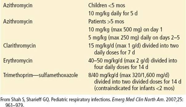
Antimicrobial agents, if given early during the course of illness, may ameliorate the symptoms. Because of delays in definitive diagnosis, empiric treatment is often started early in the illness course. However, once paroxysmal coughing is established, antibiotics have no effect on the course of illness, but are still recommended to limit transmission. Macrolide antibiotics are the first line of treatment. Erythromycin was the traditional first-line choice, but azithromycin and clarithromycin are equally effective and have fewer side effects (5). In addition, administration of erythromycin has been associated with pyloric stenosis in infants aged 1 month and younger (10,14,15). For patients allergic to macrolides, trimethoprim–sulfamethoxazole is an acceptable alternative. See Table 262.1 for dosing guidelines (14).
TABLE 262.1
Antimicrobial Treatment Guidelines for Pertussis
In addition to standard precautions, droplet precautions are recommended for 5 days after initiation of effective antimicrobial therapy. Chemoprophylaxis is recommended for all household contacts, irrespective of age and immunization status. The treatment regimen for postexposure prophylaxis is the same as that to treat active disease (14).
With the resurgence of pertussis in adolescents and adults with diminishing immunity, there has been increased focus on vaccination maintenance. Two Tdap vaccines (Tetanus toxoid, diphtheria toxoid and acellular pertussis vaccine, adsorbed), Boostrix and Adacel, were licensed in 2005. The Centers for Disease Control recommends Tdap vaccination instead of Td in adolescents and adults who require tetanus vaccination for wound management. In 2012, the Advisory Committee on Immunization Practices (ACIP) expanded their recommendation to include immunizing adults over the age of 65. Either Boostrix or Adacel is acceptable (16). The dosage of Tdap is 0.5 mL IM (1).
Cocooning, or immunization of adults and adolescents who will be in close contacts with infants <12 months, is recommended with a standard dose of Tdap. ACIP also recommends Tdap for pregnant patients during EACHpregnancy. The vaccine may be given at any time during pregnancy but is preferred between 27 and 36 weeks to maximize the level of maternal antibodies. No risk has been demonstrated to the administration of vaccine during pregnancy (17,18).
CRITICAL INTERVENTIONS
• Recognize that infants may not exhibit the classic “whoop” of pertussis.
• Recognize that infants are at risk for apnea with pertussis.
• Treat all household contacts of patients with pertussis.
• Maintain current immunization status with Tdap throughout life.
DISPOSITION
Patients with severe disease and those younger than 6 months of age with significant clinical evidence of pertussis should generally be hospitalized because of the high rate of complications in this age group. The immunization history of siblings should always be reviewed. Older children and those with milder forms of illness may be discharged home after appropriate evaluation and initiation of antimicrobials. Parents should be instructed to call if breathing difficulties occur, if there is any evidence of cyanosis or restlessness, dehydration, or if medications are not tolerated. Close follow-up is essential.
Common Pitfalls
• Failure to recognize and diagnose pertussis, especially when it presents in an atypical fashion.
• Failure to recognize that infants younger than 6 months of age are at particular risk for severe illness.
• Failure to administer chemoprophylaxis to contacts.
• Incorrect specimen collection and delay in plating secretions on appropriate media.
• Failure to immunize appropriate patients with Tdap vaccine.
REFERENCES
1. Centers for Disease Control and Prevention. Preventing tetanus, diphtheria, and pertussis among adolescents: Use of tetanus toxoid, reduced diphtheria toxoid, and acellular pertussis vaccines recommendations of the Advisory Committee on Immunization Practices (ACIP) and recommendation of ACIP, supported by the Healthcare Infection Control Practices Advisory Committee (HICPAC), for use of Tdap among health-care personnel. MMWR Recomm Rep. 2006;55(RR-17):1–37.
2. Centers for Disease Control and Prevention. Pertussis: Surveillance and reporting. Trends. Available online at: http://www.cdc.gov/pertussis/surv-reporting.html. Accessed June 3, 2013.
3. Gregory DS. Pertussis: A disease affecting all ages. Am Fam Physician. 2006;74:420–426.
4. Centers for Disease Control and Prevention. 2013 Provisional pertussis surveillance report. Available online: http://www.cdc.gov/pertussis/downloads/Provisional-Pertussis-Surveillance-Report.pdf. Accessed June 3, 2013.
5. Crowcroft NS, Pebody RG. Recent developments in pertussis. Lancet. 2006; 367:1926–1936.
6. Halperin SA. The control of pertussis—2007 and beyond. N Engl J Med. 2007; 356:110–113.
7. Klein NP, Bartlett J, Rowhani-Rahbar A, et al. Waning protection after firth dose of acellular pertussis vaccine in children. N Engl J Med. 2012;367:1012–1019.
8. Gabutti G, Rota MC. Pertussis: A review of disease epidemiology worldwide and in Italy. Int J Environ Res Public Health. 2012;9:4926–4938.
9. Hitchcock WP. Rationale for use of Tdap booster vaccines for adolescent immunization: Overview of efficacy, safety, and clinical use. Clin Pediatr (Phila). 2006;45:785–794.
10. Shah S, Sharieff GQ. Pediatric respiratory infections. Emerg Med Clin North Am. 2007;25:961–979.
11. Zimmerman RK, Middleton DB, Burns IT, et al. Routine vaccines across the life span, 2007. J Fam Pract. 2007;56:S18–S37.
12. Centers for Disease Control and Prevention. Pertussis (Whooping Cough); Diagnosis confirmation. Available online: http://www.cdc.gov/pertussis/clinical/diagnostic-testing/diagnosis-confirmation.html. Accessed December 20, 2013.
13. Centers for Disease Control and Prevention. Pertussis (Whooping Cough); Best practices for health care professionals on the use of polymerase chain re-action (PCR) for diagnosing pertussis. Available online: http://www.cdc.gov/pertussis/clinical/diagnostic-testing/diagnosis-pcr-bestpractices.html. Accessed December 13, 2013.
14. Centers for Disease Control and Prevention. Recommended antimicrobial agents for the treatment and postexposure prophylaxis of pertussis: 2005 CDC guidelines. MMWR Recomm Rep. 2005;54(RR-14):1–16.
15. Morrison W. Infantile hypertropic pyloric stenosis in infants treated with azithromycin. Pediatr Infect Dis J. 2007;26:186–188.
16. Centers for Disease Control and Prevention. Updated recommendation for use of tetanus toxoid, reduced diptheria toxoid, and acellular pertussis (Tdap) vaccine in adults aged 65 years and older – Advisory Committee on Immunization Practices (ACIP), 2012. MMWR Morb Mortal Wkly Rep. 2012;61:468–470.
17. Centers for Disease Control and Prevention. Updated recommendations for use of tetanus toxoid, reduced diptheria toxoid, and acellular pertussis (Tdap) vaccine in pregnant women– Advisory Committee on Immunization Practices (ACIP), 2012. MMWR Morb Mortal Wkly Rep. 2013;62:131–135.
18. American Academy of Pediatrics. Report of the Committee on Infectious Diseases. 29th ed. Elk Grove, IL: American Academy of Pediatrics; 2012.