
Lila O’Mahony and Rob Cloutier
Children with chronic conditions are a rapidly growing population and require emergency evaluation and care more frequently than other children (1–4). A subpopulation of these children are dependent on medical devices for a broad range of congenital problems or acquired illnesses. These devices include cerebrospinal fluid (CSF) ventricular shunts, tracheostomy tubes, enteral feeding tubes, and indwelling central catheters. Many of these children have complex chronic conditions and may require urgent or emergent transfer to a pediatric center that can provide subspecialty or critical care.
Parents and caregivers are often experts regarding their child’s medical conditions and associated equipment, and are often excellent sources of information for the emergency physician. Caregivers are often able to note the mildest of alterations in a child’s behavior or mental status and serve as a vital early warning system for illnesses or complications.
CEREBROSPINAL FLUID SHUNTS
CSF shunt placement, usually for the treatment of either congenital or acquired hydrocephalus, is the most common neurosurgical procedure performed in children (5). Untreated hydrocephalus can lead to increased ICP, brain injury, and death (6); however, complications associated with CSF shunts are relatively common and can also cause significant morbidity and even death (7).
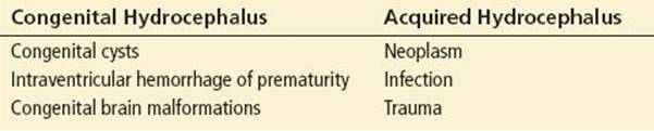
CSF shunts are catheters inserted into the ventricles or other CSF-containing structures in the brain and then threaded under the skin from the skull to the terminal location such as the right atrium, pleural cavity, or (most commonly) the peritoneum. There are three components to the shunt: (a) A proximal catheter that drains one of the cerebral ventricles or areas of CSF accumulation, (b) an extracranial one-way valve to prevent backflow of CSF, and (c) a subcutaneous tunneled distal catheter. Some shunts also have a single or double reservoir system located adjacent to the opening in the skull and can be accessed percutaneously to collect CSF. The catheters and valves have radiopaque markings to enable radiographic visualization (Table 291.1).
TABLE 291.1
Congenital Hydrocephalus and Acquired Hydrocephalus
COMPLICATIONS: SHUNT MALFUNCTION AND INFECTION
The cause of shunt malfunction may be mechanical (e.g., broken tubing) or nonmechanical (e.g., infection), or a combination of the two. Shunt malfunction occurs most commonly in the first 12 months following placement, and accounts for about 10% of all shunt malfunctions (8). After the initial year post-placement, the rate of shunt malfunction falls to about 5% (9).
Obstruction of CSF flow is the most common type of shunt malfunction. The three most common sites of obstruction are the proximal catheter, the proximal valve, and the terminal end of the distal catheter. Shunt catheters can also become disconnected at any point along their trajectory, most commonly in the neck region (10). Disconnections are often associated with improper use of connectors distal to the valve or fracture of the shunt tubing that can occur with age and calcification over time. A less common complication is migration of the tip of the shunt catheter. The catheter tip can migrate into or traverse intra- and extra-abdominal organs including the bladder, rectum, scrotum, and lungs. This is in part due to the extra length of tubing placed in the abdominal cavity to correspond to the child’s future growth. Other complications are organ specific and depend on where the catheter terminates, and may include intraperitoneal CSF collections or pseudocysts which may or may not cause peritonitis.
Obstruction, disconnection, and catheter migration often lead to complications associated with inadequate CSF drainage. However, overdrainage can also be a problem. While rare, overdrainage can lead to ventricular collapse and “slit ventricle syndrome,” which manifests as orthostatic hypotension, subdural fluid collections, craniosynostosis, ventricular compartmentalization, and cerebellar tonsillar herniation (11).
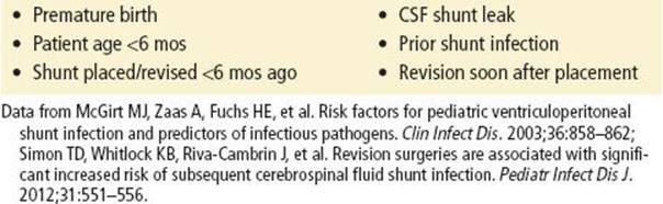
Shunt infections include the shunt hardware, the overlying tissues, or the distal terminal sites (typically within the peritoneum) and occur primarily through direct contamination or hematogenous seeding. The reported incidence averages 10% to 15% (11), the majority occurring within the first 6 months post-placement (12,13). Like other bacterial infections of the CNS, these infections can be responsible for severe neurologic injury or death (Table 291.2).
TABLE 291.2
Risk Factors for Infection
CLINICAL PRESENTATION
Children may have subtle or fulminant presentations of shunt complications. Paying close attention to the history and concerns relayed by the parents or caregivers cannot be overemphasized (14). Presenting symptoms of a shunt malfunction or obstruction may be subtle (mild irritability) or profound (signs of impending herniation and cardiopulmonary instability). Cranial nerve palsies, visual impairment, and hemiparesis represent atypical presentations (14). Headaches are a common complaint in children with shunts, and the character of the headache may help diagnose malfunction. Severe and progressive headaches are more likely to be associated with increased ICP, whereas postural headaches may be associated with slit ventricle syndrome or low-pressure conditions (Table 291.3).
TABLE 291.3
Shunt Malfunction/Obstruction

The presenting symptoms of shunt infection may be nonspecific, or suggestive of other entities, including shunt malfunction. Importantly, fever is not always present and is rarely the sole manifestation of shunt infection. Infection can include any portion of the shunt system. While seizure activity is a negative predictor of shunt malfunction, elevated ICP caused by shunt failure must still be considered (15,16). Lastly, it is important to remember that a patient can have concurrent shunt malfunction and infection and thus present with clinical evidence of both problems (Table 291.4).
TABLE 291.4
Shunt Infection

DIFFERENTIAL DIAGNOSIS
Symptoms of shunt malfunction and infection are largely nonspecific and other common pediatric illnesses, such as acute gastroenteritis, urinary tract infection, and otitis media, should be considered once shunt function has been evaluated.
ED EVALUATION
Important history includes the indication for and date of the initial shunt placement, postoperative complications, and any history of shunt malfunctions, infections, or revisions. It is important to ask about signs and symptoms that may have accompanied previous failures. The shunt itself should be examined and palpated for any signs of infection (warmth, tenderness, or erythema) or malfunction (broken tubing or subcutaneous collection of CSF). The shunt catheter can be located as it exits through a hole in the skull, travels subcutaneously just inferior to and behind the ear, and continues down the neck to the chest. For a ventriculoperitoneal shunt, the catheter travels anterior to the clavicle, passes through the subcutaneous tissue overlying the chest wall, and ultimately enters the peritoneal cavity. Of note, when inspecting shunts that have been recently placed, incision sites should be carefully examined for evidence of infection.
Imaging and laboratory studies are often indicated in suspected shunt malfunction or infection. Standard imaging should include a plain radiograph “shunt series” and either a noncontrast computed tomography (CT) or magnetic resonance image (MRI) of the head to assess ventricular size and changes from prior studies. Some shunt valves will need to be reprogrammed following an MRI and require an on-site individual trained at this procedure. The shunt series images the entire course of the shunt, including the skull, neck, chest, and abdomen, to reveal catheter fracture, kinking, disconnection, dislocation, or migration. Finally, an abdominal ultrasound or CT scan is indicated if there is concern for an intra-abdominal process such as intra-abdominal pseudocyst around the distal tip.
A complete blood count with differential, blood cultures, electrolytes, and a coagulation screen are commonly recommended for the evaluation of a potential shunt malfunction or infection. A neurosurgeon should be consulted before a diagnostic puncture is performed for aspiration (“tap”) of a ventricular shunt. For patients who are not critically ill, a shunt tap is performed to measure the opening pressure and collect CSF for laboratory analysis (cell count, protein, glucose, Gram stain, and culture) to detect possible infection. Rarely, a patient with a ventricular shunt may present to the ED with signs of cardiorespiratory failure due to elevated ICP (unilateral fixed and dilated pupil, posturing, apnea). In this unique circumstance, it may be appropriate to perform a therapeutic shunt tap without first consulting a neurosurgeon.
KEY TESTING
• Head CT or brain MRI
• Radiographic shunt series
• Laboratory studies as indicated by presentation and differential diagnosis
ED MANAGEMENT
The time between the onset of symptoms and coma from shunt failure may be only a few hours (17). A patient with a suspected ventricular shunt problem should be seen in the ED by a neurosurgeon, regardless of the results of diagnostic tests. CT scans can be notoriously unchanged from baseline despite an acute shunt malfunction. In situations where evaluation by a neurosurgeon is not possible, transferring the patient to another facility where such services are available may be the best option.
For patients with confirmed shunt infection, empiric antibiotics should be administered to cover both common gram-positive and gram-negative pathogens. A third-generation cephalosporin (e.g., ceftriaxone, cefotaxime) and vancomycin are recommended choices for initial therapy to cover typical pathogens (commonly Staphylococcus species), and penetrate well into the CNS. Patients with confirmed shunt malfunction or obstruction require a surgical revision. The timing of the revision depends on the patient’s clinical condition and the availability of the neurosurgeon; severe symptoms require immediate repair.
KEY INTERVENTIONS
• Obtain history of the reason for and date of shunt placement, prior complications, and infections.
• Obtain a shunt series and noncontrast CT.
• Transport the patient as necessary to a facility where definitive care can be provided.
DISPOSITION
For patients who require interfacility transfer, if possible, complete any invasive procedures that are likely to become necessary (endotracheal intubation, central line placement, etc.) while the patient is still in the ED, since space and personnel advantages greatly increase the chances for success.
Any child with suspected or confirmed shunt infection or malfunction requires hospital admission. If the diagnostic workup for shunt malfunction is negative and the patient is examined and cleared by a neurosurgeon, the child can be safely discharged from the ED. For children discharged home, it is important to provide clear return instructions and recommendations for outpatient follow-up.
Common Pitfalls
• Failure to recognize a shunt malfunction or infection associated with nonspecific patient complaints. Complaints such as “decreased activity” or “poor appetite” may be due to a viral illness, but warrant a focused shunt evaluation in patients with a shunt.
• Failure to review previous head CT or MRI scans.
• Failure to obtain neurosurgical consultation in the ED.
TRACHEOSTOMY TUBES
Tracheostomy tubes share two common attributes: (1) A primary cannula, and (2) an obturator that is inserted into the primary cannula for all replacements, whether routine or emergent. Parents can usually provide specific details about their child’s tracheostomy and tracheostomy tube (Tables 291.5 and 291.6).
TABLE 291.5
Tracheostomy Tubes
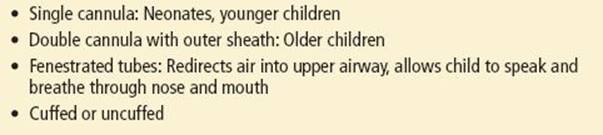
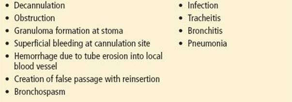
TABLE 291.6
Tracheostomy Complications
CLINICAL PRESENTATION
Children with an obstructed or dislodged tube (one of the most common complications) (18) range from those who appear well to those with impending respiratory failure. This depends on their degree of tracheostomy dependence, which can range from none to complete dependence on mechanical ventilation. Infection should be suspected if there are acute changes in tracheal secretions such as increased thickness, quantity, new color, or new odor, especially if accompanied by fever.
ED EVALUATION, MANAGEMENT, AND DISPOSITION
In the case of tracheostomy tube obstruction or displacement, it is important to recall that caregivers routinely replace tracheostomy tubes outside the hospital setting. Thus, a patient whose dislodged or obstructed tube has not been easily managed by a parent or caregiver must be considered to have a high-risk airway. Ideally, an otolaryngologist should be consulted prior to attempts at tube replacement in case the tube does not easily pass, if bleeding occurs, or other significant surgical concerns arise, especially with a recently created tracheostomy site. Patient positioning and careful preparation for emergent airway management are critical and should include tracheostomy-specific suction, oxygen delivery, and humidification systems. Place a towel roll under the child’s shoulders to hyperextend the neck for optimal visualization of the tracheostomy site and to help open the airway distally. Identify the type and size of tracheostomy needed for replacement, and if possible, have a second device, a half-size smaller, available if edema, scar tissue, or anatomic factors prevent replacement with the original size. If the type of device and tube size are not known, an appropriately sized endotracheal tube may serve as an interim solution, but it should not be advanced more than 2 to 4 cm into the stoma.
An appropriately sized endotracheal tube and airway setup should always be available in case an airway cannot be established through the tracheostomy site and emergent orotracheal intubation becomes necessary (18). Tracheal suctioning may help relieve mucus plugging, but it is important to limit suctioning to only 10 to 20 seconds to prevent hypoxemia and further respiratory compromise. Humidified oxygen should be administered. Albuterol may be administered liberally by this route if there is thought to be bronchospasm.
Prior to placement of the new tube, verify balloon integrity and lubricate the tip. Similarly, be sure to deflate the balloon on the old tracheostomy tube prior to removing it. Once the old tube has been removed, carefully place the new tube into the recently vacated stoma. To minimize the chance of creating a false passage, do not force the tube into the tract. If there is significant respiratory distress, failure to pass the tube easily should prompt consideration of orotracheal intubation.
If there is concern about tracheitis or pneumonia, tracheal aspirate samples and a chest radiograph should be obtained. Consider empiric antibiotics to cover typical pathogens: Staphylococcus species, Haemophilus influenzae B,and Moraxella catarrhalis. Disposition from the ED depends on the patient’s overall clinical status and specific tracheostomy needs. Children requiring new or increased oxygen support, intravenous antibiotics, ongoing pulmonary care, or subspecialty consultation should be hospitalized.
ENTERAL FEEDING TUBES
Enteral feeding tubes are placed for diverse indications. Some children depend on them for primary nutrition and hydration, whereas others have to be used only for supplemental nutrition or medication administration. Feeding tubes placed via the nasopharynx, such as nasogastric (NG) and nasoduodenal (ND) tubes, are typically temporary, while gastrostomy (G) and gastrojejunostomy (GJ) tubes, which are placed through the abdominal wall, are more permanent. Feeding tube complications may be categorized as those related to the anatomical location of the tube and the surrounding tissues, or those related to the tube itself (e.g., dislodgement or breakage) (Table 291.7) (19).
TABLE 291.7
Complications of Enteral Feeding Tubes

Stomal complications include dermatitis, bleeding granulation tissue, peristomal cellulitis, and superficial fungal infection.
CLINICAL PRESENTATION
The majority of children appear well, though others may complain of abdominal distention and pain. Dislodgement, especially of G tubes, is the problem most commonly seen in the ED (20).
ED EVALUATION, MANAGEMENT, AND DISPOSITION
NG and ND tubes can be replaced in the ED. ND tubes are more challenging because the tip must pass through the pylorus; placing the child on his or her right side may help with movement through the pylorus. Dislodged G tubes require urgent replacement to prevent closure of the stoma and tract. A temporary catheter (e.g., Foley catheter) may be inserted in place of the actual device to preserve the stomal tract until a more permanent replacement tube is secured. A G tube that has been placed surgically within 2 months, and especially within 2 weeks, should be replaced by the specialty service that initially placed it, because of the increased risk of creating a false tract within the incompletely healed abdominal wall (21). Analgesia and anxiolysis should be considered prior to tube replacement to help with relaxation of the abdominal wall musculature. Dilators may be required to facilitate replacing the tube, but recently created tracts (i.e., less than 30 days old) should not be dilated.
GJ tubes require interventional radiology or general surgery consultation for replacement or repair. If there is concern for an abdominal wall abscess, ultrasound is an ideal initial imaging modality. Adjunctive laboratory studies should be obtained only as clinically indicated (e.g., for suspected infection or in an ill-appearing child). Children with enteral feeding tube complications often require admission to the hospital for intravenous hydration, nutritional support, medication administration, or surgery as indicated, until the tube can be satisfactorily replaced. If the tube is replaced in the ED, the position should be verified either clinically or by a contrast-enhanced imaging study; the child can then be discharged home with routine follow-up.
INDWELLING VENOUS CATHETERS
Indwelling venous catheters, often tunneled beneath the skin, may provide access through externalized tubing (e.g., Hickman device), or a subcutaneous reservoir (e.g., Port-a-cath device). They can vary in length, diameter, and number of lumens or access ports. Special equipment and specific procedural steps are necessary to access these lines. Complications may be mechanical, including dislodgement, breakage, or obstruction, or nonmechanical, including infection and air embolism. Overall, the most common complications are obstruction and infection (22).
CLINICAL PRESENTATION
Dislodged, broken, or fractured indwelling catheters may present with bleeding; a bulge, pain, defect, or swelling at the catheter site; head or neck swelling; respiratory distress; arrhythmia; or signs of cardiac tamponade. Obstruction presents with difficulty drawing back, infusing, or flushing through the line. Families will often have tried to troubleshoot this type of complication at home prior to presenting to the ED. A catheter-related infection may present with local signs and symptoms such as erythema and swelling, or the child may have systemic symptoms such as fever and tachycardia. Immunocompromised children may not be able to mount a local inflammatory response and may present with more subtle symptoms. These patients are at high risk for disseminated infection and should be evaluated promptly. Air embolism should be suspected in patients with an acute onset of tachypnea, tachycardia, hypotension, or altered mental status.
ED MANAGEMENT AND DISPOSITION
Dislodged or broken catheters should be clamped proximally and repaired as per local institutional practices or replaced as indicated by the appropriate surgical service. Obstruction can sometimes be relieved by infusion of a fibrinolytic, but if this is unsuccessful surgical consultation is warranted. Use of these agents should follow individual institutional guidelines. If a catheter or blood infection is suspected, obtain blood cultures and initiate empiric antibiotics. Immunosuppressed children should be evaluated promptly and monitored closely. Further laboratory and imaging studies should be guided by the child’s clinical status. If air embolism is suspected, the child should be placed in Trendelenburg position on the left side while the catheter is clamped and oxygen applied. If the catheter cannot be repaired in the ED, or if infection is suspected, hospital admission is likely to be necessary until definitive repair can be scheduled.
REFERENCES
1. 2009–2010 National Survey of Children with Special Health Care Needs. www.childhealthdata.org/action/databriefs. Accessed July 10, 2012.
2. Perrin J, Bloom S, Gortmaker S. The increase of childhood chronic conditions in the United States. JAMA. 2007;297(24):2755–2759.
3. Kroner EL, Hoffmann RG, Brousseau DC. Emergency department reliance: A discriminatory measure of frequent emergency department users. Pediatrics. 2010;125(1):133–138.
4. O’Mahony L, O’Mahony DS, Simon TD, et al. Medical complexity and pediatric emergency department and inpatient utilization. Pediatrics. 2013;131(2):e559–e565.
5. Fein JA, Cronan KM, Posner JC. Approach to the care of the technology-assisted child. In: Fleisher GR, Ludwig S, eds. Textbook of Pediatric Emergency Medicine. 6th ed. Philadelphia, PA: Lippincott Williams & Wilkins; 2010.
6. Stone JJ, Walker CT, Jacobson M, et al. Revision rate of pediatric ventriculoperitoneal shunts after 15 years. J Neurosurg Pediatr. 2013;11:15–19.
7. Sood S, Ham SD, Canady AI. Current treatment of hydrocephalus. Neurosurg Q. 2001;11:36–44.
8. Tuli S, Drake J, Lawless J, et al. Risk factors for repeated cerebrospinal shunt failures in pediatric patients with hydrocephalus. J Neurosurg. 2000;92:31–38.
9. Stein SC, Guo W. Have we made progress in preventing shunt failure? A critical analysis. J Neurosurg Pediatr. 2008;1(1):40–47.
10. Ghome K, Drake J, Lamberti-Pasculli M, et al. Management of shunt disconnections and fractures in children: Experience at the Hospital for Sick Children. AANS Pediatric Section Meeting, South Beach, FL. 2007.
11. Weprin BE, Swift DM. Complications of ventricular shunts. In: Honeycutt J, Boop R, eds. Techniques in Neurosurgery. 2002;7(3):224–242.
12. Browd SR, Ragel BT, Gottfried ON, et al. Failure of cerebrospinal fluid shunts: Part I: Obstruction and mechanical failure. Pediatr Neurol. 2006;34:83–92.
13. Simon TD, Hall M, Riva-Cambrin J, et al. Infection rates following initial cerebrospinal fluid shunt placement across pediatric hospitals in the United States. Clinical article. J Neurosurg Pediatr.2009;4:156–165.
14. Barnes NP, Jones SJ, Hayward RD, et al. Ventriculoperitoneal shunt block: What are the best predictive clinical indicators? Arch Dis Child. 2002;87:198–201.
15. Johnson DL, Conry J, O’Donnell R. Epileptic seizure as a sign of cerebrospinal fluid malfunction. Pediatr Neurosurg. 1996;24:223–228.
16. Pitetti R. Emergency department evaluation of ventricular shunt malfunction. Pediatr Emerg Care. 2007;23(3):137–141.
17. Kim TY, Stewart G, Voth M, et al. Signs and symptoms of cerebrospinal fluid shunt malfunction in the pediatric emergency department. Pediatr Emerg Care. 2006;22(1):28–34.
18. Mitchell RB, Hussey HM, Setzen G, et al. Clinical consensus statement: Tracheostomy care. Otolaryngol Head Neck Surg. 2013;148:6–20.
19. Friedman JN, Ahmed S, Connolly B, et al. Complications associated with image-guided gastrostomy and gastrojejunostomy tubes in children. Pediatrics. 2004;114:458–461.
20. Saavedra H, Losek JD, Shanley L, et al. Gastrostomy tube-related complaints in the pediatric emergency department: Identifying opportunities for improvement. Pediatr Emerg Care. 2009;25:728–732.
21. Cronan KM, Fein JA, Posner JC. Acute care of technology-assisted children. In: Pediatric Emergency Medicine Secrets. 2nd ed. Philadelphia, PA: Mosby, Inc.; 2008.
22. de Jonge RC, Polderman KH, Gemke RJ. Central venous catheter use in the pediatric patient: Mechanical and infectious complications. Pediatr Crit Care Med. 2005;6:329–339.