
Jerrold B. Leikin
The history and physical examination is the cornerstone of clinical patient management, and while taking a history may be a direct method for toxin exposure identification, quite often it is not reliable. Information obtained may prove minimal in some cases and should be considered partial or inaccurate in suicidal gestures and with illegal drug or chemical use. Thus, obtaining a history of exposure from family, friends, coworkers, or prehospital personnel, may be essential to patient management.
IMMEDIATE CRITICAL INTERVENTION
An immediate decision with regard to stability needs to be made upon patient presentation. Unstable vital signs need to be assessed and addressed in the traditional manner. Supportive measures including the “ABCs” (airway, breathing, circulation) are often required before confirmation of intoxication can be obtained. Intubation is not always necessary when cough and gag reflexes are present and there is adequate spontaneous ventilation; however, when there is a concern regarding airway protection and potential clinical deterioration, it is better to secure the airway. Intubation is indicated in acute respiratory failure. Other specific indications include the need for high levels of supplemental oxygen (e.g., in carbon monoxide poisoning) and the need to protect the airway for gastric emptying or decontamination. Endotracheal intubation decreases (but does not eliminate) the risk of aspiration (which is approximately 10% in the comatose patient with drug overdose) (1).
The “coma cocktail” (dextrose, oxygen, thiamine, and naloxone) is diagnostic as well as therapeutic, and should be administered empirically to the comatose patient with a suspected toxic exposure (2,3). A rapid bedside blood glucose can determine which patients should receive dextrose, although a low normal value by blood dipstick does not always exclude low serum glucose. The dose is 50 g IV in an adult; 2 to 4 mL/kg body weight of a 25% solution in a child. Thiamine (50 to 100 mg) can treat or avoid Wernicke encephalopathy syndrome, which is particularly important in the nutritionally depleted alcoholic; there is no evidence that dextrose should be withheld until thiamine is administered.
Naloxone rapidly reverses coma, respiratory depression, and hypotension induced by opioids. An initial dose of 0.2 to 0.4 mg in adults is administered IV, subcutaneously, IM, or endotracheally. The intratrachael dosage is diluted to 1 to 2 mL in normal saline. If there is no response, an additional 1 to 2 mg can be repeated. The pediatric dose is 0.1 mg/kg, repeated every 2 to 3 minutes as needed. Use of a higher initial dose may precipitate withdrawal symptoms in opioid-dependent patients. Heroin and morphine are the most sensitive to naloxone, and while some opioid agents such as meperidine, propoxyphene, diphenoxylate, methadone, and pentazocine require larger doses. A lack of response to 10 mg of naloxone generally excludes opioid toxicity (see Chapter 305 for details about naloxone use).
Flumazenil should be considered in cases where benzodiazepine overdose alone is suspected or when reversal of therapeutic conscious sedation is desired. Case reports suggest a risk of precipitating seizures with flumazenil when there is a suspicion of benzodiazepine plus cyclic antidepressant overdose. See Chapter 308 for details on use of flumazenil. The routine use of flumazenil is discouraged, except in cases of acute benzodiazepine overdose presenting as coma or respiratory depression without any coingestants (4).
DIAGNOSTIC APPROACH
A quick and focused physical examination often leads to important clues about the nature of the toxin. These clues can be specific symptom complexes associated with certain toxins and can be referred to as “toxidromes” (Table 295.1) (5).
TABLE 295.1
Common Toxidromes

Vital Signs. Depending on the intoxication, patients may present with hypotension or hypertension, bradyarrhythmias, or tachyarrhythmias. The pathogenesis of hypotension varies and may include hypovolemia, myocardial depression, cardiac arrhythmia, or systemic vasodilation. Treatment should be individualized, but an initial strategy of rapid intravenous normal saline solution infusion is indicated in most instances. Vasopressors may be required for refractory hypotension. The vasopressor of choice depends on the type of intoxication. For example, norepinephrine appears to be advantageous over dopamine in tricyclic antidepressant-induced hypotension (6). Lipid rescue therapy can be considered in the setting of hemodynamic instability—this is discussed further in Chapter 296. Hypertension occurs with sympathomimetic drugs, anticholinergics, ergot derivatives, phenylpropanolamine overdoses, and withdrawal from nicotine, alcohol, and sedatives. The need to treat the hypertension depends on its chronicity and severity of symptoms and the inciting agent. Hypertension-induced (reflex) bradycardia generally should not be treated.
Anticholinergic and sympathomimetic substances increase heart rate, blood pressure, and temperature. This is particularly true for cocaine intoxication, where hyperthermia may be a particularly ominous sign for mortality. Important clinical signs that differentiate sympathomimetic poisoning from anticholinergic intoxication include dry skin and xerostomia along with absent bowel sounds, which usually occur in anticholinergic but not sympathomimetic poisoning. It should be noted that the antipsychotics and antidepressants with the greatest anticholinergic effects include clozapine, thioridazine, and amitriptyline. In contrast, organophosphates, opiates, barbiturates, β-blockers, benzodiazepines, alcohol, and clonidine cause hypothermia, bradycardia, and respiratory depression. Table 295.2 lists various toxins which alter temperature.
TABLE 295.2
Drugs Affecting Temperature

Sensorium. Determine whether the patient is comatose, stuporous, lethargic, or alert. Table 295.3 lists drugs that can directly affect mental status, while Table 295.4 lists toxic causes of delirium. Poisoned individuals presenting with deep coma (Glasgow Coma Scale under 7) have a mortality rate approximately seven times higher than overall mortality from acute poisoning (7). Tables 295.5 and 295.6 list clinical signs of worsening of toxin-induced coma and poor prognostic signs of coma. Table 295.7 lists drugs that can cause cyclic or relapsing coma.
TABLE 295.3
Selected Drugs Altering Mental Status
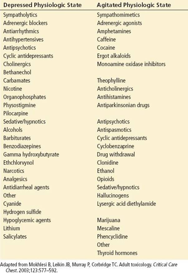
TABLE 295.4
Toxic Causes of Delirium
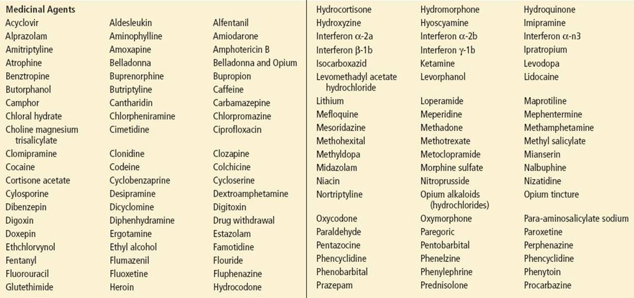

TABLE 295.5
Neurologic Signs of Worsening of Toxic Coma
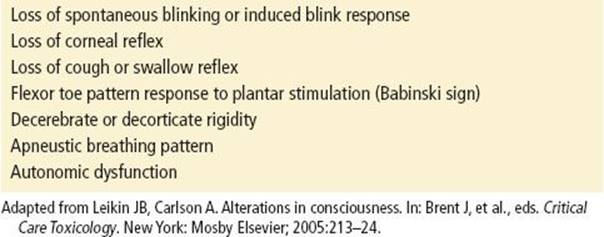
TABLE 295.6
Poor Neurologic Prognostic Signs from Recovery of Toxic Coma

TABLE 295.7
Drugs Which can Cause Cyclic or Relapsing Coma

Behavior and Hallucinations. Often the patient’s hallucinatory pattern can be specific for certain drugs. For example, with atropine the patient experiences lilliputian hallucinations; with cocaine, there is a simple visual hallucinatory pattern with objects appearing in the periphery of vision; with phencyclidine, complex hallucinations are often indistinguishable clinically from a paranoid psychosis; with LSD, the patient experiences a combination of illusions, hallucinations, synesthesia, and pseudohallucinations.
Motor Signs. Tremors, hypo- and hyperreflexes, and even the nature of seizures can be useful diagnostic tools. Like hallucinations, seizures caused by specific toxins can exhibit certain specific properties. For example, strychnine is unique in that it can cause generalized seizures while the patient is alert, often called a “spinal seizure.” Most toxin-induced seizures (including withdrawal seizures) will respond to benzodiazepines, barbiturates, or propofol. Other drug-induced seizures respond only to specific antidotal therapies and not conventional antiseizure medication. Examples include anticholinergic-induced seizures, which respond to physostigmine, and isoniazid-induced seizures, which respond to pyridoxine. Most drug-induced seizures rarely respond to phenytoin and may require multidrug therapy. Toxins resulting in isolated cranial neuropathies are rare and are listed in Table 295.8.
TABLE 295.8
Agents Causing Cranial Nerve Palsies

Ocular Findings. These can be divided into two categories: pupillary size and reactivity, and demonstration of nystagmus.
Pupillary Signs. Both atropine and cocaine can result in mydriasis, but in cocaine or other adrenergic intoxication the pupils respond to light, whereas with anticholinergics the pupils will not respond. Agents that contribute to miosis include organophosphate insecticides, narcotics, bromide, clonidine, and nicotine. Phencyclidine has been known to cause either mydriasis or miosis. It should be noted that pontine hemorrhage can result in miosis; Drug withdrawal and serotonin syndrome can cause mydriasis (see Tables 295.9 and 295.10).
TABLE 295.9
Agents Causing Miosis

TABLE 295.10
Agents Causing Mydriasis


Nystagmus. Alcohols are probably the most common etiology of horizontal nystagmus, although lithium, carbamazepine, solvents, meprobamate, quinine, and primidone can also cause horizontal nystagmus. Though phencyclidine classically causes a combination of vertical, horizontal, and even rotary nystagmus, these can also occur with phenytoin and sedative–hypnotics.
In addition to these physical signs, odors emanating from the patient may also provide important diagnostic clues. For example, a garlic odor is often caused by arsenicals, phosphorus compounds, organophosphates, and insecticides.
Laboratory evaluation. The clinical laboratory evaluation includes the evaluation of the “three laboratory gaps of toxicology”: anion gap, osmolal gap, and oxygen saturation gap. What these “gaps” have in common is that a substance is accounting for a difference between the calculated and measured determinations.
The anion gap reflects the difference between the serum or plasma measured cation and anion concentrations and is usually calculated utilizing the following formula: Anion Gap = measured Na+ concentration – [measure chloride and bicarbonate concentrations] (8). The normal range of anion gap is usually 8 to 12 mEq/L in most laboratories. An increase in anion gap (>16 mEq/L) suggests lactic acidemia, uremia, ketoacidemia, or selected intoxications (see Tables 295.11 and 295.12). A normal anion gap does not preclude intoxication because most toxins do not elevate the anion gap or there may be a coexisting condition that lowers the gap. Common among these conditions is hypoalbuminemia: for every 1 g/L decrease in the plasma albumin, the anion gap falls by 2.5 mEq/L. Clinicians should pay special attention to this correction factor to avoid missing a clinically significant anion gap. Also, in methanol or ethylene glycol poisoning, concurrent ethanol use delays the development of an elevated anion gap metabolic acidosis. In this case, an elevated osmolal gap may be the only early clue to the diagnosis.
TABLE 295.11
Common Causes of Abnormal Anion Gap

TABLE 295.12
Selected Drugs Associated with an Elevated Anion Gap Metabolic Acidosis

Low–molecular-weight drugs and toxins that are osmotically active can increase the discrepancy between measured and calculated plasma osmolality (9). Normal plasma osmolality is 285 to 295 mOsm. The calculated value is determined as follows:

in which Na+ (in milliequivalents per liter) is multiplied by 2 to account for accompanying anions (chloride and bicarbonate), and measured BUN, glucose, and ethanol (in milligrams per deciliter) is divided by the appropriate denominator. The osmolal gap must be interpreted with caution. Measurement of osmolality by vapor pressure osmometry does not detect volatile alcohols such as ethanol and methanol; however, it does detect ethylene glycol (10). Freezing point depression osmometry, the most frequently used method, measures all of these solutes. Therefore, it is important for clinicians to know the method used by their institution. With the standard formula, the normal osmolal gap is large and may range from −9 to +5 mOsm; 10 mOsm is considered the upper limit of normal. However, an osmolal gap of 10 mOsm in a patient who started at −9 mOsm may be significantly elevated and the gap may be missed. Table 295.13 lists agents that can cause an osmolal gap.
TABLE 295.13
Causes of Osmol Gap (>10 mOsm from Baseline Values)

An oxygen saturation gap is present when there is more than a 5% difference between the saturation calculated from an arterial blood gas and the saturation actually measured by co-oximetry. Co-oximetry detects the absorption of four different wavelengths, enabling it to directly measure levels of four types of hemoglobin species: oxyhemoglobin, reduced hemoglobin, carboxyhemoglobin, and methemoglobin. Because arterial blood gas analysis calculates oxygen saturation from the measured pO2 using a standard oxygen–hemoglobin dissociation curve, an oxygen saturation gap occurs in the presence of carboxyhemoglobin, methemoglobin, and sulfhemoglobin (which is not routinely measured by co-oximetry). Pulse oximetry estimates oxygen saturation by emitting a red light (wavelength of 660 nm) absorbed mainly by reduced hemoglobin, and a near-infrared light (wavelength of 940 nm) absorbed by oxyhemoglobin.
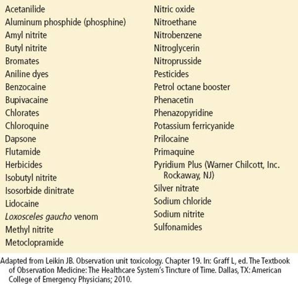
Methemoglobin absorbs almost equally at both these wavelengths. At high methemoglobin levels (over 35%), the oxygen saturation by pulse oximetry tends to regress toward 85% and plateaus at that level despite further increments in methemoglobin levels. Thus, if the actual oxygen saturation by co-oximetry is >85%, pulse oximetry underestimates it; if it is <85% by co-oximetry, it overestimates oxygen saturation. A listing of drugs/toxins causing methemoglobinemia is in Table 295.14 (11,12).
TABLE 295.14
Selected Drugs/Toxins Associated with Acquired Methemoglobinemia
Since many laboratories do not routinely use co-oximetry, a more commonly seen gap is the disparity between measured oxygen saturation by blood gas and that measured by pulse oximetry. Carbon monoxide has a wavelength absorption coefficient similar to that of oxyhemoglobin and carboxyhemoglobin may be misinterpreted as oxyhemoglobin by pulse oximetry, leading to overestimation of oxygen saturation when compared to co-oximetry. Abnormally high venous oxygen content (arterialization of venous blood) is characteristic of cyanide poisoning.
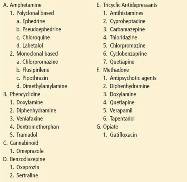
In spite of providing indirect laboratory evidence of intoxication, urine drug screening tests alter management in less than 5% of cases (13). However, toxicology urine drug screening can identify a specific toxin for which an antidote is available and in some instances quantify a toxin allowing for titrated therapy. Most institutions offer urine immunoassay–based testing for six or seven of the most commonly abused drugs. Table 295.15 lists substances that can give a false-positive urine drug screen (14,15). Results are generally available in 60 minutes. Urine confirmation with gas chromatography/mass spectrometry can usually be obtained, but frequently take an additional 2 to 3 days. Because of false positives, while mean immunoassay confirmation rates are highest for cocaine (98%) and marijuana (91%), the confirmation rate for amphetamine is only about 50% (16); in addition, the presence of a drug on a screening test does not necessarily confirm that the symptoms are from that drug. Many drugs cause a positive urine drug screen for many days. Cocaine metabolites can be seen for up to 5 days, and marijuana can be detected for up to a month. Probably the most important prognostic function for immunoassay urine drug screening is in the patient with occult barbiturate use to ascertain the potential risk for barbiturate withdrawal. More comprehensive urine screening (usually performed off-site) may take several hours or even days. Testing of gastric contents is rarely helpful in the emergency department.
TABLE 295.15
Causes of False-Positive Results in Urine Drug Screens (Immunoassay)
Serum quantification of certain toxins is useful, particularly in cases of alcohol (ethanol, methanol, ethylene glycol), acetaminophen, salicylate, phenobarbital, theophylline, digoxin, iron, and lithium intoxication. The timing of these serum specimens needs to correlate with the timing of the drug’s absorption and such an analysis is particularly useful in evaluating substances that may exhibit a delay in the onset of clinical symptoms (Table 295.16). Serum values of selected agents causing coma are listed in Table 295.17. A strong argument can be made for checking serum acetaminophen and possibly salicylate levels in all cases of medication ingestion, given the easy availability of acetaminophen and salicylate, the subtle manifestation of early poisoning, and the existence and importance of specific treatment of these potentially lethal intoxications.
TABLE 295.16
Agents that can Cause Delay in Symptoms

TABLE 295.17
Agents and Blood Concentrations Resulting in Coma

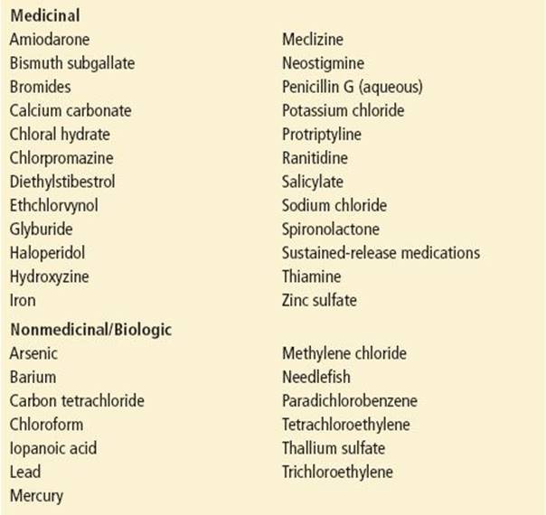
Radiography is not a reliable method for evaluating toxic exposure and should be utilized only in specific cases. Factors that influence radiodensity include molecular weight, atomic number, relative contrast to surrounding tissues, and compactness of the form of drug. Overall, only about one-third of all drug preparations would be detectable by plain abdominal x-ray, with about 2% of drugs being densely radiopaque (Table 295.18) (18). These cases include evaluation of heavy metal exposure (including leaded paint chips), concretion formation of drugs (such as salicylate, meprobamate, iron, and theophylline), suspected body packing or body stuffing, and the use of digital radiography for extended-release formulations (17).
TABLE 295.18
List of Radiopaque Substances
Common Pitfalls
• Failing to obtain collateral information regarding potential toxin exposure.
• Overlooking potential toxic causes in patients with anion gap metabolic acidosis.
• Overlooking the possibility of acetaminophen toxicity in patients presenting with gastrointestinal or hepatic complaints.
• Failing to consider drug or ethanol withdrawal in the hyper-adrenergic delirious patient.
REFERENCES
1. Marik PE. Aspiration pneumonitis and aspiration pneumonia. N Engl J Med. 2001;344(9):665–671.
2. Bartlett D. The coma cocktail: Indications, contraindications, adverse effects, proper dose and proper route. J Emerg Nurs. 2004;30:572–574.
3. Hoffman RS, Goldfrank LR. The poisoned patient with altered consciousness: Controversies in the use of a ‘coma cocktail’. JAMA. 1995;274(7):562–569.
4. Carvalho C, Walker DA. Coma cocktail: A role for flumazenil? Br J Hosp Med (Lond). 2007;68(2):112.
5. Nice A, Leikin JB, Matureen A, et al. Toxidrome recognition to improve efficiency of emergency urine drug screens. Ann Emerg Med. 1988;17(7):676–680.
6. Tran TP, Panacek EA, Rhee KJ, et al. Response to dopamine vs. norepinephrine in tricyclic antidepressant induced hypotension. Acad Emerg Med. 1997;4(9):864–868.
7. Forsberg S, Hojer J, Ludwig L. Hospital mortality among poisoned patients presenting unconscious. Clin Toxicol (Phila). 2012;50(4):254–257.
8. Kraut JA, Madias NE. Serum Anion Gap: Its uses and limitations in clinical medicine. Clin J Am Soc Nephrol. 2007;2:162–174.
9. Carstairs SD, Suchard JR, Smith T, et al. Contribution of serum ethanol concentration to the osmol gap: A prospective volunteer study. Clin Toxicol. 2013;51(5):398–401.
10. Coulter CV, Farquhar SE, McSherry CM, et al. Methanol and ethylene glycol acute poisonings – predictors of mortality. Clin Toxicol (Phila). 2011;49(10):900–906.
11. Bradberry SM. Occupational methaemoglobinaemia: Mechanism of production, features, diagnosis and management including the use of methylene blue. Toxicol Rev. 2003;22(1):13–27.
12. Wilkerson RG. Getting the blues at a rock concert: A case of severe methemoglobinaemia. Emerg Med Australas. 2010;22(5):466–469.
13. Tenenbein M. Do you really need that emergency drug screen? Clin Toxicol (Phila). 2009;47(4):286–291.
14. Leikin JB. Clinical interpretation of drug testing. Prim psychiatry. 2010;17(6):23–27.
15. Rengarajan A, Mullins ME. How often do false-positive phencyclidine urine screens occur with use of common medications? Clin Toxicol (Phila). 2013;51:493–496.
16. Swotinsky R. Ed. MRO Update. American College of Occupational and Environmental Medicine. 2004;10(12):3–4.
17. Tillman DJ, Ruggles DL, Leikin JB. Radiopacity study of extended release formulations using digital radiography. Am J Emerg Med. 1994;12:310–314.
18. Chan YC, Lau FL, Chan JCS, et al. A study of drug radiopacity by plain radiography. Hong Kong J Emerg Med. 2004;11:205–210.