
Jerry W. Snow and R. Brent Furbee
The term opiate applies to naturally occurring derivatives of opium. Opioid originally referred only to synthetic or semisynthetic compounds that exert effects through opioid receptors but increasingly, the term opioid applies to both groups. Narcotic is an outdated term to describe the effects, used before the mechanism was known. The opium poppy, Papaver somniferum, is the source of naturally occurring opioids. Opium contains morphine, codeine, and thebaine, from which natural and semisynthetic opioids are derived. Papaverine (a vasodilator) and noscapine (an antitussive) are nonanalgesics found in opium. The opioid antagonists, naloxone and naltrexone, are semisynthetic derivatives of thebaine.
Opioid prescription abuse is recognized as a growing epidemic. Increased use of opioids for treatment of chronic noncancer pain increased in the 1980s. “Chronic pain” is defined by the International Association for the Study of Pain as “pain that persists beyond normal tissue healing time, which is assumed to be three months” (1). Although it appears logical, the use of hydrocodone, oxycodone, methadone, and other opioids for chronic noncancer pain has increased dramatically despite the lack of evidence supporting its effectiveness. Several epidemiologic studies indicate an increase in methadone-related deaths in the United States. This may be associated with QTc prolongation and resultant arrhythmias (1). Sales of opioid analgesics increased fourfold between 1999 and 2010, and between 1997 and 2007 the average amount of opioids distributed to persons in the United States jumped from 96 mg of morphine equivalents/person to 700 mg/person, an increase of over 600% (2). Diversion is also a growing concern. A 2010 National Survey on Drug Use and Health (NSDUH) showed that an estimated 22.6 million, or 8.9% of Americans, aged 12 or older, were current or past month illicit drug users (3).
Since 2003, opioid analgesic deaths have been more than from cocaine and heroin combined (2) and now account for more deaths than suicide or motor vehicle accidents (3). Hydrocodone topped all opioid prescriptions with 136.7 million prescriptions in 2011. Factors that predict drug abuse, misuse, or aberrant drug behavior include personal or family history of drug or alcohol abuse, younger age, or presence of psychiatric conditions (1). Patients who are prescribed doses of <100-mg morphine equivalents/day account for nearly 80% of the at-risk population, but only 20% of prescription drug overdoses. Patients receiving high doses (>100 mg morphine equivalent/d) or those receiving prescriptions from multiple physicians accounted for about 20% of the at-risk population, but 80% of the overdoses (2). In West Virginia, and Ohio, 25% to 66% of deaths from pharmaceutical overdoses used opioids prescribed for someone else. Opioid analgesic misuse and death are highest among men, persons aged 20 to 64 years, non-Hispanic white, and poor and rural populations as well as patients with mental illness (2).
This “epidemic” is due, in part, to changing attitudes toward pain management. In the late 1990s, state medical boards began to curtail restrictions on laws controlling the prescribing of opioids for chronic noncancer pain. New pain management guidelines such as those by the Joint Commission on the Accreditation of Health Care Organizations (JCAHO) in 2000 recognizing the “right to pain relief” encouraged physicians to be more liberal in their prescribing of opioids. Manchikanti concluded in 2012 that, “It has been speculated that in the coming years, there will likely be an extensive “postmortem” on the massive opioid treatment movement and the escalating social crisis that has accompanied it. It is universally accepted that this massive treatment movement has led to huge collateral damage in terms of diversion, misuse, and abuse of opioids” (3).
MECHANISM OF ACTION
Opioids’ clinical effects are the result of complex interactions with the three primary opioid receptor types (mu, delta, and kappa). Three additional receptor types, nociception/orphanin FQ, epsilon, and zeta, are not yet well characterized in humans. The sigma receptor has been reclassified because actions mediated via this receptor are not reversed by naloxone. The mu receptor mediates the majority of clinical effects and most supraspinal analgesic effects as well as sedation and respiratory depression. Delta and kappa receptors act primarily at the spinal cord level. Euphoria is mediated by mu receptors and dysphoria by kappa receptors. These receptors are found throughout the body including the periphery, pre- and postsynaptic sites in the spinal cord, and brain to form an ascending pain transmission system and a descending initiatory system that modulates spinal cord pain transmission (4).
Upon opioid binding, G proteins are activated and use a secondary messenger system involving cyclic AMP to have several effects that produce pain inhibition through (1) blockade of calcium channels, resulting in a decrease of presynaptic neurotransmitter release, and (2) opening potassium channels, producing hyperpolarization of postsynaptic neurons and a decrease in transmission of pain (4). Alpha-2 agonists such as clonidine have some effects similar to opioid receptor binding (see Chapter 322) (5).
Opioid antagonists competitively bind to each type of the opioid receptors and reverse the clinical effect of opioids as well as altering the configuration of the receptor to decrease the availability of receptor sites. Pure antagonists (naloxone, nalmefene, and naltrexone) avidly bind to all major opioid receptor subtypes.
CLINICAL PRESENTATION
The classic opioid toxidrome includes the triad of (1) CNS depression, (2) respiratory depression, and (3) miosis. However, opioids can present with a wide range of clinical findings and atypical presentations (Table 305.1). Opioids are used through intravenous, intramuscular, intranasal, inhalational, oral, subcutaneous, intradermal, or transdermal routes. Unintentional poisoning in addicts/recreational users typically occurs in novice users, during binging, or when an experienced user obtains an unfamiliar supply of heroin and accidentally miscalculates the dose owing to differences in purity. Intentional suicidal overdose or attempted homicide is also common.
TABLE 305.1
Atypical Presentations of Opioid Poisoning
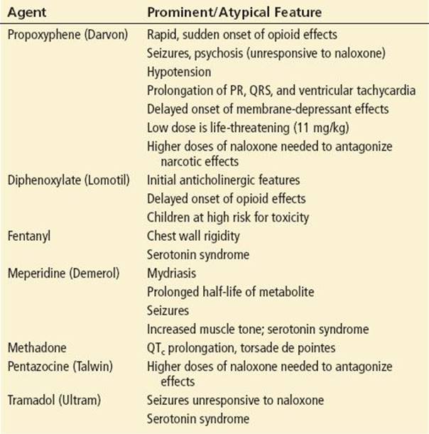
Children usually present with unintentional exposure although iatrogenic poisonings and dosing errors do occur. Overdose in children characteristically has a delayed onset, unexpected severe toxicity, and prolonged effects. Children may ingest pills or apply/ingest transdermal patches (that sometimes become stuck on the roof of the mouth). Elderly patients, particularly those on opioid therapy for chronic pain control, also represent a high-risk group for unintentional poisoning. Contributing factors include comorbidities, slower metabolism, decreased renal clearance and other age-related physiologic changes, and alterations in body composition.
The onset of symptoms is nearly immediate after injection, inhalation, and nasal insufflation. Although effects are usually noted within 30 minutes of ingestion, absorption can be erratic and delayed, particularly in children and with sustained-release formulations. Lomotil (diphenoxylate and atropine) is notorious for causing delayed toxicity, perhaps because both constituents can inhibit gastrointestinal motility. The effects of transdermal medications also begin about 30 minutes after application and can develop or progress after the patch is removed. Intravenous injection and oral ingestion of fentanyl patches delivers the entire drug content of the patch at one time rather than the ongoing absorption of small aliquots of fentanyl.
Respiratory
Respiratory depression is dose-dependent following exposure to opioids, leading to decreased respiratory rate and/or tidal volumes. The mechanism involves mu2 receptors that decrease sensitivity of the medulla respiratory center to hypercapnia. Opioids also suppress the hypoxic drive of ventilation. This combination is what eventually leads to apnea, although agonist–antagonists demonstrate a ceiling effect on respiratory depression.
Acute lung injury (ALI) from opioids has been reported since the 1880s and should be considered when hypoxia persists despite adequate respiration. Patients may develop hypoxia and pulmonary crackles within minutes or hours after regaining normal ventilation. There are several theories on the mechanism: (1) attempted inspiration against a closed glottis leading to negative intrathoracic pressure resulting in fluid shifting into to alveoli is a mechanical mechanism. In addition, (2) similar to neurogenic pulmonary edema, a sympathetic vasoactive response from catecholamines on the myocardium produces cardiogenic pulmonary edema, and (3) some feel ALI is a result of hypoxia. No single mechanism can explain all cases of opioid-induced ALI at this time. In the past, naloxone was blamed as a cause of ALI, but ALI had been clearly documented prior to the development of naloxone and is seen in fatal overdoses that never receive this antidote. In addition, pulmonary edema may not be detectable until ventilation has been restored (6).
Another complication of opioid abuse is interstitial lung disease from the injection of insoluble binding agents in oral opioid tablets such as talc, cornstarch, or cellulose. The result is microscopic pulmonary emboli and inflammation progressing to interstitial fibrosis and pulmonary hypertension. Clinically, this presents as emphysema and chronic respiratory failure. Historically, the oral opioid most frequently cited in pulmonary talcosis is pentazocine, often used in conjunction with tripelennamine or methylphenidate known as Ts (for Talwin) and Blues (for the color of the other drug). It is also seen with methadone, meperidine, codeine, and oxycodone (7). Similarly, retinopathy may develop from the injection of oral medications (8).
Neurologic/CNS
All opioids can cause CNS depression, however other manifestations of opioid CNS toxicity include seizures in acute overdoes, most likely due to hypoxia. Seizures secondary to propoxyphene, tramadol, and meperidine toxicity are well described. Serotonin syndrome occurs when opioids with serotoninergic effects (meperidine, tramadol, and dextromethorphan) are used with monoamine oxidase inhibitors (MAOIs) or other serotonin agonists; however, similar symptoms occur with large doses of those opioids in the absence of other serotonergic drugs (Chapter 343, Serotonin Reuptake Inhibitors).
A severe, acute Parkinsonism has followed illicit use of a meperidine analog synthesized in a clandestine laboratory contaminated with 1-methyl-4-phenyl-1,2,3,6-tetrahydropyridine (MPTP). Affected individuals were known as “frozen addicts.”
Miosis
The mechanism of miosis caused by opioids is controversial. But, stimulation of mu receptors in the Edginger–Westphal nucleus of CN III by morphine does produce miosis. Miosis alone is not pathognomonic for opioid overdose. Overdose with numerous other substances can produce miosis and CNS depression including clonidine, organophosphates, phenothiazines, sedative–hypnotics, anticonvulsants, olanzapine, and ethanol. In addition, normally reactive or mydriatic pupils can be seen with meperidine, propoxyphene, tramadol, and Lomotil poisoning. Numerous polysubstance ingestions or abuse may confound pupillary findings with a variety of results.
Cardiovascular
Opioids can produce mild hypotension through nonspecific histamine release. This is often orthostatic and may not be evident in the supine position. Antihistamines are effective in blocking these effects. Opioids may also produce bradycardia secondary to decreased CNS stimulation.
Propoxyphene is known to have sodium channel–blocking properties similar to class 1 A antidysrhythmics which can produce QRS widening. This can lead to wide complex tachycardias and negative contractility. Methadone and its derivative LAAM may inhibit delayed potassium rectifier currents, increasing the QT interval and the subsequent risk for developing torsade de pointes. Prolonged QT syndrome and torsade de pointes appear to be dose-dependent and are more likely with high-dose methadone.
Gastrointestinal
Nausea and vomiting are common with therapeutic use and overdose. These effects are the product of dopamine-receptor agonism at the chemoreceptor trigger zone in the medulla. Antiemetics can be effective in controlling these symptoms. Constipation secondary to decreased GI motility is very common and is mediated by mu receptors within the GI tract.
Other clinical findings
Sensorineural hearing loss has been reported with opioid exposure usually after high doses and chronic use although it has followed acute overdose. Onset is sudden and rapidly progresses to bilateral hearing loss. Vestibular symptoms are usually, but not always, absent. Hearing loss may resolve with abstinence from opioids. Cochlear implants are usually successful.
Urinary retention due to increased sphincter tone may also develop. Other complications include pressure necrosis, compartment syndrome with rhabdomyolysis, hypoglycemia, and hypothermia in patients who have been comatose.
Tolerance and Withdrawal
Tolerance may be innate (genetically determined) or acquired. Acquired forms include pharmacokinetic tolerance from repeat exposure leading to changes in metabolism or distribution. Pharmacodynamic tolerance refers to physiologic adaptation such as changes in receptor density. Learned tolerance connotes a reduction of drug effects due to learned compensatory mechanisms such as the conditioned tolerance that develops when environmental cues are consistently paired with the administration of the drug. Some opiate overdoses experienced by drug addicts may occur if the drug is taken under new circumstances, tolerance is reduced, and the drug’s effect enhanced. Numerous studies have demonstrated that altering the context of drug administration increases the lethality of many drugs, including opioids (9).
Although physical dependence and behavioral addiction is not fully understood, the mu receptor is involved in both processes and the dopamine pathway is thought to be involved in dependence. Long-term use of opioids can lead to tolerance and withdrawal. Tolerance is most pronounced with the euphoric, analgesic, and sedative effects of opioids. Respiratory tolerance has also been reported in large therapeutic doses, up to 1,000 mg/hr of IV morphine without respiratory depression (10).
CNS excitation, tachypnea, mydriasis, nausea, vomiting, diarrhea, abdominal pain, myalgias, insomnia, piloerection, yawning, lacrimation, rhinorrhea are all symptoms of opioid withdrawal. These occur when stopping use or when an antagonist is delivered. Patients may present with significant fluid and/or electrolyte abnormalities from GI symptoms. Long-term opioid use is associated with decreased levels of endogenous opioids. If exogenous opioid administration is suddenly stopped, the inadequate amount of endogenous opioids fails to provide adequate inhibition to adrenergic pathways resulting in overactivation. Unlike benzodiazepine, barbiturate, and alcohol withdrawal, opioid withdrawal is generally not life-threatening but can cause seizures in neonates. It is important to note that opioid withdrawal does not cause altered mental status.
ED EVALUATION
The history should include the type of opioid, co-intoxicants, and the amount, time, intent, and route of exposure. The physical examination should focus on assessing the airway and the adequacy of ventilation. After the patient has been stabilized, it is important to look for fentanyl patches. The exam should include the all skin surfaces, including axillae, perineum, scrotum, and oropharynx. All patches should be removed and the area cleaned with soap and water to prevent any continued absorption. All major muscle groups should be palpated to evaluate the firmness, swelling, and tenderness that might reveal concerns for compartment syndrome. Patients should have cardiac and oxygen saturation or end-tidal CO2 monitoring. Patients with altered mental status should have blood glucose testing and intravenous access established. Electrolytes, creatine kinase (CK), and urine myoglobin should be checked if patients are combative, agitated, or convulsing. A chest radiograph and ECG should be obtained for unstable vital signs, hypoxia, or an abnormal cardiopulmonary examination. Acetaminophen and aspirin, found in combination with opioids in many medications, should be excluded. Urine drug screens (UDSs) detect some opioids, but will not detect propoxyphene, meperidine, methadone, fentanyl and derivatives, buprenorphine, hydromorphone, or naloxone. Hydrocodone and oxycodone may be detected in high concentrations. Heroin will be picked up as morphine, its metabolite. To specifically determine heroin use, 6-monoacetyl morphine (6-MAM), an intermediate metabolite between heroin and morphine, should be obtained. False positives for opiates on a UDS may include poppy seeds, dextromethorphan, chlorpromazine, diphenoxylate, ephedra, and ciprofloxacin. If action such as notification of child protective services or police is going to be based on the results of a UDS, the results should be confirmed by a second method such as gas chromatography/mass spectrometry. In contrast, a lack of response to adequate doses of opioid antagonists virtually excludes this diagnosis. Although most opioid-induced CNS depression will be reversed with lower doses of naloxone, some opioids such as propoxyphene may require doses as high as 10 mg. A positive response to naloxone is usually diagnostic, but clonidine and valproate (and rarely benzodiazepines, ethanol, and chlorpromazine) have been reported to occasionally respond.
KEY TESTING
• No testing for simple cases that respond to naloxone
• Electrolytes, BUN, creatinine, creatine kinase if comatose for a long time
• ECG
• Chest x-ray in suspected ALI
ED MANAGEMENT
Supplemental oxygen and assisted ventilation should be provided, as necessary. With the expeditious use of opioid antagonists and temporary ventilation via a bag-valve-mask, endotracheal intubation can often be avoided. Although the relationship between carbon dioxide and ALI is not clear, adequately ventilating and oxygenating the patient may help prevent a catecholamine surge that could contribute to the development of pulmonary edema (6).
Naloxone is a competitive opioid receptor antagonist that reverses opioids’ clinical effects indicated primarily for the treatment of respiratory depression and may be effective for opioid-induced seizures. It can be diagnostic in patients with altered mental status of unknown etiology. The application of four-point restraints is recommended before naloxone administration, as the patient may become uncooperative and combative with reversal of opioid intoxication. Suction equipment should be set up in advance, because vomiting may occur after naloxone administration.
The onset of action of naloxone is typically less than 2 minutes when administered IV. In suspected addicts, a low initial dose of naloxone (0.04 mg IV) should be administered to avoid precipitating a severe withdrawal reaction. If there is no response, the dose should be increased every 1 to 2 minutes with escalating doses (0.5, 2, 4, 10 mg) or until respiratory depression is reversed and the patient awakens. Because some opioid overdoses (e.g., propoxyphene and methadone) are relatively resistant to naloxone, up to 15 mg may be administered. However, if the clinical findings are not reversed after the 10 mg dose, it is very unlikely that the cause is opioid toxicity. If the patient is not a suspected addict, 0.4 to 2 mg of naloxone can be given as the initial bolus. The effective dose of naloxone depends on the amount to which the patient has been exposed, the affinity of the opioid for the receptor, the patient’s weight, and the ability of the opioid to enter the CNS. Of the other two opioid antagonists, naltrexone has a slightly longer half-life and is used for abstinence maintenance. Nalmefene has a much longer half-life and is more expensive. The use of longer-acting antagonists mandates a much longer observation period after reversal of opioid effects. For that reason, naloxone is considered the best choice for use in the emergency setting.
If intravenous access cannot be obtained, naloxone may be administered via the intramuscular, intranasal, subcutaneous, or endotracheal route, but not orally because of extensive first-pass effect. The time to response after intramuscular administration is about the same as the time required to place an intravenous line and administer the drug. However, intramuscular administration decreases the risk of needlestick injury, especially in an uncooperative patient or with veins sclerosed from injection drug use.
Naloxone’s duration of action is 20 to 90 minutes, which is a much shorter time than that of many opioids, especially after oral overdose and with diphenoxylate and methadone. Repeat doses or a continuous naloxone drip of two-thirds of the initial dose of naloxone needed to reverse respiratory depression, given every hour, should maintain arousal. It may be necessary to give one-half of the original dose as an additional bolus 15 to 20 minutes after initiation of the drip. Patients that improve after a single dose of naloxone should be observed 4 to 6 hours prior to considering discharge. Patients with exposure to extended release preparations, or recurrent respiratory depression, or needing naloxone infusion warrant admission to an ICU.
A partial response to naloxone requires a careful search for other accompanying illnesses, injury, or intoxicants. Endotracheal intubation is necessary in patients with hypercarbia, hypoxia, or an inability to protect their airway. Seizures should be treated with intravenous benzodiazepines (diazepam or lorazepam) or phenobarbital. Ongoing seizures suggest a cause other than pure opioid poisoning.
Within 1 hour of ingestion, activated charcoal should be considered. However, outside of this time frame it offers no benefit and is not without risk (aspiration, impairing airway visualization). Following ingestion of a fentanyl patch, whole bowel irrigation (WBI) with polyethylene glycol have been suggested to help with elimination; however, studies of WBI have failed to show a difference in clinical outcome.
Hypotension is usually not a prominent feature of pure opioid overdose, and should be managed with crystalloid fluid and an aggressive search for another cause. Antihistamines may be helpful to block hypotension from histamine release. Propoxyphene may need vasopressors such as norepinephrine. Arrhythmias due to propoxyphene are similar to those of the tricyclic antidepressants and may require sodium bicarbonate therapy (see Chapter 339). Naloxone will not reverse propoxyphene cardiotoxicity. Although naloxone only partially inhibits tramadol-mediated analgesia, it reverses respiratory depression and coma due to this agent. Magnesium with potassium replacement may be required for methadone-poisoning, especially a prolonged QTc or torsade de pointes.
If hypoxia persists following naloxone, this may be a sign of ALI. Fortunately, most cases only require supportive care, but some patients may require positive pressure ventilation or orotracheal intubation for severe hypoxia; most cases resolve within 24 hours. These patients are typically not volume overloaded, therefore diuretics do not play a role in the treatment. Rhabdomyolysis should be treated aggressively with fluid resuscitation to help prevent renal injury. Antiemetics can be effective in controlling nausea and vomiting. If compartment syndrome is suspected, immediate consultation for fasciotomy should be obtained.
CRITICAL INTERVENTIONS
• Administer naloxone to patients with altered mental status and respiratory depression.
• Begin with small doses of naloxone in opioid addicts with unintentional poisoning to avoid withdrawal.
• Give large doses of naloxone to those with intentional overdose and to children with accidental ingestions.
• Admit all patients who require naloxone after oral overdose and all children with possible opioid ingestions.
DISPOSITION
Careful observation is required for any patient requiring naloxone. Patients with intravenous overdose can be observed for 6 hours for evidence of relapse or the development of noncardiac pulmonary edema. Complications, if they develop, are usually evident soon after presentation. In contrast, patients requiring naloxone after an oral overdose should be admitted to an intensive care unit with cardiac and respiratory monitoring (and suicide precautions, as needed). They should not be discharged until they are symptom-free (off naloxone) for 12 hours. Adults who are or become asymptomatic for 6 hours after an oral overdose may be discharged. Children, especially those under age of 3, should be admitted for 12 to 24 hours, even if ingestion cannot be confirmed. Psychiatric or social work follow-up must be arranged for all intentional or pediatric poisonings. Asymptomatic patients who have been treated with narcotic antagonists should be monitored.
The appropriate disposal of transdermal fentanyl patches is an important matter. Historically, practices were to throw them in the trash or discard them in sharp disposal containers. However, secondary to residual fentanyl in a patch after delivery, witnessed drug wastage is now recommended. For ED disposal, the recommendation is drug wastage requiring co-signatures after cutting up the patch and flushing it down the toilet. For outpatient use, an exchange program of used patches for new patches may be helpful.
Common Pitfalls
• Failure to consider opioid overdose in patients who present with altered mental status, including seizures or combativeness, and CNS depression.
• Failure to apply restraints, thereby risking injury to staff, before giving naloxone to suspected addicts.
• Failure to evaluate for pulmonary complications, aspiration, and concomitant traumatic and medical illness.
• Failure to examine thoroughly for medication patches (e.g., fentanyl).
REFERENCES
1. Chou R, Fanciullo GJ, Fine PG, et al. Clinical guidelines for the use of chronic opioid therapy in chronic noncancer pain. J Pain. 2009;10(2):113–130.
2. Centers for Disease Control and Prevention. CDC Grand Rounds: Prescription drug overdoses – A U.S. epidemic. MMWR Morb Mortal Wkly Rpt. 2012;61(1):9–13.
3. Manchikanti L, Helm S II, Fellows B, et al. Opioid epidemic in the United States. Pain Physician. 2012;15:ES9–ES38.
4. Inturrisi CE. Clinical pharmacology of opioids for pain. Clin J Pain. 2002;18(4):S3–S13.
5. Maze M, Tranquilli W. Alpha-2 adrenoceptor agonists: Defining the role in clinical anesthesia. Anesthesiology. 1991;74(3):581–605.
6. Boyer EW. Management of opioid analgesic overdose. N Engl J Med. 2012;367:146–155.
7. Padley SP, Adler BD, Staples CA, et al. Pulmonary talcosis: CT findings in three cases. Radiology. 1993;186(1):125–127.
8. O’Brien RJ, Schroedl BL. Talc retinopathy. Optom Vision Sci. 1991;68(1):54–57.
9. Seigel S, Baptista MA, Kim JA, et al. Pavlovian psychopharmacology: The associative basis of tolerance. Exp Clin Psychopharmacol. 2000;8(3):276–293.
10. Holdsworth MT, Adams VR, Chavez CM, et al. Continuous midazolam infusion for the management of morphine-induced myoclonus. Ann Pharmacother. 1995;29(1):25–29.