
Fermin Barrueto, Jr.
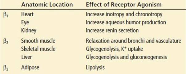
β-Blockers competitively block β-adrenergic receptors, part of the sympathetic nervous system with three main subtypes (β1, β2, and β3) that regulate physiology in various locations in the body (Table 323.1). Their blockade decreases the intracellular concentration of cyclic adenosine monophosphate (cAMP), a secondary messenger that mediates the cellular effects of β-stimulation.
TABLE 323.1
The clinical effects of β-blockers vary with regard to β-receptor selectivity, α-receptor blocking activity, lipophilicity, intrinsic sympathomimetic (β-receptor agonist) activity, and membrane-stabilizing activity (MSA) (Table 323.2). β1-Selectivity is preferred for cardiovascular therapy to avoid the detrimental effects of β2-blockade, such as bronchoconstriction. Labetalol and carvedilol are β-blockers that also antagonize α-adrenergic receptors, although not as prominently as β-receptors. These medications may exacerbate hypotension as a result of vasodilation. β-Blockers with MSA inhibit fast sodium channels, which can lead to QRS prolongation and ventricular dysrhythmias. In overdose, selective activity is often lost, and most β-blockers have similar toxicologic effects.
TABLE 323.2

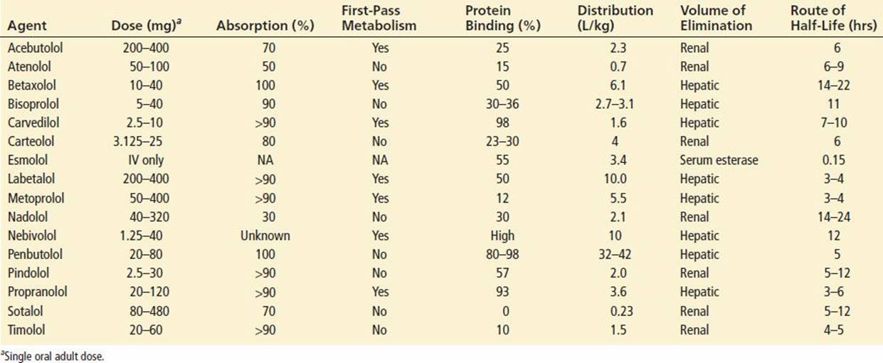
β-Blockers also vary with respect to protein binding, volume of distribution, route of elimination, and half-life (Table 323.3). They are generally well absorbed after ingestion, with peak levels and effects occurring 1 to 2 hours after a therapeutic dose. Because their duration of action is relatively short (6 hours or less), extended-release formulations were developed, to allow for less frequent dosing.
TABLE 323.3
Pharmacokinetics of α-Blockers
CLINICAL PRESENTATION
Most accidental β-blocker overdoses are relatively minor and do not result in significant toxicity, particularly in children (1,2). Large doses or the combination of a β-blocker with a calcium channel blocker can result in significant morbidity and mortality (2).
Bradycardia and hypotension are the most common effects after overdose (1). Nausea and vomiting can occur. Hypoperfusion can lead to lethargy, decreased mental status, stroke, myocardial infarction, mesenteric ischemia, renal insufficiency, with pale and clammy skin.
Highly lipophilic agents and those with MSA can affect the CNS directly to cause altered mental status despite a normal blood pressure. Delirium, coma, and seizures may occur in severe poisoning.
If the SA node is affected, sinus bradycardia may progress to sinus pauses or arrest. Negative inotropic effects add to cardiotoxicity, particularly with β-blockers that have MSA (3). β-Blockers that have MSA can cause delayed depolarization with QRS prolongation and ventricular tachyarrhythmias (3,4). Sotalol is unique in that it also blocks delayed rectifier potassium channels, which results in delayed repolarization with QT prolongation, and can cause torsade de pointes (polymorphic) ventricular tachycardia and other lethal ventricular dysrhythmias (5).
In overdose, all β-blockers can cause β2-antagonism that leads to bronchospasm. This scenario may be more prominent and dangerous in patients with asthma or chronic obstructive pulmonary disease (1). Hypoglycemia and hyperkalemia are common, although they are usually mild and not clinically significant (1). Hypoglycemia may be more common in children.
DIFFERENTIAL DIAGNOSIS
Diseases that cause bradycardia include sick sinus syndrome, inferior wall myocardial infarction, hypothermia, and carotid sinus hypersensitivity. Other drugs that can cause bradycardia include calcium channel blockers, cardiac glycosides, cholinergic agents, α2-agonists, opioids, and sedative–hypnotics.
Like β-blockers, diltiazem and verapamil have strong negative inotropic and chronotropic effects. Dihydropyridine calcium channel blockers (e.g., nifedipine, nimodipine, and amlodipine) typically cause reflex tachycardia in response to vasodilation and hypotension. Unlike β-blocker overdose, the mental status is often normal and calcium channel blockers cause hyperglycemia rather than hypoglycemia. Cardiac glycoside (e.g., digoxin) poisoning usually does not cause hypotension until a nonperfusing dysrhythmia occurs. Additionally, cardiac glycoside overdose produces characteristic ECG patterns.
Cholinergic agents (e.g., organophosphorus insecticides) cause diarrhea, lacrimation, urination, miosis, and vomiting along with bradycardia, bronchorrhea, and bronchoconstriction. Overdose with opioids, sedative hypnotics, and α2-agonists may lead to mild bradycardia and hypotension associated with a depressed mental status, but cardiac conduction disturbances are rare.
ED EVALUATION
The history should include the time and amount of drug(s) taken, treatment in the field (if any), the time of onset and nature of symptoms, known medical or psychiatric problems, and current drug therapy. Determining whether the β-blocker is a sustained-release preparation or if there were any co-ingestants with cardiovascular effects is particularly important.
The physical examination should focus on the vital signs, cardiopulmonary system, and neurologic status. Continuous cardiac monitoring and a 12-lead ECG may reveal AV nodal dysfunction with PR interval prolongation and AV block (4). A fingerstick or serum glucose concentration should be obtained, particularly with an abnormal mental status. Blood gases, chemistry panel to check for hypokalemia, complete blood count, acetaminophen level, toxicology testing, and chest radiograph (CXR) should be obtained when clinically indicated.
KEY TESTING
• ECG (which may be repeated) in patients with changes in heart rate, intervals, or dysrhythmia
• Glucose and potassium should be monitored in symptomatic patients
• Complete blood count, chemistries, CXR, and other laboratory testing as needed for supportive care
ED MANAGEMENT
Advanced cardiac life-support measures should be instituted as needed. Activated charcoal should be considered if ingestion occurred within 1 hour. If a sustained-release preparation has been ingested, activated charcoal can still be considered even later than an hour after ingestion.
Patients with bradycardia with normal blood pressure need only close monitoring. For bradycardic and hypotensive patients, a stepwise approach is recommended in which interventions, each employed to a maximal degree, are successively applied and the hemodynamic response assessed. In severely poisoned patients, multiple therapies must be instituted and maintained simultaneously. Hypotension can initially be treated with intravenous fluids. If bradycardia is also present, atropine, 1 mg intravenously (0.02 mg/kg; minimum dose, 0.1 mg in children), repeated in 5 minutes as necessary, can be given but is frequently ineffective in β-blocker overdose.
Glucagon (5 mg intravenously over 5 minutes; 50 to 100 μg/kg in children) may be effective for hypotension, even in the absence of bradycardia. If there is no effect after 10 to 15 minutes, the dose should be repeated. Patients who respond should be given an infusion of glucagon at a rate of 1 to 5 mg/hr (50 μg/kg/hr in children). Glucagon bypasses the β-blockade by activating a G-protein bound to adenylate cyclase and also activating the cascade indirectly to increase intracellular cAMP (Fig. 323.1). Because glucagon often causes vomiting, prophylactic anti-emetics may be useful. Although there are no human controlled trials, animal studies show that glucagon has a positive chronotropic effect in β-blocker overdose but a minimal effect on mean arterial pressure (6).
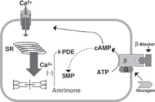
FIGURE 323.1 The effects of amrinone and glucagon on cAMP. Extracellular Ca2+ influx will cause the SR (sarcoplasmic reticulum) to release Ca2+ exponentially to bind the excitation–contraction apparatus, made of actin and myosin filaments. cAMP increases intracellular Ca2+ and is created by an adenylate cyclase that is part of the β-receptor and broken down by PDE (phosphodiesterase). Glucagon can activate the G protein, bypass the β-receptor if blocked, and increase cAMP. Amrinone inhibits PDE, thus also increasing cAMP. (FIGURE by Walter Barrueto.)
Intravenous calcium assists with inotropy in animals (7) and in reversing hypotension in humans (8,9). The dose is the same as for calcium channel blocker poisoning (see Chapter 324). Vasopressors are often necessary. There is no human data to show which pressor is more efficacious than another, so dobutamine, epinephrine, and dopamine have all been used, but the required dose is often much higher than what is traditionally used (1). Invasive cardiac monitoring or echocardiography can help to identify the agent that brings the best response and to titrate to the desired effect.
If the patient requires vasopressors, hyperinsulinemia–euglycemia (insulin and glucose) therapy has been anecdotally effective (10,11). The same protocol as in calcium channel blocker poisoning (see Chapter 324) should be used. In canine propranolol poisoning, this therapy was superior to glucagon and epinephrine (12). Hyperinsulinemia–euglycemia therapy may work by maximizing myocardial glucose utilization, augmentating cytosolic calcium concentrations, or possibly through a direct inotropic effect (12). The response may be delayed 15 to 60 minutes, so other therapies must be continued.
A phosphodiesterase inhibitor such as amrinone is of theoretical benefit but has propensity to cause vasodilation, is difficult to titrate, and often does not improve the bradycardia seen in β-blocker exposure (see Fig. 323.1) (6). This treatment is controversial and not recommended. If a lipophilic β-blocker is involved or if the patient is hemodynamically unstable or critical, one can consider using 1.5 mL/kg of 20% lipid emulsion as a single bolus with an option to repeat the bolus. This has been effective with local anesthetics and overdoses involving lipophilic drugs like verapamil and propranolol (13).
Should pharmacologic therapy be ineffective, intravenous cardiac pacing can be used to treat bradycardia; however, it is likely that there will be little improvement in cardiac output, and difficulty with capture is common. Some authors have reported a decrease in blood pressure, which may be caused by loss of atrial contraction and/or impaired ventricular relaxation. Intra-aortic balloon pump counterpulsation and partial or complete cardiac bypass pump support have been used in severe poisoning (14). Hemodialysis has been used with apparent success for atenolol and sotalol overdose; charcoal hemoperfusion might be effective for water-soluble β-blockers that have a low volume of distribution like atenolol and acebutolol (15,16). Patients who might benefit most from extracorporeal elimination measures are already hypotensive, making these procedures technically difficult and potentially hazardous. Continuous veno-venous hemodialysis is another alternative, but has never been studied.
In patients with sotalol poisoning, hypokalemia and hypomagnesemia should be corrected. The treatment of recurrent torsade de pointes includes overdrive pacing and magnesium infusion (5). Bradycardia and hypotension are treated the same as with other β-blockers.
CRITICAL INTERVENTIONS
• Treat with intravenous fluids for hypotension.
• If hypotension and bradycardia persist, use glucagon, calcium, and high-dose pressors.
• Hyperinsulinemia–euglycemia therapy should be considered if the patient remains unstable.
• Consider transvenous pacing, intra-aortic balloon pump, or extracorporeal circulatory support measures for patients who have not responded to other treatment modalities.
• Certain β-blockers (e.g., atenolol, sotalol) are amenable to hemodialysis or hemoperfusion.
DISPOSITION
Patients with β-blocker ingestions who remain or become asymptomatic 6 hours after ingestion can be discharged or medically cleared for a psychiatric disposition (2,3). If sotalol or a sustained-release preparation is ingested, a 12- to 24-hour observation period is recommended since sotalol has produced symptoms, QT prolongation, and other dysrhythmias up to 20 hours from the time of ingestion (5). With extended-release preparations, prolonged and erratic absorption may result in delayed toxicity. Patients with abnormal vital signs, mental status, or ECG findings require continuous cardiac monitoring and frequent vital signs. This usually requires admission to an intensive care unit. The hospital’s supply of glucagon should be ascertained, as many pharmacies are not prepared to administer the large doses required to treat β-blocker exposures. Transfer of the patient to a tertiary care facility should be considered in cases of severe poisoning where intra-aortic balloon pump or other advanced therapies are available. Psychiatric consultation should be obtained for patients with intentional ingestions prior to discharge. Education in poison prevention, particularly in children, should also be provided prior to discharge.
Common Pitfalls
• Failure to determine whether the drug ingested was an immediate- or extended-release formulation, potentially leading to undertreatment and inappropriate disposition.
• Failure to check a fingerstick blood glucose, as β-blockers can cause hypoglycemia.
• Failure to appreciate that β-blockers with MSA can cause delayed depolarization, cardiac conduction disturbances (QRS prolongation), and ventricular tachyarrhythmias.
• Failure to anticipate seizures with lipophilic β-blockers such as propranolol.
• Failure to appreciate that sotalol blocks potassium channels, causing a prolonged QT interval and torsade de pointes, and has late presenting symptoms.
• Failure to plan for severe poisonings that are likely to require invasive monitoring, multiple pharmacologic treatments, and mechanical hemodynamic support.
ACKNOWLEDGMENTS
The author gratefully acknowledges the contributions of Linda Kesselring and Christopher H. Linden to previous editions of this chapter.
REFERENCES
1. Bailey B. Glucagon in β-blocker and calcium channel blocker overdoses: A systematic review. J Toxicol Clin Toxicol. 2003;41:595–602.
2. Love JN, Howell HM, Litovitz TL, et al. Acute beta blocker overdose: Factors associated with the development of cardiovascular morbidity. J Toxicol Clin Toxicol. 2000;38:275–281.
3. Reith DM, Dawson AH, Epid D, et al. Relative toxicity of beta blockers in overdose. J Toxicol Clin Toxicol. 1996;34:273–278.
4. Love JN, Enlow B, Howell JM, et al. Electrocardiographic changes associated with β-blocker toxicity. Ann Emerg Med. 2002;40:603–610.
5. Hohnloser SH, Woosley RL. Sotalol. N Engl J Med. 1994;331:31–38.
6. Sato S, Tsuji MH, Okubo N, et al. Milrinone versus glucagon: Comparative hemodynamic effects in canine propranolol poisoning. J Toxicol Clin Toxicol. 1994;32:277–289.
7. Love JN, Hanfling D, Howell JM. Hemodynamic effects of calcium chloride in a canine model of acute propranolol intoxication. Ann Emerg Med. 1996;28:1–6.
8. Brimacombe JR, Scully M, Swainston R. Propranolol overdose: A dramatic response to calcium chloride. Med J Aust. 1991;155:267–268.
9. Pertoldi F, D’Orlando L, Mercante WP. Electromechanical dissociation 48 hours after atenolol overdose: Usefulness of calcium chloride. Ann Emerg Med. 1998;31:777–781.
10. Levine M, Boyer EW, Pozner CN, et al. Assessment of hyperglycemia after calcium channel blocker overdoses involving diltiazem or verapamil. Crit Care Med. 2007;35(9):2071–2075.
11. Mégarbane B, Karyo S, Baud FJ. The role of insulin and glucose (hyperinsulinaemia/euglycaemia) therapy in acute calcium channel antagonist and beta-blocker poisoning. Toxicol Rev. 2004;23(4):215–222.
12. Kerns W, Schroeder D, Williams C, et al. Insulin improves survival in a canine model of acute [beta]-blocker toxicity. Ann Emerg Med. 1997;29:748–757.
13. Cave G, Harvey M. Intravenous lipid emulsion as antidote beyond local anesthetic toxicity: A systematic review. Acad Emerg Med. 2009;16(9):815–824.
14. McVey FK, Cork CF. Extracorporeal circulation in the management of massive propranolol overdose. Anaesthesia. 1991;46:744–746.
15. Rooney M, Massey KL, Jamali F, et al. Acebutolol overdose treated with hemodialysis and extracorporeal membrane oxygenation. J Clin Pharmacol. 1996;36:760–763.
16. Saitz R, Williams BW, Farber HW. Atenolol-induced cardiovascular collapse treated with hemodialysis. Crit Care Med. 1991;19:116–119.