
Matthew D. Sztajnkrycer
Calcium is critical for cellular function, involved in diverse processes including myocyte excitation–contraction coupling, intracellular second-messenger signaling, release of neurotransmitters and hormones, and initiation and propagation of the action potential. Intracellular calcium is largely sequestered within the sarcoplasmic reticulum and released through direct receptor activation or through second-messenger–mediated systems. Calcium channels are found in the membranes of many cells, including vascular smooth muscle, the myocardium, and the specialized pacemaker and conducting fibers of the heart. Three general classes of calcium channel have been identified, based on mechanism of activation: (1) voltage-operated calcium channels (VOCCs), (2) receptor-operated calcium channels, and (3) second-messenger–operated calcium channels.
Calcium channel antagonists (CCAs) are a structurally diverse group of drugs used in the treatment of supraventricular tachyarrhythmias, hypertension, angina, heart failure, and subarachnoid hemorrhage and in the prevention of migraine headaches. Eleven CCAs are currently approved by the US Food and Drug Administration for use in the United States (Table 324.1); verapamil and amlodipine possess pediatric FDA approval. All act by depressing the activation, inactivation, and recovery of the L-type VOCC, and are thus classified as class IV antiarrhythmics. Cardiovascular (CV) responses to therapeutic doses vary considerably (see Tables 324.1 and 324.2).
TABLE 324.1
Therapeutic Doses of Calcium Channel Blockers
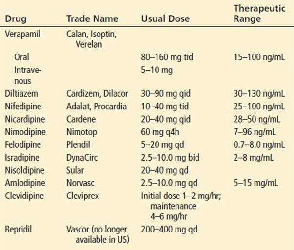
TABLE 324.2
Cardiovascular Response to Drug Therapy

CCAs are well absorbed after oral administration. With immediate-release preparations, effects are noted within 2 to 3 hours of oral administration. Effects of sustained-release preparations may be delayed 6 to 8 hours and are reported as long as 15 hours after ingestion. CCAs are 80% to 98% protein-bound, have a moderate volume of distribution (1.4 to 4.3 L/kg), are extensively metabolized by the liver, and are excreted in the urine and bile. Verapamil, diltiazem, and bepridil have active metabolites.
The CCA toxicology experience is based primarily on four agents: Verapamil, diltiazem, nifedipine, and amlodipine. Nicardipine, felodipine, nisoldipine, isradipine, and nimodipine are members of the same class as nifedipine and amlodipine (dihydropyridines) and have similar toxicity. Clevidipine, approved in 2008, is an intravenous short-acting dihydropyridine. Bepridil is a unique agent, with sodium and potassium channel antagonism in addition to CCA effects. Toxic CV effects of CCAs are in large part an exaggeration of their therapeutic effects. However, with overdose, the drug-specific effects noted in Table 324.1 are often lost (1). Though peripheral vascular resistance is always decreased, cardiac output may be normal, increased, or decreased. As only nimodipine penetrates the blood–brain barrier, central nervous system (CNS) effects are essentially due to hypoperfusion. Both hypoperfusion and impaired carbohydrate metabolism may lead to acidosis and ketosis. Impaired calcium-dependent insulin release and peripheral insulin resistance may cause hyperglycemia (2). This combination may lead to a diabetic ketoacidosis-like picture.
CCAs are considered so toxic that “one pill can kill” in the pediatric population. Death has been reported in a child after ingestion of a single tablet of nifedipine. The elderly are susceptible to toxicity at lower doses (3). Toxicity can also be seen when taken in therapeutic doses with other myocardial depressants, such as β-adrenergic antagonists and quinidine-like agents.
CLINICAL PRESENTATION
Signs and symptoms of toxicity include hypotension and bradycardia, but reflex tachycardia has been reported, especially with dihydropyridine ingestion. Inadequate perfusion may result in myocardial, cerebral, renal, and bowel ischemia or infarction. In severe cases, coma, seizures, and respiratory distress associated with cardiogenic and noncardiogenic pulmonary edema are described. Toxic effects typically last 24 to 36 hours but may be longer with bepridil, amlodipine, and sustained-release preparations.
DIFFERENTIAL DIAGNOSIS
Myocardial infarction and overdose of β-adrenergic antagonists, cardiac glycosides, clonidine, guanabenz, imidazolines, angiotensin converting enzyme inhibitors, cholinergic agents (nicotine, organophosphate and carbamate insecticides, and agents to treat myasthenia gravis), tricyclic antidepressants (TCAs), alpha-methyldopa, and veratrum alkaloids may present similarly to those with CCA ingestion.
β-Adrenergic blocking agents are more likely to cause hypoglycemia, and CCAs are more likely to cause hyperglycemia. With cholinergic agents, salivation, nausea, vomiting, diarrhea, and weakness dominate the presentation. TCA toxicity infrequently presents with heart block and bradycardia. Although both TCA and CCA ingestions may result in prolonged QRS complexes (the latter as a result of ventricular escape rhythms), TCA-intoxicated patients often manifest evidence of antimuscarinic toxidrome and CNS depression in normotensive or hypertensive patients (see Chapter 339, “Cyclic Antidepressants”).
ED EVALUATION
The history should include time of ingestion and identification of other substances ingested or available to the patient. Physical examination should focus on cardiopulmonary and CNS status. Continuous peripheral and pulmonary artery pressure monitoring may be necessary in patients with hemodynamic compromise.
Laboratory abnormalities are nonspecific and may include metabolic acidosis and hyperglycemia. Serum drug levels are not readily available and are not useful in the management of these patients. The electrocardiogram (ECG) often shows decreased rate and first-, second-, or even third-degree AV block and may have changes indicative of ischemia or infarction (Fig. 324.1) (3). QT-prolongation and torsades de pointes may be noted with bepridil ingestion. Chest radiography may show pulmonary edema.

FIGURE 324.1 Initial electrocardiogram of a patient presenting after sustained-release verapamil ingestion. Findings include complete heart block with severe bradycardia and repolarization abnormalities.
KEY TESTING
• ECG and cardiac monitoring
• CXR for pulmonary edema
• No drug levels are useful; routine laboratory studies might be helpful for general supportive care
ED MANAGEMENT
Supportive care, activated charcoal, and advanced life-support measures should be initiated as necessary. Intravenous access should be established in all patients. Whole-bowel irrigation (WBI) is recommended for ingestions of sustained-release preparations, especially in patients presenting early. Smooth muscle relaxation may result in an ileus, limiting the utility of WBI. Mild hypotension may respond to a fluid challenge with 1 to 2 L of saline solution (10 to 20 mL/kg in children). When bradycardia, with or without AV block, is also present, atropine (1.0 mg IV, 0.02 mg/kg in children) can be tried. Response is infrequent but may be improved after calcium administration (3). Patients with hypotension and concomitant symptomatic bradycardia or conduction defects should receive either calcium chloride (1 g IV slow push; 10 to 20 mg/kg in children) or calcium gluconate (3 g IV slow push; 30 to 60 mg/kg in children). This dose may be repeated every 5 to 15 minutes up to three times in patients who show incomplete, transient, or absent response (4). Calcium chloride extravasation can result in severe tissue damage, so limit calcium chloride use to central venous administration whenever possible. Owing to the short-lived effects of intravenous calcium, a continuous infusion at a rate of 0.2 to 0.4 cc/kg/hr (10% calcium chloride) or 0.6 to 1.2 cc/kg/hr (10% calcium gluconate) may be used to support heart rate and blood pressure in patients who respond to an initial bolus (5). Although calcium is frequently ineffective, proponents argue that this is a result of inadequate dosing. Doubling of serum calcium concentration was associated with significant hemodynamic improvement in both animal models and human cases. An ionized calcium concentration of 2 mmol/L has been suggested as a target level (6).
No single pressor agent has been consistently effective. Dopamine, dobutamine, norepinephrine, isoproterenol, alone or in combination, have been used with mixed success. Glucagon may also be of benefit for hypotension, but it should not be used until the effects of traditional pressors are optimized. Acidemia must be corrected, as it impairs the function of L-type VOCCs, enhances the toxic effects of some agents (e.g., verapamil), and decreases the efficacy of calcium. A swine model of verapamil toxicity demonstrated improved myocardial contractility and cardiac output with sodium bicarbonate administration (7).
The pharmacologic management of patients who fail to respond to these therapies remains controversial. Although strong evidence-based data are lacking, most toxicologists advocate the early, aggressive use of hyperinsulinemia-euglycemia (HIE) therapy in patients who show an inadequate response after initial fluid resuscitation, atropine, and calcium administration. Insulin, used as part of HIE therapy, improves hemodynamics and survival and has reversed catecholamine-unresponsive shock in both animals and in case reports (5,8,9). Insulin has positive inotropic effects in conditions of myocardial stress without increasing myocardial oxygenation demand. During myocardial stress, the metabolism of myocardial cells is shifted away from fatty acid oxidation, toward carbohydrate dependence, improving the efficiency of energy biomechanics. In cases of severe CCA toxicity, hyperglycemia is frequently noted. The etiology is multifactorial, reflecting both impaired insulin release and peripheral resistance (2). The net result is that the CCA-intoxicated myocardium is further impaired secondary to limited cellular glucose uptake at a time of glucose dependence and subsequent transition back to less-desirable fatty acid metabolism. HIE is believed to reverse this impaired energy utilization state and improve myocardial glucose utilization. HIE also likely increases intracellular calcium concentrations, although this may cause only a small improvement. The failure of catecholamines and glucagon, despite initial transient improvement, may reflect catecholamine-induced increased myocardial oxygen demand and promotion of fatty acid metabolism and ketosis (5).
Although studied dosages have varied, a reasonable approach to HIE therapy is an initial intravenous bolus of 1 IU/kg regular insulin, followed by an infusion of 1 to 10 IU/kg/hr, titrated to clinical effect (systolic BP >90 mm Hg, HR >50/min) (8,9). Patients with serum glucose levels <200 mg/dL should receive one 50-mL ampule of 50% dextrose (0.25 g/kg in children) followed by frequent glucose measurements and supplemental glucose as required. Owing to potential insulin–mediated effects on serum potassium, patients with serum potassium concentrations <2.5 mEq/L should receive 40 mEq of potassium (1 mEq/kg in children; maximum, 20 to 40 mEq/dose) as an oral supplement or IV potassium, 10 to 20 mEq (0.10 to 0.30 mEq/kg in children) administered over the course of 1 hour. Serum glucose and potassium concentrations should be regularly monitored during HIE therapy, and for up to 24 hours following discontinuation.
More recently, the successful use of intravenous fat emulsion (IFE) has been reported in the management of CCA intoxication (10), especially for the more lipophilic agent verapamil. It has been suggested that IFE works at least in part by sequestering lipophilic agents, thereby decreasing distribution to target tissues and redistributing the toxic agent from the tissue into the IFE lipid medium. While controversial in the setting of CCA toxicity, current IFE dosage recommendations for the management of local anesthetic toxicity are a 1.5 mL/kg bolus of 20% IFE followed by an infusion of 0.25 to 0.50 mL/kg/min 20% IFE until hemodynamic stability. IFE use has been associated with fat emboli syndrome and is contraindicated in the setting of acute myocardial infarction, known disorders of fatty acid metabolism and egg allergy. Concern also exists for the potential of IFE as a nonspecific sequestering agent to sequester concomitantly administered antidotes.
Hemodynamically significant bradycardia or heart block unresponsive to medical therapy may require pacemaker placement. Electrical capture occurs in only 50% of patients (Fig. 324.2) (3). Heart rate was improved more than blood pressure. In hypotensive patients unresponsive to pharmacologic therapies, intra-aortic balloon counterpulsation and cardiopulmonary bypass have been successfully employed. The specific role of enhanced elimination, other than through gastrointestinal decontamination, has not been clearly defined. Based upon pharmacokinetic properties, conventional extracorporeal elimination would be expected to be of limited effectiveness. Several reports have documented the successful use of albumin dialysis with the Molecular Adsorbents Recirculating System (MARS) (1). Full recovery can be expected unless prolonged shock has resulted in irreversible end-organ damage.
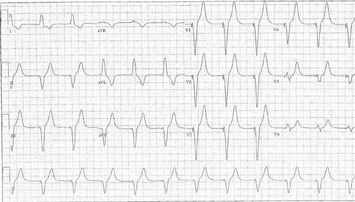
FIGURE 324.2 Follow-up electrocardiogram after emergent transvenous pacemaker placement. Note that electrical capture occurs in only 50% of patients and that blood pressure may not improve despite increased heart rate.
CRITICAL INTERVENTIONS
• Admit or observe asymptomatic patients with overdoses of sustained preparations for an extended period of time.
• Administer IV fluids, calcium, and atropine for hypotension and bradycardia.
• Initiate HIE therapy for hypotension unresponsive to fluids and calcium.
• Consider the use of IFE, especially in toxicity caused by more lipophilic agents.
• High doses of calcium may be needed for a clinical response.
• Owing to lack of consistently effective therapeutic interventions, early aggressive gastrointestinal decontamination is recommended.
DISPOSITION
Asymptomatic patients should be observed for 4 to 6 hours after ingestion of standard formulations and 18 to 24 hours after ingestion of sustained-release ones. Those who remain asymptomatic may be discharged or referred for psychiatric evaluation. Symptomatic patients should be admitted to an intensive care unit. Transfer may be necessary if an intensive care unit bed is unavailable and should be arranged as soon as the ingestion is recognized, because CV performance may rapidly deteriorate. The patient should be transferred in an advanced life-support unit with an adequate supply of CV drugs on hand. A regional poison center or a toxicologist should be consulted if the physician is unfamiliar with the management of CCA overdose.
Common Pitfalls
• Failure to consider CCA poisoning in patients with hypotension, bradycardia, or AV block, particularly when hyperglycemia is present.
• Failure to appreciate that ingestion of a single pill may be lethal in a toddler.
• Failure to consider whole-bowel irrigation and multiple-dose charcoal for patients with overdoses of sustained preparations.
• Failure to initiate aggressive HIE therapy in a timely manner.
REFERENCES
1. Pichon N, Dugard A, Clavel M, et al. Extracorporeal albumin dialysis in three cases of acute calcium channel blocker poisoning with life-threatening refractory cardiogenic shock. Ann Emerg Med.2012;59:540–544.
2. Kline JA, Raymond RM, Schroeder JD, et al. The diabetogenic effects of acute verapamil poisoning. Toxicol Appl Pharmacol. 1997;145:357–362.
3. Ramoska EA, Spiller HA, Winter M, et al. A one-year evaluation of calcium channel blocker overdoses: Toxicity and treatment. Ann Emerg Med. 1993;22:196–200.
4. Isbister GK. Delayed asystolic cardiac arrest after diltiazem overdose: Resuscitation with high dose intravenous calcium. Emerg Med J. 2002;19:355–357.
5. Shepherd G. Treatment of poisoning caused by β–adrenergic and calcium channel blockers. Am J Health Syst Pharm. 2006;63:1828–1835.
6. Lam Y-M, Lau C. Continuous calcium chloride infusion for massive nifedipine overdose. Chest. 2001;119:1280–1282.
7. Tanen DA, Ruha AM, Curry SC, et al. Hypertonic sodium bicarbonate is effective in the acute management of verapamil toxicity in a swine model. Ann Emerg Med. 2000;36:547–553.
8. Engebretsen KM, Kaczmarek KM, Morgan J, et al. High-Dose insulin therapy in beta-blocker and calcium channel –blocker poisoning. Clin Toxicol (Phila). 2011;49:277–283.
9. Espinoza TR, Bryant SM, Aks SE. Hyperinsulin therapy for calcium channel antagonist poisoning: A seven-year retrospective study. Am J Ther. 2013;20:29–31.
10. Young AC, Velez LI, Kleinschmidt KC. Intravenous fat emulsion therapy for intentional sustained-release verapamil overdose. Resuscitation. 2009;80:591–593.