
Richard D. Shih and Judd E. Hollander
Cocaine (benzoylmethylecgonine) has been used for social, religious, and medicinal purposes for many centuries. Over the past several decades, its illicit use as a drug of abuse has risen dramatically. An estimated 40 million Americans have used cocaine at least once and for ages 12 and older, 4.2 million have used cocaine in the past year, and 1.4 million have used cocaine in the past month (1), making cocaine the most commonly involved drug of abuse for visits to emergency departments (EDs).
Cocaine is well absorbed on contact with mucous membranes or by pulmonary inhalation. It is derived from the Erythroxylum coca plant, which is found in abundance in Central America, South America, the West Indies, and Indonesia. In the manufacturing process, leaves are initially dissolved in hydrochloric acid to form the cocaine hydrochloride salt which is the form most often abused via nasal insufflation or IV injection. Crack cocaine or cocaine freebase are the alkaloid forms of cocaine that are produced by an extraction process utilizing a basic solution, a solvent (usually ether), and heat. The freebase form is close to 100% purity, whereas crack is approximately 75% pure. Crack and freebase are heat stable and thus can be smoked and absorbed by the lungs.
By the IV or inhalational routes, cocaine is rapidly distributed throughout the body with peak central nervous system (CNS) effects in 3 to 5 minutes. With nasal insufflation, absorption is slower and the effects peak at approximately 20 minutes. Although not a typical route of abuse, oral ingestion has even slower absorption with peak effects occurring at 40 to 60 minutes.
Cocaine has a half-life of 0.5 to 1.5 hours. It is predominantly metabolized by hydrolysis to the active metabolites ecgonine methyl ester and benzoylecgonine (BE). These two metabolites account for approximately 80% of cocaine metabolism and these metabolites have half-lives of 4 to 8 hours and many effects similar to their parent compound, cocaine. Other metabolites include cocaethylene, ecgonine, and norcocaine. Urine drug tests typically test for BE, which is usually present for 48 to 72 hours after use. Cocaine’s effects typically last for several hours and may wear off while the urine test is still positive for urine cocaine metabolites.
Cocaine has a number of pharmacologic effects peripherally and in the CNS. It acts to augment the peripheral sympathetic system, producing tachycardia, hypertension, diaphoresis, arterial vasoconstriction, and dilated pupils. In addition, cocaine has local anesthetic effects. Because it is the only topical anesthetic agent in the amide group that has vasoconstrictive effects to control secretions and bleeding, it is often used for otolaryngology procedures and wound repair. The central effects of cocaine are very complex and not completely understood. It affects a number of neurotransmitters in the CNS; however, the net effect is augmentation of the sympathetic nervous system.
“Body packers” are individuals who have ingested containers of an illicit drug (most commonly cocaine and heroin). A packer, or “mule,” is an individual who ingests a large number of carefully wrapped packages of highly pure drug as a means of concealing and smuggling it into the United States from a foreign country or into jail. These containers can be plastic bags, plastic vials, elastic wraps, elastic condoms, or other packaging material. In contrast, a “body stuffer” is someone who hastily ingests containers just before being arrested by police so as to conceal the evidence. The body stuffer typically has the drug in a plastic baggie or loose wrapping and is more likely to experience effects of cocaine.
CLINICAL PRESENTATION
The most commonly seen manifestations in the ED involve the cardiovascular (CV) and neurologic systems. Signs and symptoms of mild cocaine intoxication include normal or minimally increased blood pressure, pulse, respiratory rate, and temperature; agitation; anxiety; euphoria; headache; hyperreflexia; nausea; vomiting; mydriasis; pallor; diaphoresis; tremors; and twitching. Moderate intoxication may result in more severe hypertension, tachycardia, dyspnea, tachypnea, hyperthermia, confusion, hallucinations, marked hyperactivity, increased muscle tone and deep tendon reflexes, abdominal cramps, formication, and generalized but brief tonic–clonic seizures. Severe intoxication can result in hypotension, tachycardia (or preterminal bradycardia), ventricular dysrhythmias, Cheyne–Stokes respirations, apnea, cyanosis, severe hyperthermia, coma, flaccid paralysis, and status epilepticus.
Chest pain is a common chief complaint associated with cocaine use in EDs. Myocardial infarction (MI) associated with cocaine use is well established (2,3) and occurs in approximately 6% of patients presenting with this complaint (4). Cocaine causes coronary ischemia through coronary artery vasoconstriction, in situ thrombus formation, platelet activation, inhibition of endogenous fibrinolysis, and triggering an increase in the myocardial oxygen demand through generation of tachycardia and hypertension (2). Chronic users develop left ventricular hypertrophy, which can further exacerbate the oxygen supply–demand mismatch (2). It was previously thought that chronic cocaine use was associated with premature atherosclerosis but that has now been disproven (5). Other less common but equally important CV complications of cocaine use include atrial and ventricular dysrhythmias, both systolic and diastolic congestive heart failure, coronary and aortic dissection, dilated cardiomyopathy, and ischemia in other vascular beds (e.g., intestinal, renal).
Euphoria, the impetus for the recreational use of cocaine, is typically short-lived and without serious sequelae. The stimulatory effects of cocaine can lead to seizures. Cocaine-induced seizures are typically single and generalized with rare status epilepticus (6). Focal neurologic events such as hemorrhagic cerebral infarction and subarachnoid hemorrhage are uncommon. Severe, persistent lethargy with an altered mental status can occur after prolonged and intense cocaine usage and has been termed the cocaine wash-out syndrome. This diagnosis should be made only after exclusion of the aforementioned neurologic catastrophes (i.e., after a normal computed tomography (CT) of the head and lumbar puncture, if indicated). The syndrome is thought to be from depletion of essential neurotransmitters following excessive cocaine usage and require 12 to 24 hours to resolve.
Rhabdomyolysis is another common manifestation of cocaine toxicity. Cocaine’s stimulatory effects lead to severe agitation and marked muscular agitation and rigidity. Ischemia of the skeletal muscle beds may also contribute. Profound elevations in creatine kinase (CK) may be seen. Acute kidney injury is a result of muscle breakdown and renal tubular precipitation of the released muscle myoglobin. Concurrent development of severe hyperthermia and acute kidney injury can be life-threatening. Hyperthermia most commonly results from a combination of environmental exposure and heat production from excessive muscle activity from agitation and is often noted to be agitated delirium.
Cocaine has a number of direct and indirect effects on the lungs. Many of these effects are a result of the inhalation rather than direct toxin effects. Asthma exacerbations are frequently reported with crack cocaine, most likely a result of particulate byproducts of combustion. Crack use is typically associated with deep Valsalva maneuvers to maximize drug delivery, which can cause pneumothorax, pneumomediastinum, and noncardiogenic pulmonary edema. Other less common effects on the lung include pulmonary infarction, bronchiolitis obliterans, pulmonary artery hypertrophy, and alveolar hemorrhage. Treatment of these conditions follows standard management protocols, whether or not they are related to cocaine.
The intestinal vascular system is very sensitive to the effects of cocaine. Acute intestinal vascular infarction has been associated with all routes of administration. Although oral ingestion is not a common route of abuse, local intestinal and systemic effects can occur when containers of cocaine leak or rupture in body stuffers and packers. Body packers are most often discovered by customs officers and present asymptomatically.
Habitual cocaine usage during pregnancy is associated with low birth weight, small head circumference, developmental problems, and a number of birth defects. Acute toxicity can also induce premature labor, eclampsia, and abruptio placentae. After delivery, neonates exposed to cocaine in utero are at risk for the development of neonatal withdrawal. This is a diagnosis of exclusion presenting with irritability, jitteriness, and poor eye contact.
DIFFERENTIAL DIAGNOSIS
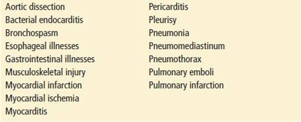
Cocaine toxicity is associated with sympathomimetic effects which can be similarly seen with hypoglycemia, environmental and malignant hyperthermia, pheochromocytoma, manic psychiatric conditions, status epilepticus, and thyroid storm. Drugs with anticholinergic or sympathomimetic (i.e., amphetamines and bath salts) properties can present with a similar picture. Patients with persistent altered mental status need to be evaluated for possible meningitis and CNS lesions. Table 345.1 lists the differential diagnosis of chest pain related to cocaine use, which also includes reasons to be short of breath. New-onset seizures, epistaxis, hypertension, MI, intracranial hemorrhage, or psychiatric illness, especially in young patients, should suggest the possibility of cocaine use as well as other causes.
TABLE 345.1
The Differential Diagnosis of Cocaine-Associated Chest Pain
ED EVALUATION
The diagnostic evaluation of patients with cocaine toxicity relies on a history of cocaine use, recognition of signs and symptoms consistent with a sympathomimetic toxidrome, and evaluation of specific organ system complaints. The history should include the total amount and time of cocaine use in relation to symptom onset. Friends or witnesses of confused patients should be questioned about a history of seizures or syncope and antecedent activities. Many patients will deny cocaine use unless approached with reassurance and compassion or confronted with a positive urine test.
The physical examination should include complete vital signs and a detailed examination of the cardiac, pulmonary, and neurologic systems. Patients should initially have continuous cardiac monitoring.
When the history is clear and symptoms are mild, laboratory evaluation is unnecessary. In contrast, if the history is absent or unreliable or the patient manifests moderate or severe toxicity, laboratory evaluation may provide evidence of the severity or identify other causes. These include a complete blood count; determination of electrolyte, glucose, blood urea nitrogen, and creatinine levels; blood gas analysis; urinalysis; CK; and cardiac troponin. Qualitative toxicologic screening of blood and urine can confirm the diagnosis and rule out other intoxicants, but toxicology testing is indicated only when confirmation of drug use would change management, counseling, or referral patterns.
An electrocardiogram (ECG) should be obtained and a chest radiograph considered in patients with chest pain or moderate to severe toxicity. Those with prolonged, unexplained pain should have serial ECG and cardiac biomarker measurements to rule out MI. Many of the clinical parameters to assess ischemia or infarction are less reliable when compared to patients with traditional coronary artery disease. The ECG is less sensitive and specific for identifying ischemia or infarction in this setting, the CK is often elevated as a result of associated rhabdomyolysis, and false elevations in the MB fraction can occur. Cardiac troponin I testing identifies myocardial injury and MI (7). Observation for a 9- to 12-hour period can be used to evaluate patients with cocaine-associated chest pain. Patients without new ischemic changes on ECG, a normal troponin test, and no cardiovascular complications during this observation (dysrhythmias, acute MI, or recurrent symptoms) can safely be sent home with follow-up and a planned outpatient workup (8).
Persistent headache despite normalization of blood pressure is an indication for head CT scan and possible lumbar puncture to rule out intracranial hemorrhage. Patients with severe abdominal or back pain should be evaluated for intestinal or renal infarction and possible aortic dissection. The urine should be inspected and the serum CK level determined, to detect myoglobinuric renal failure.
Occult infections should be excluded in patients with fever, even though fever can be due solely to cocaine toxicity. A brief seizure clearly related temporally to cocaine use in an otherwise healthy person should be evaluated with CT to exclude serious underlying pathology, but does not require further workup, provided the patient is alert and coherent, has no headache, and has a normal neurologic examination. Patients suspected of body packing or stuffing should be evaluated by abdominal imaging and cavity searches (digital or visual examination of the rectum or vagina). Toxicity lasting for more than 4 hours suggests continued drug absorption and should prompt a similar workup.
The route of administration may influence the patient’s chief complaint or which organ system is affected. Intravenous users may present with fever and malaise secondary to infectious complications such as cellulitis, endocarditis, hepatitis, pneumonia, and the acquired immunodeficiency syndrome. Chronic nasal use may lead to rhinitis, septal perforation, and epistaxis. Inhalational use may produce dyspnea, cough, or hemoptysis from reactive airway disease, “crack lung” pneumonitis, or pulmonary edema or may result in pulmonary barotrauma (e.g., pneumothorax, pneumomediastinum, pneumopericardium) from a Valsalva maneuver or from blowing smoke into the mouth of a partner. Patients with barotrauma may also complain of neck and chest pain with tachypnea, subcutaneous emphysema, or Hamman sign.
Behavioral disorders (e.g., agitation, combative behavior), headache, back pain (renal infarction or aortic dissection), abdominal pain (mesenteric ischemia), altered level of consciousness, or cardiopulmonary arrest are other presentations that may follow the use of cocaine by any route. Patients with excited delirium, severe agitation, an elevated temperature, and metabolic acidosis deserve immediate attention. Patients may present as victims of trauma because of the violent, irrational, and risk-taking behavior associated with drug use. Delayed CV complications can sometimes occur and MI has been reported in the first few days after cocaine use but has also been reported 1 to 2 weeks after last use.
Manifestations of chronic cocaine abuse include anorexia, insomnia, formication, depression, impotence, weight loss, paranoia, and psychosis. Halo vision (lights around objects) and “snow lights” (flashes in the peripheral fields) have also been described.
KEY TESTING
• Mild Toxicity
None necessary
• Moderate to Severe Toxicity
CBC, electrolytes, glucose, BUN, UA
CK for rhabdomyolysis
CXR for persistent SOB and chest pain
ECG and troponin for suspected ischemia
CT (head/chest/abdomen) for persistent complaint
ED MANAGEMENT
Management depends on the specific complaint and presentation. Patients presenting with a sympathomimetic toxidrome are at risk for hyperthermia and rhabdomyolysis. After initial attention to the airway and CV status, management should focus on lowering any elevated core body temperature, halting further muscle agitation and heat production, and giving IV fluids to ensure a good urinary output. The agents of choice for muscle relaxation in this setting are benzodiazepines and in severely agitated individuals, control should be obtained with rapid use and use of supranormal cumulative doses may be necessary.
Patients with severe hypertension or tachycardia needing pharmacologic measures can usually be safely treated with benzodiazepines. When large doses of benzodiazepines are not effective, intravenous nitroprusside or phentolamine should be considered (4). β-Antagonists or compounds with partial β-blocking effects are contraindicated as the use of β-antagonists in the setting of cocaine intoxication can lead to unopposed α-stimulation, with resultant marked increases in hypertension and worsening coronary vasoconstriction (2,4).
Patients with suspected cocaine-induced ischemia or MI should be treated similarly to those with traditional acute coronary syndromes (ACSs), with some notable exceptions (2,4). Aspirin, nitroglycerin, and heparin remain important initial therapies. Intravenous benzodiazepines should be provided as early management (2,4,9). They will decrease the central stimulatory effects of cocaine, thereby indirectly reducing the CV toxicity of cocaine (2,4). β-Antagonists are contraindicated, as they exacerbate cocaine-induced coronary artery vasoconstriction (2,4). Percutaneous interventions (angioplasty) are preferred over fibrinolysis, which has no proven efficacy in the setting of cocaine-associated MI and a possible reduced safety profile. There is an increased likelihood of interventions being performed in patients who are not sustaining an acute infarction because young patients have a high prevalence of early repolarization and “false-positive” ST-segment elevations on the ECG. Such a therapy should, therefore, be used with caution. Finally, there are anecdotal reports of the safety and efficacy of phentolamine, an α-antagonist, for treatment of cocaine-associated ACS (2,4). Verapamil reverses cocaine-induced vasoconstriction, but several animal experiments suggest that it exacerbates CNS toxicity. Thus, it may have a role in patients with continued ischemia who do not have signs of central stimulation from cocaine.
Supraventricular dysrhythmias due to cocaine toxicity may be difficult to treat. Adenosine can be administered, but its effects may be only temporary. Use of calcium channel blockers in association with benzodiazepines appear to be most beneficial. β-Blockers should be avoided (2,4).
Ventricular dysrhythmias may be a result of excess adrenergic tone or cocaine’s sodium channel blocking effects. Management with benzodiazepines, lidocaine, and/or sodium bicarbonate appears to be most useful (2,4). Bicarbonate is preferred when patients have dysrhythmias directly after the use of cocaine. In this setting, the dysrhythmias are presumably related to the type I antiarrhythmic effects of cocaine. Bicarbonate reverses cocaine-associated QRS widening. Lidocaine can be used when dysrhythmias appear to be related to cocaine-induced ischemia (10). ECG evidence for the type I antiarrhythmic effects of cocaine has often disappeared by the time symptomatic ischemia develops. There is no data regarding the use of amiodarone.
Cocaine-induced seizures are typically brief and self-limited. For refractory cases, benzodiazepines and phenobarbital are the first- and second-line agents, respectively. Phenytoin is not recommended.
Patients with suspected cerebrovascular infarctions and hemorrhage should be treated the same as other patients with these conditions. Intracranial and systemic hypertension necessitating treatment is accomplished using standard therapies, except that β-blockers should not be utilized.
The main objective with asymptomatic body packers is to eliminate the packages out of the gastrointestinal system. Whole-bowel irrigation with subsequent radiologic verification of passage of all drug-filled containers may be warranted.
Body stuffers who show clinical signs of toxicity should be treated similar to other cocaine-exposed individuals. In addition, activated charcoal should be administered liberally.
Symptomatic body packers should be treated aggressively, because rapid deterioration and severe toxicity can result from leakage from a single package causing potentially massive exposures. Immediate surgical removal of the ruptured package(s) may be lifesaving. Aggressive supportive care with activated charcoal and benzodiazepines is warranted as preparations are made for surgery.
CRITICAL INTERVENTIONS
• Rapidly cool and sedate the hyperthermic and agitated patient.
• Assess patients with cocaine toxicity for myocardial ischemia/MI, dysrhythmias, hyperthermia, and rhabdomyolysis.
• Administer intravenous benzodiazepines for CNS and cardiovascular toxicity.
DISPOSITION
Patients with severe agitation, hyperthermia, and possible rhabdomyolysis need to be admitted. Patients with cocaine-associated chest pain need to be assessed for risk of CV events. Criteria for patients with a low risk of CV events that are safe for ED discharge after a brief observation period include lack of ischemic changes on ECG; normal serial cardiac troponin I values; and no dysrhythmias, or recurrent symptoms during a 9- to 12-hour observation period (8). Most patients with ED cocaine-associated chest pain (∼70%) will meet these criteria (8). Coronary computerized tomographic imaging of the coronary arteries can be used to identify low-risk individuals and shorten the duration of observation (11).
The disposition of patients with neurologic complications is the same as for other patients with these conditions. The likelihood of compliance with outpatient follow-up should be considered when contemplating the discharge of patients who may need further evaluation. If definitive care cannot be provided at the site of presentation, transfer may be necessary. Personnel with advanced life-support training should accompany patients requiring transfer. Substance abuse counseling and treatment should be discussed with all patients with referral if possible.
Body stuffers who remain asymptomatic for 4 to 6 hours may be discharged after treatment with activated charcoal. Body packers need to be admitted until packets have been removed.
Common Pitfalls
• Failure to aggressively treat patients with agitated delirium from cocaine.
• Failure to consider cocaine toxicity in the differential diagnosis of patients with chest pain, seizures, agitation, altered mental status, and dysrhythmias.
• Failure to recognize the limitations of the ECG in the evaluation of cocaine-related chest pain.
• Failure to appreciate the differences between the management of cocaine-related chest pain and traditional therapy for ACS.
• Failure to recognize the dangers of using β-adrenergic antagonists to treat cocaine toxicity.
• Failure to ensure the rectal passage of cocaine-filled packages in body packers prior to discharge.
REFERENCES
1. Results from the 2011 National Survey on Drug Use and Health: Summary of National Findings. http://store.samhsa.gov/home
2. Hollander JE. Management of cocaine-associated myocardial ischemia. N Engl J Med. 1995;333:1267–1272.
3. Hollander JE, Hoffman RS, Burstein J, et al. Cocaine associated myocardial infarction. Mortality and complications. Cocaine Associated Myocardial Infarction Study Group. Arch Intern Med.1995;155:1081–1086.
4. McCord J, Jneid H, Hollander JE, et al. Management of cocaine associated chest pain and myocardial infarction. A scientific statement from the American Heart Association Acute Cardiac Care Committee of the Clinical Cardiology Council. Circulation. 2008;117(14):1897–1907.
5. Chang AM, Walsh KM, Shofer FS, et al. Relationship between cocaine use and coronary artery disease in patients with symptoms consistent with an acute coronary syndrome. Acad Emerg Med.2011;18:1–9.
6. Majlesi N, Shih RD, Fiessler F, et al. Cocaine-associated seizures and incidence of status epilepticus. West J Emerg Med. 2010;11:157–160.
7. Hollander JE, Levitt MA, Young GP, et al. Effect of recent cocaine use on the specificity of cardiac markers for diagnosis of acute myocardial infarction. Am Heart J. 1998;135:245–252.
8. Weber JE, Shofer FS, Larkin GL, et al. Validation of a brief observation period for patients with cocaine-associated chest pain. N Engl J Med. 2003;348:510–517.
9. Baumann DM, Perrone J, Hornig SF, et al. Randomized, double-blind, placebo-controlled trial of diazepam, nitroglycerin, or both for treatment of patients with potential cocaine-associated acute coronary syndromes. Acad Emerg Med. 2000;7:878–885.
10. Shih RD, Hollander JE, Hoffman RS, et al. Clinical safety of lidocaine in cocaine associated myocardial infarction. Ann Emerg Med. 1995;26:702–706.
11. Walsh KM, Chang AM, Perrone J, et al. Coronary computerized tomography angiography for rapid discharge of low risk patients with cocaine associated chest pain. J Med Toxicol. 2009;5:111–119.