
Fiona M. Garlich
Anticholinergic toxicity is a common side effect of many pharmaceutical agents, natural remedies, and plants in both the therapeutic and overdose settings. These agents are often abused recreationally for the desired effects of anticholinergic delirium and hallucinations. Anticholinergic symptoms result from inhibition of normal transmission of the neurotransmitter acetylcholine (ACh) in the peripheral and the central nervous system (CNS). ACh is released from cholinergic neurons and then acts upon two types of postsynaptic ACh receptors: nicotinic and muscarinic. Anticholinergic toxicity is more accurately referred to as antimuscarinic toxicity since it results from blockade of muscarinic ACh receptors, which are located in the brain and in organs innervated by parasympathetic nerve endings, such as the heart (via the vagus nerve), gastrointestinal (GI) tract, skin, lungs, bladder, salivary glands, lacrimal glands, and pupillary constrictor muscles.
Signs and symptoms of anticholinergic toxicity range from mild (e.g., dry mouth and blurred vision) to severe delirium with agitation, hallucinations, aggressive behavior, seizures, and vital sign abnormalities.
Numerous pharmaceuticals and toxins have anticholinergic activity (see Table 344.1). Muscarinic antagonists are commonly used therapeutically to treat parkinsonism, urinary incontinence, and motion sickness. Often overlooked exposures include ophthalmic mydriatic and cycloplegic drops such as scopolamine, and anticholinergic drops used to treat infantile colic (1). Anticholinergic toxicity may be seen with overdose and as an iatrogenic complication of therapy with these medications, particularly in susceptible populations such as infants and the elderly (2). Agents such as tricyclic antidepressants (TCAs) and some antipsychotics have antimuscarinic activity in addition to their effects on norepinephrine, dopamine, and serotonin transmission. Anticholinergic symptoms can be a prominent part of the toxicity seen with overdose of these agents, contributing primarily to altered mental status and sinus tachycardia.
TABLE 344.1
Causes of Anticholinergic Toxicity

The most commonly encountered pharmaceuticals with anticholinergic properties are the antihistamines, specifically the “first-generation” H1-receptor antagonists. These agents are competitive antagonists of histamine at H1-receptor sites, but also have significant antimuscarinic activity. They are also more lipophilic and thus more sedating than the newer “second-generation” antihistamines. H1-receptor blockers are widely available in both prescription and over-the-counter (OTC) formulations, and are used for the treatment of allergic conditions, as sleep aids, and as antiemetics. These agents are often combined with analgesics, sympathomimetics, caffeine, or anticholinergic compounds which can significantly alter the clinical presentation in overdose (3). Many OTC antihistamines are coformulated with acetaminophen, phenylephrine, or dextromethorphan. Abuse or overdose of these combination products can result in mixed symptomatology and concurrent poisoning by acetaminophen. Children and the elderly may develop anticholinergic toxicity or encephalopathy following exposure to topical antihistamines, especially if applied to broken skin (4).
First-generation antihistamines have other toxic effects in overdose, the most significant of which is sodium channel blockade. This can result in prolongation of the cardiac action potential, leading to widening of the QRS interval, as seen in TCA poisoning. Wide-complex tachycardia and cardiac arrest have been reported, especially with diphenhydramine and dimenhydrinate (4,5). The first-generation antihistamines astemizole and terfenadine were withdrawn from the US market due to prolongation of the QT interval and reports of torsade de pointes. Mild QTc prolongation has been reported with diphenhydramine (4).
Agents with anticholinergic side effects are often abused recreationally for their ability to produce sedation, hallucinations, or an altered mental state. Diphenhydramine is the most commonly abused medication in this group, but recreational use of benztropine, chlorpheniramine, dimenhydrinate, promethazine, and pheniramine is also reported. Abuse of OTC medications, especially sedating antihistamines and cough and cold preparations, has become increasingly popular among adolescents (6). Significant toxicity can occur with recreational abuse and is complicated by the presence of coformulants.
Several plant species of the Solanacea family have potent anticholinergic properties and are used recreationally to induce hallucinations. The most common of these is Datura stramonium, or Jimson weed, which contains the tropane alkaloids atropine, scopolamine, and hyoscyamine. All parts of the plant are toxic, but the highest concentration of anticholinergic alkaloids are found in the seeds, which are harvested from locally grown plants or ordered online, and then eaten or brewed into teas. Recreational ingestion of D. stramonium can result in severe prolonged anticholinergic toxicity and death (7). Other plants with anticholinergic properties are listed in Table 344.1. See Chapter 355.
Another cause of anticholinergic toxicity is the adulteration of illicit drugs. Scopolamine is used as an adulterant in heroin and cocaine, leading to outbreaks of agitated anticholinergic delirium in drug users (8). Scopolamine can also be added surreptitiously to drinks, often in liquid form as so-called “knock-out drops,” with the intent to facilitate sexual assault or robbery. See Chapter 309.
CLINICAL PRESENTATION
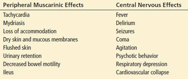
The patient presenting with anticholinergic toxicity may exhibit peripheral or central manifestations, or, more commonly, a combination of both (See Table 344.2). The classic anticholinergic toxidrome is characterized by mydriasis, dry skin and mucous membranes, flushing, fever, altered mentation, decreased bowel sounds, and tachycardia. These features provide the basis for the mnemonic “blind as a bat, dry as a bone, red as a beet, hot as a hare, mad as a hatter.” Although this mnemonic describes the majority of anticholinergic symptoms, it ignores tachycardia, one of the most consistent and reliable clinical findings. However, in patients taking β-blockers or calcium channel blockers, infants, the elderly, and alcoholics with autonomic neuropathy, tachycardia may be absent. Hence, the absence of tachycardia does not rule out anticholinergic toxicity. Central anticholinergic effects range from mild confusion and delirium to profound agitation, hallucinations, sedation, and coma. The severity and duration of central symptoms vary with lipid solubility, which affects the ability of an agent to penetrate the CNS. Pronounced, prolonged, and relatively isolated CNS effects have been described in patients exposed to lipophilic scopolamine and benztropine. At the extremes of age, and when anticholinergic agents are combined, delirium may persist for several days. Of note, the peripheral features of the toxidrome may be absent in patients who present to the hospital several hours following poisoning with an anticholinergic agent (2).
TABLE 344.2
Anticholinergic Symptoms and Signs
While the majority of patients with pure anticholinergic toxicity demonstrate mild to moderate symptoms, severe complications and death have been reported (7). Significant morbidity can result from prolonged agitation, hyperthermia, acidosis, cardiovascular collapse, and the consequence of dangerous behavior due to impaired perception and judgment.
Patients with severe toxicity from overdose of H1-receptor blockers can present with seizures, hypotension, rhabdomyolysis, hyperthermia, wide-complex tachycardia, and respiratory or cardiac arrest (4,5). It is likely that these complications result from the combination of anticholinergic effects with other pharmacologic properties of the agents involved. In adults with less severe overdoses of first-generation antihistamines, CNS depression (drowsiness, ataxia, coma) is common due to central antihistamine effects. CNS excitation in children is more common as a result of their sensitivity to the anticholinergic effects (4). After antihistamine exposures, coma and seizures may occur within 30 minutes of ingestion. In children <2 years old, seizures have occurred with ingestion of 150-mg of diphenhydramine. Fatal diphenhydramine doses in adults may range from 20 to 40 mg/kg, but in infants as little as 12 mg/kg may result in death (4).
Overdose of TCAs often causes early anticholinergic signs such as sinus tachycardia and altered mental status, but toxicity following large ingestions is dominated by hypotension, seizures, and cardiac conduction delay resulting from other mechanisms (see Chapter 339).
DIFFERENTIAL DIAGNOSIS
Anticholinergic toxicity should be considered in any patient who presents with altered mental status, fever, urinary retention, tachycardia, or seizures. Anticholinergic symptoms may easily be misdiagnosed as a CNS infection, dehydration, heat illness, psychiatric disorder, or sepsis. Intoxications with salicylates, sympathetomimetics, hallucinogens, TCAs, antipsychotics, and drug withdrawal should also be considered in the differential diagnosis. The anticholinergic and sympathomimetic toxidromes can appear quite similar, but a patient with anticholinergic toxicity can be distinguished by the absence of diaphoresis and a diminished pupillary light reflex. Serotonin syndrome should also be considered, but should demonstrate neuromuscular findings that are typically absent in anticholinergic toxicity.
ED EVALUATION
A detailed history and complete physical examination are essential in the evaluation of anticholinergic toxicity or antihistamine poisoning. The physical examination should specifically search for anticholinergic features including pupil size, skin dryness, flushing, bladder distention, and bowel sounds. Dry skin, which may not be overtly obvious on physical examination, can be assessed by placing a gloved hand in the patient’s axilla. The absence of any moisture indicates pathologic dryness. Bladder distention can be assessed with a bedside ultrasound or placement of a foley catheter. Bowel sounds are typically absent or significantly diminished, though auscultation for bowel sounds is often impractical in the ED setting. Flushing of the skin may be present, but this occurs infrequently. A neurologic examination, including assessment of mental status, should be performed. Cardiac monitoring and an electrocardiogram (ECG) should be obtained to assess for prolongation of the QRS or QT intervals, particularly in the presence of tachycardia.
If seizures or altered mentation are present, further diagnostic testing such as computed tomography (CT) scan or lumbar puncture (LP), should be considered, especially if the history is unclear. Administration of physostigmine may obviate the need for further investigations if complete normalization of mental status occurs, confirming anticholinergic toxicity (9). In patients with significant symptoms, routine laboratory evaluation such as electrolytes, blood urea nitrogen, creatinine, glucose, and creatine kinase can direct supportive care. Checking serum concentrations of acetaminophen and salicylate can detect coingestion of these agents, especially from OTC products. Delayed absorption of acetaminophen may occur because of anticholinergic-induced slowed gastric emptying, making interpretation of the Rumack–Matthew nomogram difficult.
Toxicology screens are rarely useful. None of the medications or plants that cause anticholinergic toxicity are detected by standard immunoassay tests for drugs of abuse. Comprehensive testing with thin-layer and gas chromatography may assist in identifying an unknown or suspected xenobiotic, but delayed results rarely impact clinical management. As a result, the diagnosis must be made predominantly by the history and physical examination.
KEY TESTING
• Routine laboratory analysis including electrolytes, creatinine, and glucose
• Acetaminophen and salicylate concentrations with ingestion of OTC medication
• Consider head CT or LP for altered mentation or seizure
ED MANAGEMENT
The mainstay of therapy for poisoning with anticholinergic agents is supportive care, with the institution of advanced life support measures as necessary. Most patients with symptoms require a safe environment in which they cannot do themselves any harm. Comatose or hypoventilating patients should have appropriate airway intervention. Hypotension should be treated initially with intravenous (IV) crystalloid boluses followed by the use of pressor agents such as norepinephrine as needed. Agitation and seizures can be controlled with parenteral benzodiazepines. Occasionally large doses are required, resulting in impaired airway reflexes and necessitating endotracheal intubation.
GI decontamination with appropriate airway protection should be considered in patients presenting shortly after an ingestion of anticholinergic medications. A single dose of activated charcoal is adequate in most instances, but more aggressive interventions such as gastric lavage should be considered in patients with large ingestions of potentially toxic medications such as TCAs or diphenhydramine. An anticholinergic-induced delay in gastric emptying may result in the prolonged presence of pills in the stomach. There is no benefit for hemodialysis or multiple doses of activated charcoal to enhance elimination.
Physostigmine rapidly reverses central and peripheral signs of anticholinergic toxicity (9). It is a short-acting reversible inhibitor of the enzyme acetylcholinesterase, physostigmine allows ACh to accumulate in neuronal synapses and to overcome blockade of muscarinic ACh receptors. In pure, isolated anticholinergic poisoning, dramatic resolution of symptoms occurs within minutes of physostigmine administration, allowing the patient to verbally confirm the history of poisoning with an anticholinergic agent, discuss coingestants, and deny symptoms consistent with alternative diagnoses (e.g., preceding fever, headache, and neck stiffness). This may prevent the need for escalating doses of benzodiazepines to control agitation, and obviate further diagnostic workup.
The use of physostigmine is not without risks and should be undertaken with caution. Case reports document asystole following physostigmine administration to critically ill patients with TCA overdose and wide QRS intervals (10; see Chapter 339). While controversial, physostigmine can be a safe and effective therapy when used in the appropriate clinical setting, that is, in a patient with a clear anticholinergic toxidrome, a supporting history of exposure, lack of coingestion of TCAs, and no ECG evidence of cardiac conduction delay (QRS prolongation or terminal r-wave in lead AVr). Other contraindications include bowel obstruction, significant cardiac or peripheral vascular disease, asthma, chronic obstructive pulmonary disease, and pre-existing urinary tract obstruction.
Physostigmine should be administered with continuous cardiac monitoring and atropine immediately available at the bedside. One to 2 mg is given slowly over at least 5 minutes (0.02 mg/kg in a child to a maximum of 0.5 mg). The onset of action is typically within minutes. If the response is incomplete, a repeat dose can be administered after 10 to 15 minutes. Clinical effects may last from 30 to 120 minutes, and dosing may be repeated as needed for recurrence of symptoms. If excessive cholinergic symptoms such as bradycardia, diaphoresis, bronchorrhea, or hypersalivation occur following physostigmine administration, atropine should be administered and titrated to effect (an atropine dose of half the administered dose of physostigmine is typically recommended).
When the diagnosis of anticholinergic toxicity is uncertain, or physostigmine administration is not safe or feasible, agitation may be controlled with parenteral benzodiazepines. Neuroleptic agents such as phenothiazines should be avoided because of their associated anticholinergic effects.
Wide-complex tachycardia associated with massive overdose of first-generation H1-receptor antagonists (e.g., diphenhydramine) should be treated with a bolus of sodium bicarbonate to overcome sodium channel blockade, with consideration of serum alkalinization with sodium bicarbonate infusion (5). Class IA antiarrhythmics (e.g., procainamide) and class III agents (e.g., sotalol) should be avoided, as they can promote dysrhythmias as a result of prolongation of cardiac muscle cell repolarization.
CRITICAL INTERVENTIONS
• Rapidly control agitation with IV benzodiazepines to prevent rhabdomyolysis, hyperthermia, and injury to the patient or staff.
• Consider IV physostigmine to reverse agitation and coma after obtaining a normal ECG without cardiac conduction delay.
• Treat wide-complex tachycardia from first-generation H1-receptor antagonist poisoning with IV sodium bicarbonate.
Patients with anticholinergic toxicity should be observed until they have normal vital signs, normal mental status, and all anticholinergic signs have resolved. This observation often requires admission to a monitored bed. Patients with agitation, coma, seizures, or cardiovascular instability should be admitted to an intensive care unit. The duration of anticholinergic delirium may be from 12 hours to several days depending on the agent and dose ingested. Patients with antihistamine poisoning who become or remain asymptomatic after a 4- to 6-hour observation period may be medically cleared. Suicide risk should be evaluated in patients with intentional overdoses.
Common Pitfalls
• Failure to consider the diagnosis of anticholinergic toxicity in patients with altered mental status (particularly agitated delirium), tachycardia, urinary retention, fever, or seizures.
• Failure to appreciate that anticholinergic symptoms may present as isolated central or peripheral manifestations.
• Use of physostigmine in patients with a history of TCA ingestion, with ECG evidence of cardiac conduction delay, or without adequate monitoring and resuscitation capability.
• Use of neuroleptics to treat anticholinergic agitation and hallucinations.
• Failure to obtain acetaminophen and salicylate serum concentrations in the patient who has ingested OTC medications.
ACKNOWLEDGMENT
The author gratefully acknowledges the contributions of Andis Graudins to this chapter in previous editions.
REFERENCES
1. Myers JH, Moro-Sutherland D, Shook JE. Anticholinergic poisoning in colicky infants treated with hyoscyamine sulfate. Am J Emerg Med. 1997;15:532–535.
2. Feinberg M. The problems of anticholinergic adverse effects in older patients. Drugs Aging. 1993;3:335–348.
3. Cetaruk EW, Aaron CK. Hazards of non-prescription medications. Emerg Med Clin North Am. 1994;12:483.
4. Scharman EJ, Erdman AR, Wax PM, et al. Diphenhydramine and dimenhydrinate poisoning: An evidence-based consensus guideline for out-of-hospital management. Clin Toxicol (Phila). 2006;44(3):205–223.
5. Clark RF, Vance MV. Massive diphenhydramine poisoning resulting in a wide-complex tachycardia: successful treatment with sodium bicarbonate. Ann Emerg Med. 1992;21:318–321.
6. Cooper RJ. Over-the-counter medicine abuse - a review of the literature. J Subst Use. 2013;18(2):82–107.
7. Centers for Disease Control and Prevention (CDC). Jimson weed poisoning–Texas, New York, and California, 1994. MMWR Morb Mortal Wkly Rep. 1995;44(3):41–44.
8. Hamilton RJ, Perrone J, Hoffman R, et al. A descriptive study of an epidemic of poisoning caused by heroin adulterated with scopolamine. J Toxicol Clin Toxicol. 2000;38(6):597–608.
9. Burns MJ, Linden CH, Graudins A, et al. A comparison of physostigmine and benzodiazepines for the treatment of anticholinergic poisoning. Ann Emerg Med. 2000;35:374–481.
10. Pentel P, Peterson CD. Asystole complicating physostigmine treatment of tricyclic antidepressant overdose. Ann Emerg Med. 1980;9(11):588–590.