
Carrie D. Tibbles
The genitourinary (GU) system is involved in approximately 10% of acutely injured patients (1). The presentation of GU trauma may be occult and is often overshadowed by more dramatic and immediate life-threatening injuries. However, if these injuries are not identified and properly treated, the patient may experience delayed complications and significant long-term morbidity. Clues to suggest the possibility of GU trauma lie in the mechanism of injury, a constellation of associated injuries, and physical examination findings identified during the secondary survey. For diagnosis and management, GU trauma is generally divided into upper tract injuries (kidney and ureter) and lower tract injuries (bladder, urethra, and external genitalia). A systematic approach and thorough understanding of the appropriate diagnostic modalities are necessary for emergency physicians to effectively manage these injuries.
CLINICAL PRESENTATION
Upper Tract Injury
The kidneys and ureters lie securely in the retroperitoneum, beneath the lower ribs and musculature of the back. Therefore, injuries to the upper urinary tract typically occur in the setting of high-energy, multisystem trauma. Blunt force to the back, flank, or abdomen; falls from height; or, less commonly, rapid deceleration may result in injuries to the kidneys or ureters (2). Upper tract injuries may occur in isolation. However, more commonly, trauma that results in renal injuries produces associated bony, vascular, and visceral injuries that directly contribute to the observed morbidity and mortality (1). Penetrating injury, which accounts for 6% to 11% of renal injuries, is suggested by transaxial wounds or wounds in anatomic proximity to the kidneys and ureter. Although nearly all significant blunt renal injuries will present with either gross or microscopic hematuria, about 25% to 30% of ureteral injuries will not have associated hematuria. The presence of hematuria is a less reliable indicator of upper tract injury with penetrating trauma and the location of the wound is used to guide subsequent management decisions (3). A vascular pedicle injury may present as refractory hypotension following severe blunt trauma; this injury usually results from a deceleration mechanism.
The American Association for the Surgery of Trauma (AAST) has established a classification system for renal injuries based on anatomic location and severity (Table 35.1; Fig. 35.1) (4). Grade I and grade II injuries consist of contusions and lacerations confined to the cortex respectively and are considered minor injuries. Eighty-five percent of renal injuries are classified as minor injuries. Grade III injuries are deeper lacerations that extend into the corticomedullary junction. Lacerations into the collecting system or those involving segmental arteries are grade IV injuries. The most severe injuries are classified as grade V and include a shattered kidney, thrombosis of the main renal artery, or avulsion of the renal hilum. A review of the National Trauma Data Bank found that the AAST injury scale predicts the need for nephrectomy in both blunt and penetrating trauma and mortality in blunt trauma patients (5). Recently investigators from Parkland recommended a substratification of grade IV injuries into high risk and low risk based on three predictors of needing operative intervention, specifically, intravascular contrast extravasation, hematoma size greater than 3.5 cm, and a medial renal laceration site (6,7).
TABLE 35.1
Grading System for Renal Injuries

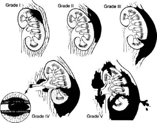
FIGURE 35.1 Grading system of renal injuries. (From Santucci RA, McAninch JW, Safir M, et al. Validation of the American Association for the Surgery of Trauma organ injury severity scale for the kidney. J Trauma. 2001;50:195–200, with permission.)
Lower Tract Injuries
Blunt trauma to the lower urinary tract (bladder and urethra) is usually associated with pelvic rami fractures and symphyseal diastasis. Anterior urethral injuries (penile and bulbous urethra, below the urogenital diaphragm) are associated with self-instrumentation, falls, and straddle injuries. Posterior urethral injuries (membranous and prostatic urethra, above the urogenital diaphragm) commonly accompany pelvic fractures (Fig. 35.2). As the urinary continence mechanism and autonomic innervation responsible for erection are contained in the posterior urethra, posterior urethral injuries may result in permanent incontinence and erectile dysfunction. Signs of anterior urethral injury depend on the integrity of Buck fascia. If this fascial layer is torn, blood and urine may pass into the penis, scrotum, and abdominal wall. Clinical signs of urethral injury are blood at the urethral meatus, high-riding prostate, scrotal or perineal hematoma, and difficulty voiding. However, these signs can be absent in some patients with urethral injury.

FIGURE 35.2 Normal anatomy of the male urethra. (From Schneider RE. Genitourinary system. In: Marx JA, Hockberger RS, Walls RM, eds. Rosen’s Emergency Medicine Concepts and Clinical Practice. 5th ed. St. Louis, MO: Mosby; 2002:438, with permission.)
Bladder rupture is classified as extraperitoneal or intraperitoneal. Extraperitoneal bladder injuries are most commonly associated with pelvic fractures and bladder laceration by bony fragments. There is leakage of urine into the perivesicular space. Intraperitoneal bladder injuries result from compressive forces in the presence of a full bladder, and rupture occurs at the dome of the bladder through the peritoneum into the abdominal cavity. The presence of a pelvic fracture and gross hematuria is virtually diagnostic of bladder rupture. Bladder injury is more likely in pelvic fractures with a widened pubic symphysis and sacroiliac disruption (8). Ninety-eight percent of patients with bladder rupture will have gross hematuria. Other signs of bladder injury are suprapubic or lower abdominal pain and tenderness, inability to void, and blood at the urethral meatus.
As with penetrating trauma to the upper GU tract, penetrating injuries to the lower tract are suggested by location of wounds. Any penetration of the lower abdominal wall in the region of the pelvis raises the possibility of urinary tract injury. Given the variability of the projectile tract, any gunshot wound in the region should include consideration of GU tract injury.
External Genitalia Trauma
Testicular injuries are often the result of a fall or direct trauma. They include lacerations, contusions, dislocations, or fractures. Symptoms include pain, nausea, lightheadedness, and acute urinary retention. A swollen, tender testicle or a hematoma may be seen on physical examination. Penile injuries range from small lacerations and contusions to complete amputations. Penile fracture, which involves rupture of the tunica albuginea, most commonly occurs during overzealous vaginal intercourse, although several other mechanisms have been reported (9). Any constricting object placed around the penis can result in marked edema and eventual incarceration and necrosis distal to the constriction. Entrapment of the penis in a zipper occurs most often in uncircumcised young boys.
Vaginal injuries are frequently complications of pelvic fractures but may also be seen following sexual assault or other penetrating injuries. The majority of patients present with vaginal bleeding. A careful vaginal examination is required in any female patient suspected of having a pelvic fracture. According to one series, up to one-third of patients with vaginal trauma unrelated to childbirth will have concurrent urethral injuries (10).
ED EVALUATION
Upper Tract Injury
The emergency department (ED) evaluation of a multitrauma patient begins with the standard advanced trauma life support assessment and resuscitation. In this manner, immediately life-threatening injuries are identified and treated. In the unstable patient requiring emergency laparotomy for other reasons, gross hematuria and signs of a pelvic fracture should be noted during the secondary survey. Definitive evaluation of the GU tract can be performed once the patient has been stabilized. In the stable patient, the emergency physician determines the necessity of further evaluation and diagnostic imaging.
The first task is to identify those patients at risk for significant renal injuries. Mee et al. published the classic article establishing criteria for the evaluation of blunt renal trauma. This 10-year prospective study of 1,007 consecutive blunt trauma patients established markers associated with increased risk of major renal lacerations. Gross hematuria, microscopic hematuria (>5 red blood cells [RBCs]/high power field [HPF]) with shock either in the prehospital setting or in the ED, or, less commonly, a history of significant deceleration, are associated with renal injuries. The authors concluded that if radiographic evaluation were limited to those patients, no significant renal injuries would be missed (11). These guidelines have been supported by subsequent studies and have become the criteria defining the population of patients who require further radiographic evaluation (2,3,12). While children are more prone to renal injuries than adults because the pediatric kidney is proportionally larger and has less protection from surrounding tissues, the same criteria screening used to image adult patients may be appropriately applied to pediatric patients (13,14). Most authors advocate radiologic imaging in the setting of penetrating trauma to evaluate for upper urinary tract injuries, as a substantial percentage of patients may have significant injury in the absence of hematuria (2,3).
Computed tomography (CT) scanning is the preferred diagnostic modality for evaluating the hemodynamically stable patient with blunt or penetrating trauma. CT clearly defines parenchymal lacerations, hematomas, and the presence of urinary extravasation; may identify renal vascular injuries; and can identify associated injuries (Fig. 35.3). Significant findings on CT scan include a devitalized renal segment, a nonfunctioning kidney, actively expanding hematoma, or extravasation of urine. Nonfunction suggests extensive trauma to the kidney, renal artery thrombosis, or a severely shattered kidney. Extravasation of contrast material implies trauma involving the capsule, parenchyma, or collecting system.

FIGURE 35.3 Renal lacerations with perirenal hematoma.
Arteriography has a very limited role in the evaluation of renal injuries and is reserved for stable patients requiring further clarification of pedicle injuries. Arteriography may demonstrate intimal tears and complete or partial disruption of the renal vasculature. CT findings requiring further evaluation by arteriography include large perirenal hematomas, major renal fractures, or segmental areas of nonenhancement. Nonenhancement of the kidney on CT scan is diagnostic of renal artery thrombosis and is sufficient indication for surgery without additional studies (15).
Ureteral injury remains a diagnostic challenge. Approximately one-third of ureteral injuries are not identified on initial evaluation. If a ureteral injury is suspected, a contrast enhanced helical CT with delayed images should be obtained. Extravasation of contrast visualized on the delayed images may be the only sign of ureteral injury (Fig. 35.4).
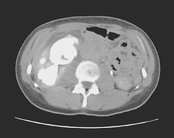
FIGURE 35.4 Delayed CT image demonstrating extravasation of contrast diagnostic of ureteral disruption following blunt trauma.
Lower Tract Injury and External Genitalia Injury
Lower tract GU injuries should be evaluated in a retrograde fashion. Once the potential for injury has been identified based on mechanism of injury, physical findings (i.e., scrotal hematoma, blood at the urethral meatus, or a high-riding prostate), or radiographic evidence of associated high-risk injuries (i.e., pelvic fracture), the urethra should be evaluated first. The presence of urethral injury should be ruled out prior to Foley catheter placement, because catheter placement may potentially convert a partial urethral tear to a complete urethral disruption. A retrograde urethrogram (RUG) is the diagnostic modality for evaluating the urethra. However, many trauma patients may require urgent CT scan for the evaluation of multiple injuries. The performance of the RUG and subsequent Foley catheter placement can often be delayed until after more urgent CT scans are performed. High resolution CT scan may demonstrate signs of urethral trauma, typically extravasation of urinary contrast material, but is not adequately sensitive to rule out a urethral injury or to fully evaluate or characterize specific injuries.
A RUG is performed with the patient supine. An initial KUB (kidneys, urethers, bladder) is obtained for comparison. A Foley catheter is then passed approximately 1 to 2 cm to the fossa navicularis, and the balloon is inflated with 1 to 2 mL of saline. Alternatively a 60-cc Toomey syringe with a Christmas tree or Cooke adapter may be gently inserted until snug. Lateral traction is applied to the penis, and 60 mL of water-soluble contrast material is injected over 30 to 60 seconds. The radiograph is exposed as the last 10 mL of contrast material is injected. Extravasation of contrast material or failure of contrast material to reach the bladder is diagnostic of urethral injury, mandates urologic consultation, and precludes the passage of a urethral catheter by the emergency physician (Fig. 35.5).
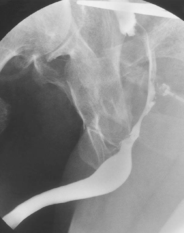
FIGURE 35.5 Retrograde urethrogram with partial tear of membranous urethra.
If a team member inadvertently places a urinary catheter prior to adequate evaluation for urethral injury, it should not be removed. It is not necessary to remove the catheter to perform a RUG. Instead, the presence or absence of urethral injury should be determined by passing a small feeding tube beside the catheter and performing a modified RUG.
A diagnostic workup for bladder injury is indicated for patients with gross hematuria, inability to void, or a pelvic ring fracture in association with microscopic hematuria. After exclusion of urethral injury and placement of catheter, a CT cystogram may be performed. Importantly, while a routine CT scan of the abdomen and pelvis may demonstrate evidence of a bladder injury, it is not sufficient to fully exclude these injuries. A CT cystogram is performed after retrograde instillation of 400 mL of dye through a Foley catheter into the bladder to cause bladder distension. CT cystogram is 95% sensitive and 100% specific for the detection of bladder rupture (16).
Clinical examination of the scrotum after blunt trauma is often difficult because of severe pain and swelling. If there is a clinical suspicion of a testicular injury, further evaluation with a CT scan should be performed. Any patient with significant external genitalia trauma with hematuria, urinary retention, or pain with urination should have a RUG to evaluate for a urethral tear.
KEY TESTING
Hemodynamically stable patients with any of the following require a contrast enhanced CT scan for upper tract injury:
Penetrating trauma
Blunt trauma with gross hematuria
Blunt trauma with microscopic hematuria and hemodynamic instability (hypotension or tachycardia)
High-energy deceleration mechanism or suspected associated intra-abdominal injuries
Patients with any of the following signs require imaging to assess for a lower tract injury:
Indications for a RUG:
High-riding prostate
Gross hematuria
Blood at the meatus
Trauma to the external genitalia
Indications for a CT cystogram:
Gross hematuria
Inability to void
Pelvic ring fracture
Lower abdominal pain following significant trauma
ED MANAGEMENT
Upper Tract Injury
The principles of care for the patient with multisystem trauma apply to the patient with trauma to the upper GU tract. Hemodynamic stabilization with crystalloid and blood products is the initial priority. Once immediately life-threatening injuries are addressed, evaluation of the GU tract and associated injuries can occur as outlined in the previous section.
In a review of more than 2,500 trauma patients, Santucci et al. (17) demonstrated a very strong correlation between AAST renal injury grade and the subsequent need for surgical intervention. In general, the majority of blunt renal trauma may be successfully managed nonoperatively. Expectant management of grade I, II, and III renal injuries is widespread. In many centers, hemodynamically stable patients with grade IV and potentially grade V injuries are also initially managed with a trial of expectant management. Overall injury severity, renal grade, hemodynamic instability, and transfusion requirements are predictors of subsequent nephrectomy (18). CT scan findings that predict need for operative intervention include extravasation of contract, a perirenal hematoma greater than 3.5 cm, and a medial location of the renal laceration (6). Delayed interventions may be necessary in a subset of patients managed nonoperatively who develop complications, including persistent bleeding, formation of urinoma, or persistent urine leak. As expected, these complications are more common in patients with higher grade injuries. Patients with the combination of a devitalized renal segment and extravasation of urine on the initial CT scan are particularly at risk. Most complications can be managed by either endoscopic or percutaneous techniques (19). Following traumatic nephrectomy or conservative management of high grade lesions, the majority of patients will have full recovery of their renal function and a normalization of their creatinine (20).
The management of penetrating upper tract trauma has evolved in recent years. There is a lower threshold for operative exploration and intervention after penetrating trauma, but many of these injuries are now managed nonoperatively as well. According to the EAST Practice Guidelines, nonoperative treatment of penetrating renal lacerations is appropriate in hemodynamically stable patients without associated injuries who have been staged completely with CT scan (12). Expectant management of stab wounds is more widely accepted, although small series of successful nonoperative management of renal gunshot wounds have also been published. Delayed bleeding can be seen in up to 20% of patients following nonoperative management of renal stab wounds, particularly when the patient increases activity. This bleeding may be managed by therapeutic angiography and rarely requires intervention (19). Figure 35.6 outlines a potential algorithm for the conservative management of renal injuries. Ureteral injuries and injuries to the renal pelvis are repaired surgically or stented. Renovascular injuries are typically managed with an immediate nephrectomy or they are repaired primarily.
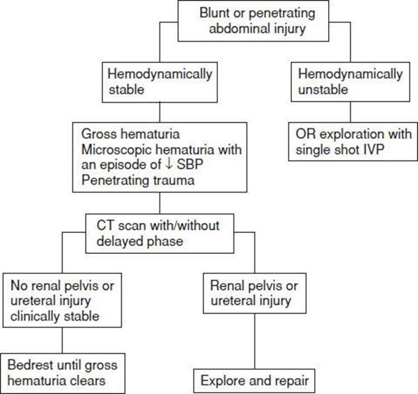
FIGURE 35.6 Algorithm for the management of renal parenchymal injuries. (Adapted from Santucci RA, Fisher MB. The literature increasingly supports expectant (conservative) management of renal trauma—a systematic review. J Trauma. 2005;59:
493–503.)
Lower Tract Injuries
If a urethral disruption is identified, urologic consultation should be obtained and a suprapubic tube should be placed to allow drainage of the bladder. This may be performed in the resuscitation bay if the patient is stable. If the patient is unstable and requires operative intervention for associated injuries, it can be performed in the operating room. If a catheter was placed prior to identifying the injury, it should be left in place. Occasionally, a catheter may be placed through a partial urethral tear. Once the partial disruption is identified, however, any gentle and limited attempts at urethral catheterization should be done by a urologist.
The acute management of bladder rupture is bladder drainage via Foley catheter. Patients with intraperitoneal bladder rupture undergo primary surgical repair. Those patients with extraperitoneal bladder rupture are typically managed nonoperatively, with simple urinary drainage by Foley catheter or a suprapubic catheter. In general, in the absence of urethral injury, a transurethral catheter is preferred over a suprapubic catheter as it is associated with fewer complications and days of catheterization (12).
External Genitalia Injury
Diagnosis of testicular rupture or dislocation should prompt urgent urologic consultation, as surgical repair within 72 hours dramatically improves the salvage rate.
There have been several reported cases of microvascular reimplantation following amputation of the penis. Successful repair depends on the condition of the severed part and the ischemia time. In the prehospital and emergency setting, the amputated penis should be preserved in a manner similar to that for other amputated parts. Prompt urologic and microvascular consultation is imperative for successful reimplantation (21). Penetrating trauma to the external genitalia mandates early surgical exploration and conservative debridement; this is followed by primary repair in most cases (22). Some authors have also advocated operative repair of penile fractures, proposing that it decreases the incidence of erectile deformity and may speed recovery (9). For patients with penile zipper entrapment, Nolan and associates described the successful atraumatic removal by cutting the median bar of the zipper with bone cutters. The upper and lower shields of the zipper device separate, releasing the skin with minimal injury (23). Penile incarceration may be managed by the string technique, similar to that described for removal of rings from fingers.
CRITICAL INTERVENTIONS
• Address immediately life-threatening injuries first. If the patient is hemodynamically unstable, evaluation of the GU system can be performed after operative intervention for other injuries (e.g., intraperitoneal injuries).
• Rule out a urethral injury by clinical examination, and RUG if necessary, before placement of a Foley catheter.
• Obtain an urgent urology consultation if testicular disruption is diagnosed.
DISPOSITION
The majority of patients with blunt renal injuries require admission to the hospital. Selected patients with microscopic hematuria in the absence of any hemodynamic instability, and no associated injuries, may be discharged with a follow-up repeat urinalysis in 1 week to ensure resolution. All patients with penetrating renal or ureteral trauma require admission or transfer to a facility capable of providing definitive diagnostic and therapeutic care. Disposition following trauma to the external genitalia depends on the complexity and severity of the injury as well as any associated injuries. Many of these patients will require a urology consultation in the ED or close outpatient urology follow-up.
Common Pitfalls
• Failing to consider upper tract injury in the absence of hematuria. Patients with renal pedicle injury resulting from a deceleration mechanism, or ureteral injury resulting from penetrating trauma, may not have hematuria.
• Delaying an urgent laparotomy to perform further diagnostic imaging of the GU tract.
• Ruling out significant injury following penetrating trauma to the flank based on a suboptimal local wound exploration. If the wound is in anatomic proximity to the GU tract, the patient requires further diagnostic evaluation.
• Failure to obtain a RUG prior to attempting to place a Foley catheter in the presence of a potential urethral injury.
• Attempting to rule out bladder injury with routine abdominal CT without adequate distension of the bladder (i.e., instillation of 400 mL of contrast).
REFERENCES
1. McGeady JB, Breyer BN. Current epidemiology of genitourinary trauma. Urol Clin North Am. 2013;40:323–334.
2. Shenfeld OZ, Gnessin E. Management of urogenital trauma: State of the art. Curr Opin Urol. 2011;21(6):449–454.
3. Santucci RA, Bartley JM. Urologic trauma guidelines: A 21st century update. Nat Rev Urol. 2010;7(9):510–519.
4. Moore EE, Shockford SR, Pachter HL, et al. Organ injury scaling: Spleen, liver and kidney. J Trauma. 1989;29(12):1664–1666.
5. Kuan JK, Wright JL, Nathens AB, et al. American Association for the Surgery of Trauma Organ Injury Scale for kidney injuries predicts nephrectomy, dialysis, and death in patients with blunt injury and nephrectomy for penetrating injuries. J Trauma. 2006;60:351–356.
6. Dugi DD 3rd, Morey AF, Gupta A, et al. American Association for the Surgery of Trauma grade 4 renal injury substratification into grades 4 a(low risk) and 4b (high risk). J Urol. 2010;183(2):592–597.
7. Hardee MJ, Lowrance W, Brant WO, et al. High grade renal injuries: Application of Parkland Hospital predictors of intervention for renal hemorrhage. J Urol. 2013;189(5):1771–1776.
8. Aihara R, Blansfield JS, Millham FH, et al. Fracture locations influence the likelihood of rectal and lower urinary tract injuries in patients sustaining pelvic fractures. J Trauma. 2002;52:205–209.
9. El-Taher AM, Aboul-Ella HA, Sayed MA, et al. Management of penile fracture. J Trauma. 2004;56:1138–1140.
10. Goldman HB, Idom CB, Dmochowski RR. Traumatic injuries of the female external genitalia and their association with urological injuries. J Urol. 1998; 159(3):956–959.
11. Mee SL, McAninch JW, Robinson AL, et al. Radiographic assessment of renal trauma: A 10-year prospective study of patient selection. J Urol. 1989;141:1095–1098.
12. Holevar M, Ebert J, Luchette F, et al. EAST practice management guideline: The management of genitourinary trauma. Available online at http://www.east.org/tpg.asp. Accessed November 21, 2013.
13. Santucci RA, Langernburg SE, Zachareas MJ. Traumatic hematuria in children can be evaluated as in adults. J Urol. 2004;171:822–825.
14. Tasian GE, Bagga HS, Fisher PB, et al. Pediatric genitourinary injuries in the United States from 2002 to 2010. J Urol. 2013;189(1):288–293.
15. Hotaling JM, Sorensen MD, Smith TG 3rd, et al. Analysis of diagnostic angiography and angioembolization in the acute management of renal trauma using a national data set. J Urol. 2011;185(4):1316–1320.
16. Deck AJ, Shaves S, Talner L, et al. Computed tomography cystography for the diagnosis of traumatic bladder rupture. J Urol. 2000;164(1):43–46.
17. Santucci RA, McAninch JW, Safir M, et al. Validation of the American Association for the Surgery of Trauma organ injury severity scale for the kidney. J Trauma. 2001;50(2):195–200.
18. Davis KA, Reed L 2nd, Santaniello J, et al. Predictors of the need for nephrectomy after renal trauma. J Trauma. 2006;60:164–169.
19. Mani NB, Kim L. The role of interventional radiology in urologic tract trauma. Semin Intervent Radiol. 2011;28(4):415–423.
20. Dozier KC, Yeung LY, Miranda MA Jr, et al. Death or dialysis? The risk of dialysis- dependent chronic renal failure after trauma nephrectomy. Am Surg. 2013;79(1):96–100.
21. Kunkle DA, Lebed BD, Mydlo JH. Evaluation and management of gunshot wounds of the penis: 20-year experience at an urban trauma center. J Trauma. 2008;64(4):1038–1042.
22. Bjurilin MA, Kim DY, Zhao LC, et al. Clinical characteristics and surgical outcomes of penetrating external genital injuries. J Trauma Acute Care Surg. 2013; 74:839–844.
23. Nolan JF, Stillwell TJ, Sands JP Jr. Acute management of the zipper-entrapped penis. J Emerg Med. 1990;8(3):305–307.