
Maria E. Moreira and Peter T. Pons
Prompt recognition and management of peripheral vascular injuries is crucial to limb salvage and preservation of function. A vascular injury should be suspected in any patient with a penetrating injury or a major fracture or dislocation secondary to blunt trauma. Penetrating trauma accounts for approximately 85% of vascular injuries in the United States; blunt trauma and iatrogenic causes are responsible for the rest.
Blunt trauma can cause spasm, external compression, mural contusion, intimal tear, thrombosis, or aneurysm formation. A high index of suspicion is necessary in these cases as injury can be very subtle in presentation. Stretching, tearing, or shearing forces can damage a vessel while leaving very little external injuries. The classic example is dislocation of the knee with associated popliteal artery injury in which there are very few visual clues to indicate vascular injury.
In penetrating trauma, the penetrating object can directly injure vascular structures when traversing the path of a vessel. Injuries can include partial or complete vessel transection, contusion, laceration, or arteriovenous fistula. The most commonly involved vessels are the brachial and axillary arteries in the upper extremity and the femoral and popliteal arteries in the lower extremity.
The emergency physician must obtain a complete history, perform a careful physical examination, and be aware of the mechanics that produce vascular injury, even if there are minimal or absent physical findings that suggest vessel damage.
CLINICAL PRESENTATION
Evaluation of extremity pulses distal to the injury is essential to the identification of vascular injuries. The following pulse findings are indicative of a vascular injury: diminished or absent pulses, thrill or bruit suggestive of an arteriovenous fistula, a pulsatile mass or rapidly expanding hematoma. Other etiologies for the pulse difference, such as shock should also be considered. Pulse differences due to shock should correct with resuscitation. The examiner must consider that the presence of a pulse does not completely exclude a vascular injury. Collateral circulation or transmitted pressure waves through a small clot or intimal flap can produce a distal pulse.
A Doppler can be used to further assess quality of pulses. The three levels of Doppler signal are monophasic, biphasic, and triphasic. Triphasic is the normal signal in an intact vessel. Changes in the signal can provide an indication of vascular compromise. When a triphasic signal changes to a monophasic signal the provider needs to consider loss of flow. This signal is evaluated in conjunction with other physical examination evidence of perfusion (1).
Determining the arterial pressure index (API) is an important adjunct to the physical examination. A Doppler is used to determine the systolic blood pressure distal to the penetrating injury. The systolic pressure is then measured in the same location in the contralateral uninjured extremity. The ratio of the injured to the uninjured extremity is the API. A normal API is greater than or equal to 0.9. A ratio of less than 0.9 is indicative of a vascular injury and necessitates further evaluation (1–3). The sensitivity and specificity of API of less than 0.9 is 87% and 97%, respectively. In one study when insignificant injuries were excluded the sensitivity of API less than 0.9 increased to 95% (4).
Ankle-brachial index (ABI) is often considered synonymous with API. The ABI is calculated as follows: systolic blood pressure at dorsalis pedis or posterior tibialis divided by the brachial systolic blood pressure. An ABI less than 0.9 is considered abnormal. Typically, ABI has been used in the diagnosis of peripheral vascular disease, although it is not uncommonly used for trauma patients as well. In a patient with underlying peripheral vascular disease obtaining both an ABI and an API may be useful in identifying a new injury due to trauma.
The other “P”s of arterial compromise leading to ischemia include pallor, pain, poikilothermia, paresthesias, and paralysis. Additional findings may include tenseness of the extremity, coolness to touch, and delayed capillary refill (>3 seconds). Unfortunately, some of these signs require a patient who can relate these complaints, and many multiply injured patients cannot. In addition, concomitant neurologic injury of the extremity may present with similar findings. Careful examination is necessary to differentiate the paresthesias of vascular insufficiency from those of neurologic injury. As many as 70% of upper- and 30% of lower-extremity vascular injuries have associated nerve damage, of which 40% to 45% result in permanent deficit.
Many vascular injuries appear relatively innocuous at first glance. Penetrating wounds of an extremity can present with a small entrance site that harbors significant underlying vascular damage. Blunt injury may be quite misleading because of minimal obvious external signs. Thus, repeat examination of the patient is important to look for evolving signs of vascular injury.
Finally, the structural integrity of the skeletal system must be evaluated. About 18% of patients with peripheral vascular injuries have an associated fracture from either blunt or penetrating trauma.
ED EVALUATION
Initial physical examination findings will dictate further evaluation. Patients with hard signs of vascular injury (Fig. 36.1) require immediate intervention. Hard signs include absent pulses, active hemorrhage, rapidly expanding hematoma, and palpable thrill/bruit. In such cases, operative intervention should not be delayed for other diagnostic studies or radiographic imaging, unless the location of the vessel injury cannot be determined clinically (5). In the absence of hard signs in a hemodynamically stable patient, further evaluation can be undertaken. Patients with an API <0.9 require further testing. Those with soft signs of injury (diminished pulses, large nonexpanding hematoma, peripheral nerve injury, or delayed capillary refill) and proximity wounds can be followed with serial physical examinations, tracking the API (3,6–9). Color-flow duplex Doppler ultrasonography can also be used to assess patients with soft signs (10–12). Color-flow duplex Doppler ultrasonography interprets the sound of blood flow and shows it as red and blue colors based on the speed and direction of the flow relative to the ultrasound probe. This sonographic technique will easily demonstrate absence of arterial flow but is less valuable for more subtle injuries such as intimal flaps or pseudoaneurysms that may not interrupt blood flow in the damaged vessel (10).
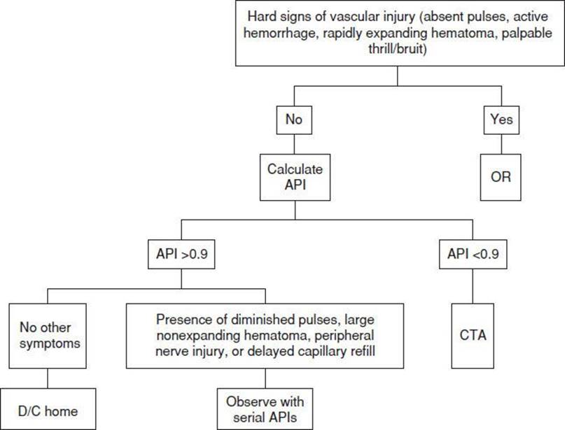
FIGURE 36.1 Peripheral vascular injury clinical algorithm.
The mainstay of evaluation and diagnosis has been contrast arteriography, with a sensitivity of 97% to 99% for detecting arterial injury. Magnetic resonance angiography (MRA) and spiral computed tomographic angiography are other options for evaluating vascular injuries (13–18). CTA is currently the preferred study for initial evaluation of the vasculature due to its ready availability and use of only intravenous contrast. Several studies demonstrated CTA to have a 100% sensitivity and specificity for detecting clinically significant arterial injuries (4,19,20).
Shotgun wounds to an extremity have a much higher incidence of vascular injury owing to the nature of the weapon (i.e., multiple missiles and multiple trajectories) (12). In addition, victims of blast injury, high-velocity weapons, and military munitions have a higher incidence of vascular injury (21). Therefore, angiography is indicated in these patients unless the physical examination, including API, suggests that the potential for vascular injury is remote. If angiography is not performed, a period of observation is warranted.
Each institution should determine in advance how it plans to evaluate patients with potential peripheral vascular trauma by having protocols in place agreed upon by the emergency medicine, surgery, and radiology departments.
KEY TESTING
• Determine the arterial pressure index (API)
• CTA should be performed in patients with API <0.9
• Patients with soft signs can be followed by serial examinations monitoring the API
ED MANAGEMENT
Prehospital and emergency department (ED) management priorities include control of hemorrhage, fluid resuscitation, and assessment and management of other life threats (9).
Hemorrhage should be controlled by direct pressure over the bleeding site. Blind clamping of bleeding vessels deep in a wound is to be condemned, as adjacent structures such as nerves and tendons may be crushed in the attempt to stop hemorrhage. If a bleeding vessel can be identified and isolated, it may be clamped using a noncrushing vascular clamp. Recent work in animals and experience in the conflicts in Iraq and Afghanistan suggest that hemostatic agents may be of benefit in controlling exsanguinating hemorrhage secondary to proximal vascular injury such as femoral artery or vein injury in the groin (22). If compression or hemostatic agents are unsuccessful at stopping the bleeding, tourniquets can be used as a temporizing method (3). The key is to minimize the tourniquet time.
Fluid restoration is accomplished by initiating one or more intravenous lines (as the trauma mechanism and vital signs dictate) and infusing crystalloid. After bleeding is controlled and resuscitation started, a careful examination for other life-threatening injuries is performed. As with all trauma patients, a patent airway and adequate ventilation must be ensured while circulation is being addressed. Vascular injuries are treated after other potential life threats have been assessed and managed.
Angulated fractures associated with pulse deficits should be anatomically repositioned and splinted. This may restore perfusion to an ischemic extremity by alleviating kinking of the blood vessel.
Blood for type and crossmatch should be sent to the laboratory and appropriate preparations made for transfusion if blood loss or anticipated surgical needs indicate. Unless indications for immediate surgery are present, complete evaluation for possible vascular injury should follow.
Tetanus immunization status should be ascertained and the patient immunized, if necessary. Prophylactic antibiotics are indicated when operative repair or vascular grafts are to be undertaken and if there is an open fracture. Anticoagulants should generally not be given until surgical consultation has been obtained, the absence of other major injuries has been determined, and the patient is ready to undergo arterial repair.
CRITICAL INTERVENTIONS
• Control hemorrhage by using direct pressure over the bleeding site.
• Resuscitate with crystalloid infusion and blood when indicated.
• After bleeding is controlled, perform a general examination for other life-threatening injuries.
• Measure the arterial pressure index in all patients with penetrating extremity trauma.
• Reduce angulated fractures associated with pulse deficits immediately.
DISPOSITION
Surgical consultation must be obtained as early as possible, as the amount of time elapsed from injury until operative repair is a crucial determinant of successful repair and decreased morbidity in the ischemic extremity. Endovascular embolization may be an option for treatment of nonessential vessels in select situations. Endovascular stent placement is an alternative to surgery for the treatment of vascular injuries and is gaining increasing acceptance and use (23). Observation for a period of 12 to 24 hours is appropriate for vascular proximity wounds in patients without obvious signs of vessel injury and a normal API. In fact, one study suggests that asymptomatic patients with an API ≥1 may be safely discharged from the ED (24). In addition, clinically occult arterial injuries have been shown to have a benign course. Therefore, patients meeting the following criteria are candidates for observation: <5-mm intimal disruption, adherent intimal flaps, intact distal circulation, and no active hemorrhage (6,9). The medical team taking care of the patient will need to make the assessment of whether operative repair versus amputation is the optimum treatment for the patient given other potential injuries (25).
Common Pitfalls
• Failure to realize that vascular injuries may be subtle in origin and misleading on physical examination.
• Failure to appreciate injury mechanics or vessel proximity to a penetrating wound.
• Failure to recognize that delays in diagnosis of vascular compromise may lead to permanent deficit or loss of the limb.
• Blind clamping of bleeding vessels deep in a wound.
REFERENCES
1. Stovall RT, Pieracci FM, Johnson JL. Perioperative management of peripheral vascular trauma. Semin CardiothoracVasc Anesth. 2012;16(3):133–141.
2. Graves M, Cole PA. Diagnosis of peripheral vascular injury in extremity trauma. Orthopedics. 2006;29:35–37.
3. Kragh JF, Walters TJ, Baer DG, et al. Survival with emergency tourniquet use to stop bleeding in major limb trauma. Ann Surg. 2009;249:1–7.
4. Fox N, Rajani RR, Bokhari F, et al. Evaluation and management of penetrating lower extremity arterial trauma: An Eastern Association for the Surgery of Trauma practice management guideline. J Trauma Acute Care Surg. 2012;73:
S315–S320.
5. Gonzalez RP, Falimirski ME. The utility of physical examination in proximity penetrating extremity trauma. Am Surg. 1999;65:784–789.
6. Dennis JW, Frykberg ER, Veldenz HC, et al. Validation of nonoperative management of occult vascular injuries and accuracy of physical examination alone in penetrating extremity trauma: 5- to 10-year follow-up. J Trauma.1998;44:
243–253.
7. Francis H III, Thal ER, Weigelt JA, et al. Vascular proximity: Is it a valid indication for arteriography in asymptomatic patients? J Trauma. 1991;31:512–514.
8. Frykberg ER, Dennis JW, Bishop K, et al. The reliability of physical examination in the evaluation of penetrating extremity trauma for vascular injury: Results at 1 year. J Trauma. 1991;31:502–511.
9. Modrall JG, Weaver FA, Yellin AE. Diagnosis and management of penetrating vascular trauma and the injured extremity. Emerg Med Clin North Am. 1998; 16:129–144.
10. Bynoe RP, Miles WS, Bell RM, et al. Noninvasive diagnosis of vascular trauma by duplex ultrasonography. J Vasc Surg. 1991;14:346–352.
11. Gagne PJ, Cone JB, McFarland D, et al. Proximity penetrating extremity trauma: The role of duplex ultrasound in the detection of occult venous injuries. J Trauma. 1995;39:1157–1163.
12. Schwartz M, Weaver F, Yellin A, et al. The utility of color-flow Doppler examination in penetrating extremity arterial trauma. Am Surg. 1993;59:375–378.
13. Grainger ML, Wilson CH, Augustine SJ, et al. Radiologic case study. Traumatic dislocation of the knee. Orthopedics. 2003;26:351–354.
14. James CA. Magnetic resonance angiography in trauma. Clin Neurosci. 1997;4:
137–145.
15. Soto JA, Munera F, Morales C, et al. Focal arterial injuries of the proximal extremities: Helical CT arteriography as the initial method of diagnosis. Radiology. 2001;218:188–194.
16. Hiatt MD, Fleischmann D, Hellinger JC, et al. Angiographic imaging of the lower extremities with multidetector CT. Radiol Clin North Am. 2005;43:1119–1127.
17. Rieger M, Mallouhi A, Tauscher T, et al. Traumatic arterial injuries of the extremities: Initial evaluation with MDCT angiography. AJR Am J Roentgenol. 2006;186:656–664.
18. Willmann JK, Wildermuth S. Multidetector-row CT angiography of upper- and lower-extremity peripheral arteries. Eur Radiol. 2005;15(suppl 4):D3–D9.
19. Inaba K, Branco B, Reddy S, et al. Prospective evaluation of multidetector computed tomography for extremity vascular trauma. J Trauma. 2011;70:808–815.
20. Seamon MJ, Smoger D, Torres D, et al. A prospective validation of a current practice: The detection of extremity vascular injury with CT angiography. J Trauma. 2009;67:238–244.
21. Fox CJ, Gillespie DL, O’Donnell SD, et al. Contemporary management of wartime vascular trauma. J Vasc Surg. 2005;41:638–644.
22. Alam HB, Uy GB, Miller D, et al. Comparative analysis of hemostatic agents in a swine model of lethal groin injury. J Trauma. 2003;54:1077–1082.
23. Piffaretti G, Tozzi M, Lomazzi C, et al. Endovascular treatment for traumatic injuries of the peripheral arteries following blunt trauma. Injury. 2007;38:1091–1097.
24. Conrad MF, Patton JH Jr, Parikshak M, et al. Evaluation of vascular injury in penetrating extremity trauma: Angiographers stay home. Am Surg. 2002;68:
269–274.
25. Scalea TM, DuBose J, Moore EE, et al. Western Trauma Association critical decisions in trauma: Management of the mangled extremity. J Trauma. 2012;72:86–93.