
Scott C. Sherman
The shoulder joint is composed of three bones (humerus, clavicle, and scapula) and three joints (sternoclavicular [SC], acromioclavicular [AC], and glenohumeral). Figure 38.1 demonstrates the essential osseous and ligamentous anatomy of the shoulder. The rotator cuff surrounds the glenohumeral joint and is composed of the teres minor, infraspinatus, and supraspinatus muscles, which insert on the greater tuberosity of the humerus; and the subscapularis muscle, which inserts on the lesser tuberosity of the humerus. Superficial to these muscles is the deltoid muscle.
The proximal humerus articulates with the glenoid, forming the glenohumeral joint. Because the articular surface ends at the anatomic neck, fractures located proximal to this location are considered articular surface fractures. The surgical neck is the narrow portion of the proximal humerus distal to the greater and lesser tuberosities.
The clavicle is anchored to the scapula by the AC and coracoclavicular ligaments. The SC and costoclavicular ligaments anchor the clavicle medially. The scapula lies over ribs 2 through 7 posteriorly. It is covered with thick muscles over its entire body and spine. The scapula is connected to the axial skeleton solely by the AC joint.
The glenohumeral joint and scapulothoracic articulation function as a unit in abducting the humerus. The ratio of scapular to glenohumeral movement is 1:2; therefore, for every 30 degrees of abduction of the arm, the scapula moves 10 degrees and the glenohumeral joint moves 20 degrees. If the glenohumeral joint is completely immobilized, the scapulothoracic articulation can provide 65 degrees of abduction.
DIFFERENTIAL DIAGNOSIS
The differential diagnosis of shoulder injuries includes tendonitis, rotator cuff tears, bursitis, fractures of the clavicle, proximal humerus and scapula, shoulder dislocations, and injuries to the AC and SC joints.
ED EVALUATION
The clinician should ask the patient about the mechanism of injury, as this frequently aids in diagnosis. In atraumatic conditions, inquire about overuse and activities that worsen pain. A history of prior shoulder injury or surgery is also useful. Finally, the clinician should consider the possibility that the source of pain is not due to shoulder pathology and may represent referred pain from a more severe underlying condition (e.g., myocardial ischemia, perforated viscus).
The examination of the shoulder should be systematic to avoid missing injuries. The examiner starts by inspecting and noting how the patient holds the shoulder and also the presence of any gross deformities or ecchymosis. Palpation begins at the SC joint immediately lateral to the suprasternal notch. The examiner then moves laterally along the clavicle, noting any areas of deformity or tenderness. The AC joint is palpated by pushing in a medial direction against the distal end of the clavicle as it protrudes above the flattened acromion process.
Next, the examiner palpates the proximal humerus. The greater tuberosity of the humerus lies lateral to the acromion process and can easily be felt by following the acromion process to its lateral edge and then sliding the fingers inferiorly. Absence of the proximal humerus in its typical location and a squared-off appearance of the shoulder are consistent with an anterior shoulder dislocation. The bicipital groove, which contains the biceps tendon, is located anterior and medial to the greater tuberosity and is bordered laterally by the greater tuberosity and medially by the lesser tuberosity. Tenderness in this location is consistent with bicipital tendonitis. External rotation places the groove in a more exposed position for palpation and permits the examiner to palpate the greater tuberosity first, then the bicipital groove, and finally the lesser tuberosity by moving from a lateral to a medial position. Lastly, the attachment of the supraspinatus tendon on the greater tuberosity is palpated. Tenderness in this location is seen in patients with supraspinatus tendonitis and subacromial bursitis.
As with any extremity injury, the clinician must consider injury to nerves and blood vessels and perform a thorough neurovascular examination. The subclavian vessels and the brachial plexus lie in close proximity to the clavicle, so displaced clavicle fractures can be associated with injuries to these structures, but this is rare. The brachial plexus, axillary nerve, and axillary artery lie in close proximity to the proximal humerus, and injuries to these structures may accompany proximal humerus fractures.
The examiner should test function of the associated muscles and range of motion of the shoulder joint. Inability to initiate abduction or hold the arm in a 90-degree position despite a slight pressure on the distal forearm suggests a significant tear to the rotator cuff muscles. Strength of abduction is proportional to the size of the tear. Inability to externally or internally rotate the shoulder suggests the possibility of a shoulder dislocation.
Radiographs are indicated in most traumatic situations. Routine shoulder radiographs consist of an anteroposterior (AP) view in internal and external rotation as well as a scapular “Y” view. The scapular “Y” view is obtained by placing the radiographic film along the lateral side of the humerus, with the beam projected along the length of the scapula. The “Y” consists of the scapular spine, body, and coracoid process, with the humeral head positioned at the junction of the “Y.” This radiograph is useful to identify dislocations of the glenohumeral joint and scapular fractures. When the location of the humeral head is in question, it is important to obtain an axillary view. The axillary view requires abduction of the shoulder and the beam is projected superiorly through the axilla with the plate on top of the shoulder.
Clavicle radiographs include an AP view of the upper thorax to define the structures. An AP view with the tube directed 45-degree cephalad (apical lordotic) is useful to detect medial-third fractures or SC joint dislocation (1). Multidetector CT will aid in diagnosing occult fractures or better delineate complex proximal humerus fractures, but is not routinely indicated in the ED (2).
KEY TESTING
• Routine shoulder radiographs that include AP views in internal and external rotation and a scapular “Y” view.
• An axillary view should be obtained when a dislocation is suspected on routine views.
• SC joint dislocation will be occult on routine views of the clavicle. Computed tomography (CT) scan or apical lordotic view should be ordered when clinically suspected.
ED MANAGEMENT
Clavicle Fractures
Clavicle fractures account for 5% of all fractures. They are the most common childhood fracture and may be encountered in newborns secondary to birth trauma. Two mechanisms are responsible: (i) Force applied directly to the clavicle and (ii) force applied to the outer end of the clavicle, which can push the shoulder inward toward the chest, causing a shearing force that fractures the clavicle. Most patients have swelling and tenderness over the fracture site. Frequently, the shoulder is held in downward and inward positions due to loss of support.
Middle-third clavicle fractures are most common, occurring in 80% of all clavicle fractures (eFig. 38.1). Nondisplaced middle-third fractures in adults have an intact periosteum. These heal well and require only a sling for support and ice to reduce swelling. For displaced fractures, a figure-of-eight harness can be used for reduction and maintenance of position, but it provides no more benefit in pain control or fracture reduction than a simple sling. Surgical fixation is frequently recommended in adults with displaced clavicle fractures, necessitating referral to an orthopedist. Complications of middle-third clavicle fractures include malunion; excess callus formation, resulting in cosmetic defect; and neurovascular compromise.
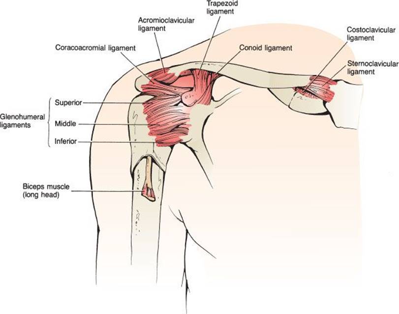
FIGURE 38.1 The ligamentous and bony anatomy of the shoulder.

eFIGURE 38.1 Clavicle fracture. The attachment of the sternocleidomastoid muscles pulls the medial segment superiorly.
Distal-third clavicle fractures account for 15% of all clavicle fractures. Nondisplaced fractures of the distal clavicle are splinted by the surrounding intact ligaments and muscles. They are usually treated symptomatically with ice, analgesics, a sling, and early motion. Displaced fractures of the distal clavicle may require surgical intervention because of rupture of the coracoclavicular ligament (3). Complications of displaced fractures treated conservatively include nonunion. Degenerative arthritis may occur when there is intra-articular involvement.
Medial-third clavicle fractures represent only 5% of all clavicle fractures. Considerable force is required to fracture the medial clavicle, so associated intrathoracic injuries should be considered. Management includes ice, analgesics, and a sling for support. Displaced fractures require orthopedic referral for reduction. Degenerative arthritis frequently complicates these fractures.
Proximal Humerus Fractures
Proximal humeral fractures account for approximately 2% to 3% of upper extremity fractures. They are most common in elderly osteoporotic women. Anatomically, proximal humeral fractures include all humeral fractures proximal to the surgical neck. Two mechanisms of injury are commonly responsible: (i) a direct blow on the lateral aspect of the arm and (ii) a fall on an outstretched arm. Patients present with pain and swelling over the upper arm and shoulder. Most commonly, the arm is held adducted.
The Neer classification system for proximal humerus fractures is widely used and has both prognostic and therapeutic implications. In this system, the proximal humerus is divided into four segments: greater tuberosity, lesser tuberosity, humeral head, and humeral shaft (4). Depending on the displacement and angulation of fractures of these four components, the injury is divided into one-, two-, three-, and four-part fractures (eFig. 38.2).

FIGURE 38.2 Anterior shoulder dislocation. A: A “rounded off” appearance of the affected shoulder is typical clinically. B: Characteristic radiographic appearance of a subcoracoid dislocation.

eFIGURE 38.2 Proximal humerus fracture. This image is of a surgical neck fracture with complete displacement. This is an example of a two-part fracture.
One-part fractures are nondisplaced (<1 cm) fractures without significant angulation (<45 degrees). These fractures are the most common type of proximal humerus fracture and represent more than 85% of cases. The humeral fragments are held in place by the periosteum, rotator cuff, and joint capsule. Therapy for one-part fractures includes immobilization in a sling and swathe and orthopedic referral within 4 to 5 days. Circumduction exercises should begin as soon as tolerated, and should be followed by passive exercise of the elbow and shoulder at 2 to 3 weeks. Shoulder motion exercises can usually be started within 3 to 4 weeks.
Two-part fractures have a fragment with more than 1 cm of displacement or over 45 degrees of angulation. These fractures require immobilization with a sling and swathe and orthopedic referral. Surgery is recommended for two-part fractures of the greater and lesser tuberosity, angulated surgical neck fractures, and in unstable surgical neck fractures in which there is disengagement of the shaft from the humeral head.
Three- and four-part fractures result from severe forces. Three-part fractures involve two fragments that are individually displaced from the humeral head. Four-part fractures involve three fragments individually displaced from the remaining humeral head. These fractures are associated with shoulder dislocations, rotator cuff injuries, and neurovascular compromise. Therapy includes sling immobilization and emergent orthopedic referral or admission. Virtually all of these fractures require surgical repair.
Scapular Fractures
Scapular fractures are rare and represent only 1% of all fractures and 5% of fractures involving the shoulder. Treatment, associated injuries, and complications depend on the portion of the scapula that is fractured.
Body or spine fractures are significant because a great deal of force is usually required to cause these fractures; therefore, they are sometimes associated with other more serious injuries to thoracic structures. Typically, there is little displacement, due to the support of the investing muscles and periosteum. Treatment includes sling and swathe immobilization, early limited exercise, and orthopedic referral.
Acromion fractures result from a direct downward blow to the shoulder. Due to the large amount of force required to produce these fractures, they are often accompanied by brachial plexus injuries, rotator cuff tears, and shoulder dislocations. Nondisplaced fractures are treated with sling immobilization in conjunction with an elastic circular dressing that pushes the elbow up and the lateral clavicle down. Displaced fractures require internal fixation.
Glenoid rim fractures are associated with two mechanisms of injury. A direct blow may result in a stellate fracture. An upward force, secondary to a fall on the flexed elbow, may cause a displaced glenoid rim fracture. Glenoid rim fractures also occur with shoulder dislocations and are referred to as Bankart lesions. Therapy for fractures with small fragments consists of sling immobilization, ice, and analgesia. Large or widely displaced fragments and stellate fractures require surgical fixation.
Coracoid process fractures occur from a direct blow to the superior part of the shoulder, or from violent contraction of one of the inserting muscles. The patient presents with tenderness to palpation over the coracoid process. Radiographs should include an axillary view for delineation of any displacement of the fragment. Coracoid fractures are usually treated symptomatically with a sling, ice, and analgesics.
Anterior Shoulder Dislocation
Anterior shoulder dislocation is one of the most common dislocations, accounting for half of all dislocations presenting to the ED. This injury occurs when a blow to an abducted, externally rotated arm disrupts the anterior capsule and the glenohumeral ligaments (5). Less commonly, a force to the posterior humerus or a fall on an outstretched arm is the cause.
The patient presents with the affected arm held to the side and resistant to all movements. The patient will be unable to touch the uninjured shoulder with the hand of the dislocated shoulder. In thin individuals, the acromion is prominent, and there is loss of the normal rounded contour of the shoulder (Fig. 38.2A). Upon palpation lateral to the acromion, there is a noticeable gap where the humeral head used to be positioned.
Axillary nerve injury is the most common associated neurologic injury in anterior shoulder dislocations. The axillary nerve is assessed by testing pinprick sensation over the lateral aspect of the deltoid in the “shoulder badge” distribution or deltoid muscle weakness. Unfortunately, sensory loss is not reliably present and an accurate motor examination is impractical in a patient with an acute dislocation. While nerve dysfunction is common, it is seen in up to 42% of patients, most patients recover without intervention.
On AP radiographs, the humeral head is in one of three locations: subclavicular, subcoracoid, or subglenoid (Fig. 38.2B). The physician should determine the position of the humeral head to ascertain if there are associated injuries. The typical position is subcoracoid. If the humeral head is in a subglenoid or subclavicular position, the patient has either a greater tuberosity fracture or a rotator cuff tear. The scapular Y view, true AP (Grashey view—beam directed at a 45-degree angle from a medial to lateral direction with the plate parallel to the scapular body), and axillary view are used in equivocal cases.
Clinically important fractures are seen in approximately 25% of cases and are more common in patients over age 40, first-time dislocations, and after a traumatic mechanism. In patients with none of these three factors present, a prereduction radiograph is unlikely to be beneficial when the diagnosis is obvious clinically (6,7). The most common associated fracture is an impaction fracture on the posterior lateral portion of the humeral head known as the Hill–Sachs deformity. It is present in 40% of dislocations. Other fractures include Bankart fractures of the anterior glenoid rim (5%) and greater tuberosity fractures (10%). A nondisplaced fracture of the surgical neck is a rare associated injury, but important to note, as attempts at closed reduction of the shoulder dislocation are contraindicated. Attempts at reduction may displace the fracture and worsen the prognosis.
Reduction should begin after obtaining informed consent. Procedural sedation is not necessary in all cases, especially in recurrent dislocators who present promptly. Patient preference should be considered. Reduction without procedural sedation is unlikely to be successful in a patient who is anxious, in pain, and unable to assist in the reduction by relaxing their musculature. Some have used intra-articular lidocaine with good results (8).
There are numerous reduction techniques described, but most techniques use or combine scapular movement, external rotation, shoulder abduction, and traction of the arm. The best technique is one that is quick, simple, associated with few complications, and requires the smallest amount of force. One method that meets these requirements is scapular manipulation with downward traction on the arm by an assistant. Success rates with this maneuver range from 80% to 100% (9–11). This technique is attempted without procedural sedation in most cases. If this is unsuccessful, the external rotation technique can be used, followed by traction–countertraction, if necessary. Other acceptable alternative techniques include the Milch, Stimson, Spaso, Fares, and Cunningham methods.
Scapular Manipulation Technique
The head of the stretcher is placed at 90 degrees. The patient sits upright with the unaffected shoulder leaning against the head of the stretcher and the legs hanging off the edge (no procedural sedation). The physician stands behind the patient and pushes the tip of the scapula medially and the superior aspect of the scapula laterally (11). This scapular rotation allows the humeral head to disengage from the glenoid. An assistant, standing in front of the patient, gently applies a downward force (Fig. 38.3A) (18).

FIGURE 38.3 Methods of reducing an anterior shoulder dislocation. A: Scapular manipulation with downward traction on the arm for anterior shoulder dislocation. B: External rotation technique for anterior shoulder dislocation. C:Traction–countertraction technique for reducing anterior shoulder dislocations.
External Rotation Technique
This technique is also simple and may permit the shoulder muscles to reduce the dislocation with minimal analgesia (13). The patient lies supine with the shoulder adducted and elbow flexed to 90 degrees. The patient is encouraged to allow their arm to fall to the side. One of the physician’s hands supports the elbow, and the other hand slowly guides the patient’s arm (Fig. 38.3B). When pain or spasm is experienced, movement is stopped and the muscles are allowed to relax. Over the course of 5 to 10 minutes, the arm is externally rotated to 90 degrees. The rotation must be performed gradually, and the patient must remain relaxed. After reaching 90 degrees, the shoulder reduces spontaneously with success rates ranging from 80% to 90% (14).
If the shoulder is not reduced, an additional maneuver (Milch technique) can be added. The externally rotated arm is slowly elevated above the patient’s head, and slight traction is applied. If the patient is unable to tolerate the external rotation technique because of pain and anxiety, procedural sedation will frequently allow the necessary muscle relaxation to reduce the shoulder.
Traction–Countertraction
With the patient lying in the supine position, an assistant applies countertraction with a folded sheet in the axilla while the examiner applies traction to the arm (Fig. 38.3C). Additional lateral traction may be added with a second sheet applied around the proximal portion of the humerus. To prevent avulsion injuries when using this maneuver, the patient must have good muscle relaxation.
Reduction in the ED is unsuccessful in 5% to 10% of cases. If the dislocation cannot be reduced, general anesthesia should be considered and reduction attempted in the operating room. Irreducible dislocations are usually due to soft tissue interposition.
Upon successful reduction, the arm is immobilized and the patient is sent for radiographs. Postreduction radiographs are not necessary if the prereduction radiographs did not reveal a fracture, the reduction was performed smoothly, and the postreduction physical examination is not suspicious for an unsuccessful reduction.
There is controversy regarding whether the shoulder should be immobilized in internal (sling and swathe or commercially available shoulder immobilizer) or external rotation (15–17). Immobilization in external rotation has been postulated to put the detached labrum in a better position to heal and consequently reduce the rate of recurrent dislocation. Study results are mixed, but immobilization of these patients in internal rotation is recommended until more conclusive evidence becomes available.
The patient is referred to an orthopedic surgeon within 1 week. The older the patient, the earlier mobilization should be instituted to avoid stiffness. For patients younger than 30 years of age, the shoulder is immobilized for 3 weeks, followed by gentle active range-of-motion exercises. The patient should avoid abduction and external rotation for an additional 3 weeks. For patients older than age 30, the shoulder is immobilized for 1 week, followed by active range-of-motion exercises, avoiding abduction and external rotation.
Complications of anterior shoulder dislocations include tears of the rotator cuff, avulsion of the greater tuberosity, axillary nerve injury, and fracture of the humeral head. The most common complication is recurrent dislocation due to labral and ligamentous injury. This complication occurs in 50% to 90% of patients younger than age 20. Prevention of recurrent dislocation is achieved by internal rotation exercises that strengthen the subscapularis muscle. Operative repair is indicated in patients with recurrent dislocations and occasionally in young, first-time dislocators engaged in highly demanding physical activities.
Posterior Shoulder Dislocation
Posterior dislocations are less common and more subtle on examination and radiographs than their anterior counterpart, making delayed and misdiagnosis more likely. The common mechanisms involved in this injury include violent internal rotational force occurring during a fall on the forward-flexed, internally rotated arm or a violent muscle contraction following a generalized tonic–clonic seizure or electrocution.
On physical examination, the arm is held in adduction and internal rotation, with abduction severely limited. External rotation of the shoulder is blocked, a critical feature of the examination that should raise clinical suspicion. Additional physical examination findings include prominence in the posterior aspect of the shoulder accompanied by flattening of the normal anterior shoulder contour. Neurovascular compromise is uncommon.
Evidence of a posterior shoulder dislocation on a standard AP view is subtle and may be undiagnosed in 50% of cases (18). Some clues to the radiographic diagnosis include the light bulb, trough line, and rim signs (Fig. 38.4A). The light bulb sign refers to the circular appearance of the humeral head caused by internal rotation that changes the projection of the tuberosities. The trough line sign is a linear appearance within the humeral head that represents the “trough” of the reverse Hill–Sachs impaction fracture. The rim sign is present when the distance between the medial aspect of the humeral head and the anterior glenoid rim is abnormal, usually with additional overlap between the two. Whenever there is a question of a posterior shoulder dislocation on AP views, an axillary view should be obtained (Fig. 38.4B). Isolated fractures of the lesser tuberosity are commonly associated.

FIGURE 38.4 Posterior shoulder dislocation. A: AP view. Notice there is additional overlap of the glenoid and humeral head (rim sign), a rounded appearance of the humeral head (light bulb sign), and a linear opacity medial within the humeral head (trough line sign). B: Axillary view.
Consultation with an orthopedic surgeon is recommended for posterior shoulder dislocations. Closed reduction is attempted only if the anterior surface defect (“reverse Hill–Sachs deformity”) is <20% of the total humeral head surface and the duration of dislocation is <3 weeks (19). Posterior shoulder dislocations may be reduced by hanging the arm with weights (Stimson method), or other techniques that push the posteriorly displaced humeral head forward. If there is severe pain or muscle spasm, general anesthesia may be necessary. Surgical intervention is needed when the articular surface defect is >20% of its surface area, when the dislocation is irreducible using closed techniques, or when a lesser tuberosity fracture remains displaced after joint reduction.
Inferior Shoulder Dislocation (Luxatio Erecta)
Inferior dislocations of the shoulder are uncommon, representing approximately 0.5% of all shoulder dislocations. The mechanism of injury is forceful hyperabduction. Because of its dramatic presentation, this dislocation is rarely missed.
On physical examination, the patient is in severe pain and holds the arm upright, as if asking a question. The humeral head can be palpated along the lateral chest wall. Compression of the axillary artery and brachial plexus may occur as the humeral head tears through the inferior capsule. Vascular injury occurs in approximately 3% of patients, but it is more common with inferior dislocations than with any other form of shoulder dislocation (11,20).
Radiographically, the humeral head is seen beneath the coracoid or glenoid with the humerus abducted. Associated fractures of the acromion, inferior glenoid rim, and greater tuberosity may be present (20).
To reduce the dislocation, the physician applies traction in the longitudinal axis of the humerus while an assistant applies countertraction with a folded sheet wrapped around the supraclavicular region. With traction maintained, the arm is rotated inferiorly in an arc toward a position of complete adduction. Closed reduction is successful in most cases. After reduction, the shoulder is immobilized for 2 to 4 weeks. Rotator cuff tears are frequently present. Occasionally, general anesthesia may be required to reduce the dislocation and repair the torn rotator cuff.
AC Joint Injury
The AC joint functions to elevate and abduct the arm. Stability is provided by two ligaments—the AC and the coracoclavicular. Injury to these ligaments, commonly referred to as a “shoulder separation,” accounts for 10% of all dislocations. Most injuries of the AC joint are caused by a direct force applied to the point of the shoulder. The therapy for AC joint injuries depends on the amount of ligamentous injury—first-, second-, or third-degree injuries (21).
First-degree injuries are a simple sprain of the AC ligament and involve an incomplete tear of that structure. The patient complains of pain and has tenderness over the AC joint. Swelling is minimal, no subluxation is present, and radiographs are normal. Therapy includes rest, ice, and a sling, with early range of motion.
Second-degree injuries involve disruption of the AC ligament, with sprain of the coracoclavicular ligament. There is a partial subluxation of the distal end of the clavicle from the acromion. The patient experiences tenderness to mild palpation and moderate swelling is present. Radiographic findings are subtle and include an increased width of the AC joint of 3 mm or more or greater than a 50% increase compared to the opposite side. The distal clavicle is elevated no more than half its diameter from the acromion. Stress films, previously recommended to help distinguish between second- and third-degree injuries, are no longer routinely recommended because they do not affect treatment decisions (22). Second-degree injuries are treated conservatively with ice, rest, and immobilization in a sling. Commercially available clavicle braces (Kenny-Howard sling) may be used but have not been shown to produce superior long-term function over a sling.
Third-degree injuries result in complete dislocation at the AC joint, with disruption of both the AC and coracoclavicular ligaments. The diagnosis of an AC joint injury is made by examining the patient in the upright position and looking for a deformity at the tip of the shoulder (eFig. 38.3A). On radiographs, the inferior border of the distal clavicle is above the midpoint of the acromion (eFig. 38.3B). Treatment is controversial. In the past, third-degree injuries were treated surgically in young patients. However, conservative treatment has been shown to be equivalent to surgery. Patients with these injuries should be immobilized in a sling and referred to an orthopedist within 1 week (23,24).

eFIGURE 38.3 Acromioclavicular separation—Third degree. A: Clinical picture. B: Radiograph of the same injury.
SC Joint Injuries
The SC joint is stabilized by the SC ligament and the costoclavicular ligament. The most common mechanism of injury to this joint is a force that thrusts the shoulder forward. In the absence of trauma, an infectious process within the joint should be considered. Traumatic injuries are divided into mild and moderate sprains and SC joint dislocation.
Mild SC joint sprain involves incomplete tears of the SC and costoclavicular ligaments. The patient experiences minimal swelling and has tenderness at the joint. Therapy includes ice, analgesics, and a sling for 3 to 4 days.
Moderate SC joint sprain involves subluxation of the clavicle from its manubrial attachment. Complete rupture of the SC ligament and partial rupture of the costoclavicular ligament occur. There is pain on abduction of the arm, and swelling is noted over the joint. Treatment includes 6 weeks of immobilization with a figure-of-eight clavicle strap and a sling to permit ligamentous healing.
SC joint dislocation involves complete rupture of the SC and costoclavicular ligaments, permitting the clavicle to dislocate from its manubrial attachment. Anterior dislocations are much more common, but posterior dislocations have the potential to result in much more serious injuries. Patients experience severe pain, which is increased by any motion of the shoulder. Posterior dislocations may constitute a true emergency because respiratory embarrassment can occur secondary to tracheal compression, pneumothorax, or venous congestion. These associated injuries may necessitate emergency reduction by the emergency physician, whereas reduction in the operating room is recommended in all other cases (1). Reduction is accomplished by placing a folded sheet between the shoulders, with the patient supine. This serves to separate the clavicle from the manubrium. The arm is abducted and traction is applied. For posterior dislocations, following the instillation of local anesthesia, the clavicle should be grasped near its medial border with a towel clip and pulled out of its posterior position. Unstable SC joint dislocations may require surgical intervention (3).
Complications of anterior SC dislocation are cosmetic, with chronic swelling around the joint. Although it has been reported that up to 25% of posterior dislocations are associated with complications—including pneumothorax, laceration of the superior vena cava, occlusion of the subclavian artery or vein, and rupture or compression of the trachea—this may be an overestimate (1).
Rotator Cuff Injury
Tears of the rotator cuff are more common in the elderly and may occur after minimal trauma. The mechanism of injury following an acute injury, which is more common in younger patients, is typically a sudden forceful elevation of the arm against resistance. The most common area of cuff injury is at the anterosuperior portion, at the attachment of the supraspinatus muscle. The tendon at this point is worn down by the compressive forces between the humeral head and the coracoacromial arch.
Patients complain of pain that radiates to the anterior aspect of the arm and is aggravated by activity. Abduction and external rotation of the arm are painful and weak. Tenderness is present over the insertion at the greater tuberosity. Thirty percent or more of the tendon must be ruptured to produce a significant reduction of strength. The extent of the tear is directly related to the limitation of shoulder abduction.
The drop arm test is positive in patients with significant tears. This is performed by horizontally elevating the arm to the 90-degree position and asking the patient to continue to hold the arm in this position. Slight pressure on the distal forearm will cause the patient to drop the arm suddenly. In addition, the patient cannot bring the arm from the abducted position to the side in a slow fashion, but drops it precipitously.
Emergency treatment includes ice, immobilization, and orthopedic referral within 4 to 5 days. Passive range-of-motion exercises should be instituted as soon as possible in elderly patients. Magnetic resonance imaging is diagnostic of full-thickness tears with 100% sensitivity, but is not indicated in the ED (25). Conservative therapy results in a good outcome in 50% of patients. In the young, early surgical repair is indicated for complete tears of the rotator cuff. Anterior tears have the best outcome, while patients with posterior tears are poor candidates for arthroscopy (26). In the elderly patient with a more sedentary life, repair may not be indicated.
CRITICAL INTERVENTIONS
• Evaluate patients with scapular fractures for associated thoracic injuries, with careful physical examination, radiographs, and further testing when indicated.
• In patients who have had a grand mal seizure, check external rotation of both shoulders to evaluate for posterior shoulder dislocation.
• Use the towel clip method to reduce posterior SC dislocations when they are associated with respiratory distress due to tracheal compression.
Common Pitfalls
• Failure to obtain an axillary view when assessing the shoulder for dislocation.
• Failure to remember that lesser tuberosity fractures are often associated with posterior shoulder dislocations.
• Attempting to reduce an anterior shoulder dislocation in a patient with a concomitant surgical neck fracture.
REFERENCES
1. Beecroft M, Sherman SC. Posterior displacement of a proximal epiphyseal clavicle fracture. J Emerg Med. 2007;33(3):245–248.
2. Haapamaki VV, Kiuru MJ, Koskinen SK. Multidetector CT in shoulder fractures. Emerg Radiol. 2004;11(2):89–94.
3. Anderson K. Evaluation and treatment of distal clavicle fractures. Clin Sports Med. 2003;22(2):319–326, vii.
4. Neer CS 2nd. Displaced proximal humeral fractures. I. Classification and evaluation. J Bone Joint Surg Am. 1970;52(6):1077–1089.
5. Flatow EL, Cuomo F, Maday MG, et al. Open reduction and internal fixation of two-part displaced fractures of the greater tuberosity of the proximal part of the humerus. J Bone Joint Surg Am.1991;73(8):1213–1218.
6. Hendey GW. Necessity of radiographs in the emergency department management of shoulder dislocations. Ann Emerg Med. 2000;36(2):108–113.
7. Hendey GW, Chally MK, Stewart VB. Selective radiography in 100 patients with suspected shoulder dislocation. J Emerg Med. 2006;31(1):23–28.
8. Miller SL, Cleeman E, Auerbach J, et al. Comparison of intra-articular lidocaine and intravenous sedation for reduction of shoulder dislocations: A randomized, prospective study. J Bone Joint Surg Am.2002;84-A(12):2135–2139.
9. Anderson D, Zvirbulis R, Ciullo J. Scapular manipulation for reduction of anterior shoulder dislocations. Clin Orthop Relat Res. 1982;(164):181–183.
10. Kothari RU, Dronen SC. Prospective evaluation of the scapular manipulation technique in reducing anterior shoulder dislocations. Ann Emerg Med. 1992;21(11):1349–1352.
11. McNamara RM. Reduction of anterior shoulder dislocations by scapular manipulation. Ann Emerg Med. 1993;22(7):1140–1144.
12. Sagarin MJ. Best of both (BOB) maneuver for rapid reduction of anterior shoulder dislocation. J Emerg Med. 2005;29(3):313–316.
13. Leidelmeyer R. Reduced! A shoulder, subtly and painlessly. Emerg Med. 1977; 9:233–234.
14. Eachempati KK, Dua A, Malhotra R, et al. The external rotation method for reduction of acute anterior dislocations and fracture-dislocations of the shoulder. J Bone Joint Surg Am. 2004;86-A(11):2431–2434.
15. Iannotti JP, Zlatkin MB, Esterhai JL, et al. Magnetic resonance imaging of the shoulder. Sensitivity, specificity, and predictive value. J Bone Joint Surg Am. 1991; 73(1):17–29.
16. Miller BS, Sonnabend DH, Hatrick C, et al. Should acute anterior dislocations of the shoulder be immobilized in external rotation? A cadaveric study. J Shoulder Elbow Surg. 2004;13(6):589–592.
17. Murrell GA. Treatment of shoulder dislocation: Is a sling appropriate? Med J Aust. 2003;179(7):370–371.
18. Gor DM. The trough line sign. Radiology. 2002;224(2):485–486.
19. Cicak N. Posterior dislocation of the shoulder. J Bone Joint Surg Br. 2004; 86(3):324–332.
20. Mallon WJ, Bassett FH 3rd, Goldner RD. Luxatio erecta: The inferior glenohumeral dislocation. J Orthop Trauma. 1990;4(1):19–24.
21. Bannister GC, Wallace WA, Stableforth PG, et al. The management of acute acromioclavicular dislocation. A randomised prospective controlled trial. J Bone Joint Surg Br. 1989;71(5):848–850.
22. Bossart PJ, Joyce SM, Manaster BJ, et al. Lack of efficacy of “weighted” radiographs in diagnosing acute acromioclavicular separation. Ann Emerg Med. 1988; 17(1):20–24.
23. Bradley JP, Elkousy H. Decision making: Operative versus nonoperative treatment of acromioclavicular joint injuries. Clin Sports Med. 2003;22(2):277–290.
24. Kwon YW, Iannotti JP. Operative treatment of acromioclavicular joint injuries and results. Clin Sports Med. 2003;22(2):291–300, vi.
25. Itoi E, Hatakeyama Y, Kido T, et al. A new method of immobilization after traumatic anterior dislocation of the shoulder: A preliminary study. J Shoulder Elbow Surg. 2003;12(5):413–415.
26. Lyons AR, Tomlinson JE. Clinical diagnosis of tears of the rotator cuff. J Bone Joint Surg Br. 1992;74(3):414–415.