
Andrew D. Perron and Carl A. Germann
Musculoskeletal injury has been cited as the second most common reason for emergency department (ED) visits in the United States (1). In addition, missed fracture has been cited as a common reason for medical malpractice lawsuits against emergency physicians (2). A coherent and systematic approach to the assessment and management of patients with orthopedic injuries and other musculoskeletal complaints is essential (3). Musculoskeletal injuries range from minor (tendinosis, sprains, strains, and contusions) to potentially life- and limb-threatening trauma (pelvis fracture, femur fracture, knee dislocation). Most of the time, acute trauma is responsible for the presenting complaints, and usually the primary impairment is an inability to move a part of the body owing to pain or discomfort. The mechanisms of trauma include sports injuries, assaults, motor vehicle collisions, falls in the home, and industrial catastrophes. When acute trauma is absent, overuse syndromes or repetitive movements are often the culprits.
The immediate tasks of the emergency physician evaluating a patient with a musculoskeletal complaint include the following:
• Accurately determining those injuries that need only symptomatic care and distinguishing them from ones that require more complex, timely specialist intervention.
• Differentiating injuries that can be handled solely by an emergency physician (the vast majority of musculoskeletal injuries) from those that require emergent consultation or timely referral to an orthopedic surgeon (e.g., open fractures, compartment syndrome, growth plate injuries, and flexor tendon injuries).
• Formulating a logical, cost-effective algorithm for the diagnosis and management of each patient.
• Realizing the limitations of diagnostic modalities and the potential for “missed” injuries.
• Aggressively managing the pain associated with these injuries early and effectively, recognizing that “a mere sprain or contusion” can be the source of exquisite pain. Also paying particular attention to analgesia in those patients at the extremes of age (very young and very old), where it has been shown that oligoanalgesia is the rule, rather than the exception.
• Recognizing that care for many musculoskeletal injuries is not complete when emergency care has been delivered. Successful management of the patient is complete only when appropriate follow-up has been arranged, as a large percentage of acute musculoskeletal injuries require reevaluation and ongoing care (e.g., physical therapy and rehabilitation) for optimal recovery.
CLINICAL PRESENTATION
Sprains
A sprain is a ligamentous injury. Ligaments link bone to bone. Damage to the fibers of the ligament ranges from microscopic damage to complete disruption. A sprain can result from an abnormal (nonbiomechanical) force vector applied to a joint, an excessive force applied along a normal vector, or a combination of the two mechanisms. It is possible to sustain a sprain that is significantly more severe than an analogous fracture. For example, a complete lateral ankle ligament rupture and a transverse avulsion fracture of the distal fibula cause the same pattern of instability. However, because bone regenerates without scarring, the outcome for the latter injury can be significantly better.
The grading of sprains is based on the severity of the damage, recognizing that with an acute injury there may be too much pain and spasm to accurately determine the grade of the sprain.
Grade I Sprain
A grade I sprain (in older terminology, “first-degree”) represents microscopic tears of the ligament, which lead to localized hemorrhage and inflammation as the healing process evolves. Active range of motion is usually preserved but painful. Abnormal movement of the joint stabilized by the sprained ligament is not elicited by stress maneuvers, but these maneuvers may produce focal pain, and the area overlying the sprained ligament is tender to palpation.
Grade II Sprain
In a grade II sprain (“second-degree”) or partial tear, the overall macroarchitecture of the ligament is preserved, with the majority of fibers still in continuity, but clearly some of them have been completely ruptured. Hemorrhage is more extensive, and swelling is often pronounced. Loss of function secondary to decreased and painful range of motion is the hallmark. When the joint that the ligament stabilizes is stressed, there may be some gapping of the joint or abnormal motion. However, with a grade II sprain, any abnormal laxity is limited, and frank instability does not occur.
Grade III Sprain
A grade III sprain (“third-degree”) denotes a complete tear of the ligament and is synonymous with a complete rupture. Hemorrhage is more extensive, and healing involves the formation of scar tissue in the area between the torn fragments of the ligament. Stressing the joint reveals greater gapping than in the case of a grade II sprain, and frank instability without a well-defined end point may be elicited. This frank instability can be summed up by the examiner’s thought, “I should stop stressing this joint, as it isn’t going to resist my efforts.” This is tempered by the knowledge that with acute injury, the physical examination may be severely limited owing to acute pain and muscular spasm.
Common ligament sprains seen in the ED include lateral ankle sprains, injuries to the anterior cruciate and collateral ligaments of the knee, and ligamentous injuries of the shoulder, associated with either glenohumeral dislocations or acromioclavicular separations.
The clinical signs of a sprain typically include point tenderness to palpation, swelling, painful or decreased range of motion, and ecchymosis. With a complete (grade III) sprain, with no fibers in continuity, the joint may be completely pain free when stressed. Often, athletes will continue to play after a grade III sprain of the medial collateral ligament of the knee, but it is rare that they continue activities after a grade II sprain. In fact, pain may be inversely proportional to the severity of the injury. A grade I sprain is extremely painful when stressed, as there are many intact but damaged and inflamed fibers that will be stretched, triggering the discharge of pain fibers as the ligament is loaded with force. With a grade III sprain, the same maneuver does not cause any stretching of the ligament, as it cannot be loaded by force, and the only pain fibers triggered by the stress maneuver will lie in surrounding soft tissue. Bone can be injured at the same time as a ligament is damaged. For example, grade III sprains of the medial, deltoid ligament of the ankle are often accompanied by fibula fractures. Bony evidence of sprains on x-ray may include small avulsion fractures at the point of insertion of the ligament into the bone, impaction injuries, or joint space widening.
Strains
Tendons connect muscles to bone. A strain is an injury to this musculotendinous unit, which can range from mild “pulled muscles” to complete tendon ruptures. The mechanisms of injury are twofold: (a) Stretching a muscle beyond its normal range of motion by forced extension (“overstretching injury”) and (b) tearing of muscle or tendon fibers by a violent, uncontrolled contraction or repetitive, prolonged contractions, often in attempted compensation for an unexpected change in direction of movement, unanticipated variations in terrain, or a sudden force applied to the antagonist muscle group.
Strains are graded from first-degree to third-degree, reflecting the severity of damage:
• In a first-degree strain, microscopic damage occurs to the musculotendinous unit, with associated localized hemorrhage, inflammation, and tenderness to palpation. Active range of motion is often unrestricted, but pain can be elicited by resisted contraction or forced extension of the muscle group.
• A second-degree strain involves more severe damage, with rupture of some fibers of the muscle or tendon. Hemorrhage is usually visible, swelling more severe, and range of motion limited by pain.
• In a third-degree strain, the muscle or tendon is completely ruptured. The rupture can occur in the muscle belly, at the musculotendinous junction, within the tendon itself, or at the point of insertion of the tendon into bone.
Common strains seen in the ED include those to the quadriceps, hamstring, rotator cuff, thigh adductor (groin), and Achilles tendons.
The clinical signs of a strain include pain, ecchymosis, edema, and decreased or painful range of motion. With complete disruption of the muscle or tendon, a palpable defect is often present. Physical examination should include inspection of the injured area, observation of active and passive range of motion, palpation of the affected area, and provocative maneuvers (either resisted contraction or passive stretching) to elicit the location and extent of injury. In addition, with certain suspected injuries, specific tests should be performed. For example, if an Achilles tendon rupture is suspected, then a Thompson test is performed. While the patient lies prone with the knee flexed at 90 degrees, the examiner squeezes the calf on the affected side and observes whether the foot plantar flexes. The presence of plantar flexion indicates an Achilles tendon that is at least partially intact. Absent plantar flexion often represents complete Achilles tendon rupture.
Contusions
A contusion is a bruise caused by a direct blow to a body part. Contusions can occur to bone, with significant pain from periosteal bruising and bone edema, or, more commonly, to soft tissue, especially fat and muscle. In these crush injuries, the degree of injury is proportional to the force of the blow and the surface area of impact. Tissue damage occurs as the energy transmitted by the impact dissipates into or is absorbed by surrounding structures.
The diagnosis of contusion is one of exclusion, with fracture, dislocation, sprain, and strain being the most important diagnoses that must be ruled out. Common contusions seen in the ED include bruised quadriceps muscles, rib cage contusions, contusions of the ulnar aspect of the forearm in “defensive maneuvers” during an assault, and direct blows to the anterior tibia.
Either the blow itself or the body’s reaction to it causes signs and symptoms that prompt the patient to seek emergency care. Typically, they include pain, swelling, ecchymosis, and decreased range of motion of the affected part.
Usually, the entities that are ruled out (e.g., fracture, dislocation, sprain, and strain) are potentially more serious than the diagnosis of contusion itself. However, the clinician must remember that contusions can lead to serious complications, including severe pain, rhabdomyolysis, compartment syndrome, and myositis ossificans.
One of the most common reasons that patients seek reevaluation in an emergency room is inadequate analgesia following a soft tissue injury. Initial attention to adequate analgesia ensures patient comfort and decreases the need for reevaluation. A number of studies have confirmed that physicians notoriously undertreat painful conditions (4–6). This is particularly true in both the pediatric and geriatric patient populations.
Severe muscle breakdown can occur after strenuous exercise without an acute, isolated injury or after a significant contusion to a muscle body. Rhabdomyolysis results from the destruction of myocyte cell membrane integrity. The subsequent release of intracellular contents, including potassium, myoglobin, uric acid, phosphate, and the enzyme creatine kinase, into the bloodstream can result in a number of systemic complications, including metabolic abnormalities and acute renal failure. The emergency physician must assess the extent of tissue involvement, the degree of tissue destruction, and the potential for systemic complications. If rhabdomyolysis is suspected, urinalysis should be performed to look for evidence of myoglobinuria (dipstick positive for blood without microscopic evidence of erythrocytes), and the serum creatine kinase level should be checked (see Chapter 213, “Rhabdomyolysis” for details).
Compartment syndrome occurs when pressure within soft tissues in a fixed body compartment increases to the point at which it compromises venous blood flow and limits capillary perfusion. This leads to muscle ischemia and necrosis. Most commonly associated with long bone fractures, it can occur in the setting of severe contusion, crush injury, or excessive exertion and must be considered in any patient with pain out of proportion to physical findings or with swelling causing obvious tautness in a nonexpansible compartment, such as the anterior tibial compartment. When this diagnosis is considered, it must be objectively ruled out by manometry (see Chapter 48, “Acute Compartment Syndrome,” for details).
Severe hemorrhage into a muscle can initiate a cascade of “healing,” which can lead to the formation of ectopic bone within the muscle, or myositis ossificans. A large number of these cases have occurred in athletes treated with early, aggressive nonsteroidal anti-inflammatory drugs (NSAIDs), heat, massage, and physical therapy in the hope of facilitating early return to athletic competition.
This is of concern to the emergency physician for a number of reasons. If severe quadriceps tear and resultant hemorrhage and ecchymosis are suspected, the initial management should include ice, a brief period (24 hours) of immobilization in a stretched position (knee flexed to 120 degrees for severe quadriceps contusion), and acetaminophen, instead of NSAIDs, for pain control (7). This regimen limits the ongoing bleeding and inflammation in the area. These patients must be referred for appropriate long-term follow-up care. In addition, a decreased range of motion, ongoing pain, and a mass or firmness in soft tissue weeks to months after sustaining a contusion should suggest the possibility of myositis ossificans, as noted above. A soft tissue x-ray is the best way to evaluate for heterotopic bony formation. If heterotopic bone is present, the patient should be referred to an orthopedic surgeon for close follow-up care and potential resection of the bone when it has matured (usually at 9 to 12 months).
Fractures
Most fractures require urgent medical attention. Without appropriate emergency care, many will not heal properly or healing will be accompanied by unnecessary complications. A fracture is defined as a partial or complete break in a bone. Bone is unique in that it is the only tissue in the human body, other than the liver, that heals by regeneration, not by scarring (8–10). For regeneration to occur, however, the bone must be immobilized to allow uninterrupted formation of new bone. Ensuring appropriate immobilization is one of the fundamental tasks of the emergency physician when dealing with musculoskeletal problems.
The process of new bone formation begins with hematoma bridging the realigned fracture fragments. Hematopoietic cells in the hematoma secrete growth factors, which stimulate the formation of granulation tissue at the fracture ends, resulting in slow resorption of the hematoma. Over the next several weeks, a primary callus is formed, beginning as a soft callus and progressing to a hard callus. The final phase of healing is remodeling, during which the bone reassumes its original architecture as it is exposed to the normal stresses of weight bearing and movement (9,10).
Fractures also require immediate attention for several other reasons. Virtually all fractures are painful. The vast majority of fractures result from high-energy trauma (relative to sprains, strains, and contusions) and, because of force vectors, are accompanied by well-known associated injuries (e.g., 15% of calcaneal fractures sustained in a fall from height have associated lumbar vertebral fractures). In addition, many fractures place the patient at risk for specific complications (e.g., compartment syndrome in long bone fractures, blood loss in pelvic fractures, avascular necrosis [AVN] in scaphoid fractures of the hand, fat embolism with femur fracture).
A working knowledge of the nomenclature of fractures is essential to the successful management of fractures in the ED and to ensure clear communication with radiologists and consulting orthopedic surgeons. Adequate description of a fracture includes the bone(s) involved, whether it is open or closed, the location and direction of the fracture within the bone, the number of fragments, the alignment of the fracture fragments, the displacement of the fragments, and notation of associated complications (e.g., compartment syndrome, neurologic or vascular deficits).
The description of the injury should include the side of the body, the specific bone, and the location within the bone. By convention, long bones are divided into proximal, middle, and distal thirds, and these divisions are used to describe the fracture location. Alternatively, long bone injuries can be described as diaphyseal (shaft), metaphyseal (flared ends), or epiphyseal (rounded ends). If the fracture lies at the junction of any two-thirds of the bone, it should be described as such. Fractures in children deserve special mention. In this group, fractures occur frequently through the physis (growth plate), as this is the weakest point in the bone or investing soft tissue. Fractures to the physis can disrupt future bone growth if not recognized and adequately treated. These injuries are described with the Salter–Harris classification system (Fig. 37.1).
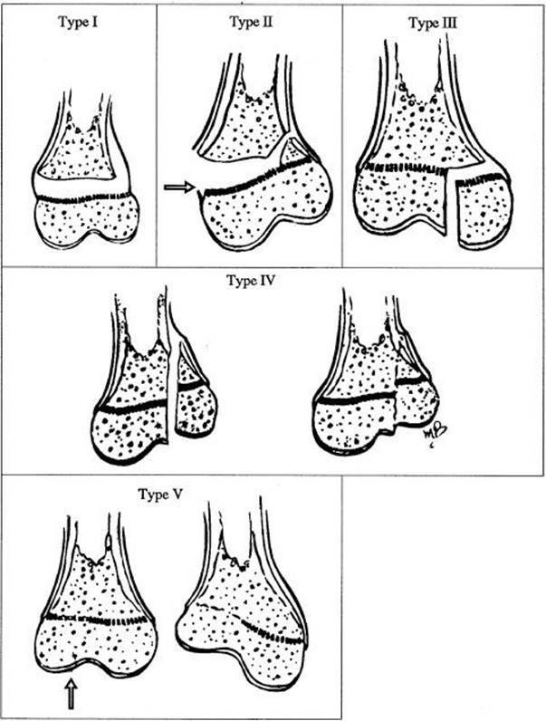
FIGURE 37.1 Salter–Harris fracture classification. (From Rang M. The growth plate and its diseases. In: Rockwood CA Jr, Wilkins KE, King RE, eds. Fractures in Children. 3rd ed. Philadelphia, PA: Lippincott-Raven Publishers; 1991:128, with permission.)
Closed Versus Open Fractures
The old terminology of simple (closed) and compound (open) fractures should be abandoned, as “simple” linear fractures may be open, and comminuted fractures may be closed. In a closed fracture, the skin and soft tissue overlying the fracture have not been violated. Therefore, no communication exists between the outside environment and the fracture fragments. Any break in the skin overlying a fracture must be considered an open fracture until proven otherwise. It is frequently difficult to tell whether there has been actual communication between the bone and the environment or whether the patient has simply sustained an abrasion to the skin overlying the fracture.
Open fractures are traditionally classified into three types based on the size of the wound and the amount of soft tissue injury. Type I wounds are <1 cm long, the energy dissipated by the mechanism of injury is low, and there is minimal soft tissue trauma. Type II wounds are <10 cm long, the mechanism of injury has dissipated a low or moderate amount of energy, and there is moderate soft tissue trauma but no gross contamination of the wound, major crush injury, or soft tissue defects or flaps. Type III wounds are >10 cm long. These include high-velocity gunshot wounds, close-range shotgun wounds, open fractures with major vascular injuries, fractures more than 8 hours old, or open fractures sustained in environments where exposure to fecal contents is a concern (e.g., open pelvic fractures with perineal exposure), or extensive soft tissue or periosteal stripping of bone, or evidence of gross contamination or significant crush injury (11).
Open fractures are orthopedic emergencies. These injuries frequently require immediate control of hemorrhage, which can usually be accomplished with appropriate splinting. In addition, open fractures may require emergent reduction if neurovascular compromise exists. Principles of care include irrigation, early administration of sufficient analgesia and appropriate antibiotics, tetanus prophylaxis, and emergent consultation with orthopedic surgery for definitive irrigation, debridement, reduction, and fracture repair in the operating room. Appropriate early intervention is the best way to prevent future complications, as the incidence of infection following open fracture is directly proportional to the time from injury to definitive irrigation and debridement.
Direction and Description of Fractures
The direction of a fracture refers to the configuration of the fracture—the way the fracture disrupts the bone. By convention, this descriptive modifier communicates the direction of the fracture in relation to the long axis of the involved bone (Fig. 37.2). A transverse fracture disrupts the bone perpendicular to its long axis; an oblique fracture lies at an angle to the long axis of the bone; a spiral fracture results from a rotational force and spirals around the center of the long axis of the bone. Given the relative flexibility of their bones, children may present with a greenstick fracture, or a partial fracture of the cortex with deformity of the intact bone.
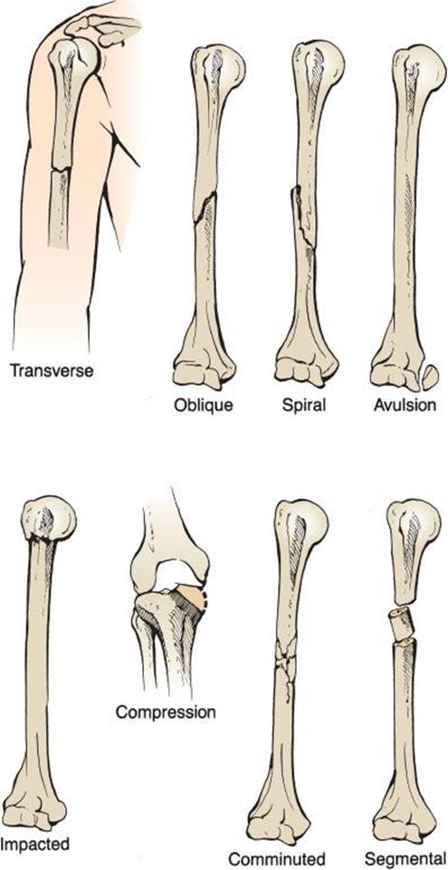
FIGURE 37.2 Fracture configuration.
Additional terms are used to describe the configuration of a fracture. A comminuted fracture has three or more fragments. Segmental refers to the presence of two or more fractures in a bone, creating at least one segment of bone that is not connected to either end of the bone. An avulsion fracture occurs when a piece of bone is pulled off at the site of insertion of a tendon or ligament. Avulsion fractures are usually the result of traction injuries. Their presence indicates potential instability at the joint, which is caused by the loss of stabilization normally provided by the avulsed ligament or tendon. An impacted fracture occurs when the fractured surfaces of the bone are forced into each other, foreshortening the bone. On computed tomography (CT) scan, impacted fractures are often comminuted with numerous small fragments wedging across the main fracture line. Compression refers to a fracture in which the trabeculae of the bone are compressed, decreasing the overall volume of the affected part of the bone.
Fracture Alignment
A fracture is also described in terms of its alignment—the relationship of the fracture fragments to each other. The fracture may be displaced, angulated, or rotated.
A fracture is displaced if the fragments are offset in a transverse or longitudinal plane. The position of the distal fragment in relation to the proximal fragment is described. The distal fragment may be displaced in an anterior, posterior, medial, lateral, radial, ulnar, proximal, or distal direction. The amount of displacement is commonly measured in terms of the width of the fractured shaft, using fractions of the width in the description.
A fracture is angulated if there is a deviation from the normal relationship of the bony fragments along the longitudinal axis of the bone (eFig. 37.1). The direction of angulation may be expressed in two ways. It may be described in terms of the direction of the apex of the angle formed at the fracture site by the two main fracture fragments. The apex may point in an anterior, posterior, lateral, or medial direction. The direction of the apex may also be described by other adjectives, including ulnar, radial, dorsal, or volar, depending on the involved bones. A second way to describe the angulation of the fracture is in terms of the relation of the distal fragment to the proximal fragment. If the distal fragment is angled away from the midline of the body, there is valgus angulation (the fracture is in valgus). If the distal fragment angles toward the midline, there is varus angulation to the distal fragment (the fracture is in varus).

eFIGURE 37.1 Fracture alignment.
Rotation refers to twisting of the distal fragment in relation to the proximal fragment around the center of the long axis of the fractured bone. When viewed along the long axis of the bone from the most distal point on the bone, if the distal fracture fragment has rotated toward the midline in relation to the proximal fragment, it is medially rotated; if it has rotated away from the midline, it is laterally rotated.
Other Descriptive Modifiers
An incomplete fracture involves only one cortex of the bone; a complete fracture disrupts both cortices. A pathologic fracture occurs in an area of abnormal bone, weakened by disease or prior trauma, and is often the result of apparently trivial trauma or a biomechanically routine force applied along a natural vector. Pathologic fractures may occur at sites of old fractures, through primary osteosarcomas, at sites of tumor metastases, near cysts, and in bone weakened by Paget disease, rickets, osteomalacia, tuberculosis, osteomyelitis, chronic steroid therapy, obesity, anorexia, and many other disease processes. A stressfracture is the result of the bony cortex becoming fatigued by repeated low-level loading rather than disrupted by one acute episode of trauma. The repeated stress to the cortex causes bone resorption and may result in overt fracture if adequate healing is not allowed. Clinically, patients have pain with activity and tenderness to palpation over the affected bone. Eating disorders and amenorrhea predispose to stress fractures in female athletes. Stress fractures are most common in the proximal tibia but have also been described in the spine, pelvis, femur, ribs, tarsal navicular, and metatarsal bones, among other sites (12).
COMPLICATIONS OF FRACTURES
No assessment and description of a fracture is complete without a thorough assessment for, and identification of, possible associated complications (eTable 37.1). Well-recognized complications of fractures are suggested by the site of the fracture and the mechanism of injury.
TABLE 37.1
Orthopedic Emergencies

eTABLE 37.1
Complication of Fractures and Dislocations

Blood Loss and Shock
Bone has a generous blood supply, and fractures can disrupt the blood vessels, causing localized bleeding. If this bleeding results in the loss of a significant percentage of the patient’s blood volume, hypovolemic shock can result. Rarely, exsanguination occurs. An isolated long bone fracture is rarely the cause of shock, with a unilateral tibia and fibula fracture typically resulting in loss of approximately 500 mL of blood. Certain femur fractures can result in the loss of 3 or 4 units of blood into the thigh, with subsequent development of shock. Hemorrhagic shock can occur with pelvic fractures, in which typical blood loss is between 1,500 and 3,000 mL (10). If a bleeding disorder such as hemophilia A, thrombocytopenia, or marked anemia is present, a fracture that typically results in modest blood loss may be the source of life-threatening hemorrhage. This is also true in patients on anticoagulation therapy. In these patients, isolated long bone fractures may require administration of vitamin K, fresh frozen plasma, and transfusion of packed red blood cells.
Vascular Injuries
Vascular injuries occur most commonly in open fractures, widely displaced fractures, or fracture-dislocations and at sites where the vessels lie close to the bone or are held in a relatively fixed position. Vulnerable sites include the popliteal artery at the knee, which may be injured in supracondylar fractures of the distal femur or in fracture-dislocations of the knee; the axillary artery at the shoulder, injured in fractures of the proximal humerus and humeral head; and the brachial artery at the elbow, which is at risk in supracondylar fractures of the distal humerus (eFig. 37.2). Even at the most vulnerable sites, these injuries are rare, but if they are not diagnosed in a timely fashion, the consequences can be devastating.
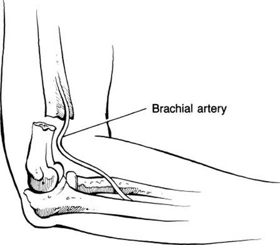
eFIGURE 37.2 Fracture-associated vascular injury.
The classic signs of vascular injury are the “Five Ps”: Pain, pallor, pulselessness (or diminished pulse), paresthesia, and paralysis. The Five Ps are present only in a complete injury to the vessel. A partial injury can have subtle clinical signs and symptoms or be completely asymptomatic. In addition, collateral circulation may maintain distal flow around a vascular injury. Vascular compromise may also be delayed, as in the case of an initial vascular intimal injury leading to progressive thrombosis. Therefore, the location of the fracture and the mechanism of injury determine the need to assess for a potential vascular injury in the asymptomatic patient. Initial evaluation should involve assessment of pulses, evaluation with a Doppler stethoscope if palpable pulses are diminished or absent, observation of the color of the distal extremity, and assessment of capillary refill. With extremity injuries, this assessment should be compared to that of the opposite limb. The ankle-brachial index may be assessed to help evaluate patients where there is concern after the foregoing examination. The systolic pulse is assessed in the brachial artery and in the dorsalis pedis artery in the foot with a Doppler stethoscope. The ratio of the pressure in the ankle is then compared to that in the arm. A ratio of >0.9 is reassuring, although it does not completely rule out partial arterial injury (13,14).
If vascular injury is suspected, further evaluation with angiogram, CT angiogram, color-flow Doppler ultrasound, magnetic resonance angiography, or surgical exploration is usually warranted. Complications of fracture-associated vascular injuries include in situ thrombosis, false aneurysms, true aneurysms, compartment syndrome, arteriovenous fistulas, and distal limb ischemia with mechanical dysfunction.
Nerve Injuries
Nerves are more frequently injured than vessels in association with fractures. They can be damaged by blunt trauma, along the trajectory of penetrating trauma causing an associated fracture, or by the fracture fragments themselves. Nerves are at increased risk of injury when they are superficial, lie close to the bone, or span the joint, making them susceptible to stretch injury.
The most frequently injured nerves in association with specific fractures include the axillary and musculocutaneous nerves in shoulder fracture-dislocations; the radial nerve in fractures of the distal third of the humerus; the radial, median, and ulnar nerves in supracondylar elbow fractures; the median nerve in fracture-dislocations about the wrist; the peroneal and tibial nerves in knee fracture-dislocations; and the peroneal nerve in fractures of the fibular head or proximal fibula (eFig. 37.3).

eFIGURE 37.3 Peroneal injury associated with a knee fracture.
Nerve injuries are classified based on the amount of damage to the nerve according to Seddon staging system:
• Neuropraxia: The least severe injury, neuropraxia involves a contusion to the nerve, with minor injury to the myelin sheath. Sensory loss and transient paralysis may occur, but spontaneous and complete recovery occurs over a period of several days to weeks (occasionally, a few months).
• Axonotmesis: In axonotmesis, more severe crush injury to the nerve occurs with axonal breakdown followed by distal wallerian degeneration. The epineurium and the continuity of Schwann tubes are maintained. Prolonged paresthesias and muscle weakness are the clinical hallmarks. Recovery can be complete but takes months to years. The axon has to regenerate proximally to distally and, for functional conduction to occur, the myelin sheath must be fully repaired. Recovery is often incomplete.
• Neurotmesis: The most severe form of nerve damage is neurotmesis disruption of the axon, epineurium, and Schwann cell sheath (complete severance of the nerve). When complete disruption occurs, all functions controlled by the injured nerve are interrupted.
The emergency evaluation of a nerve injury should involve assessment of motor strength, deep tendon reflexes, light touch, and two-point discrimination. Formal testing can be performed using electromyogram; however, this is not necessary in the ED.
Compartment Syndrome
Compartment syndrome (fully discussed in Chapter 48) should be suspected in long bone fractures and fractures associated with significant vascular injuries or pronounced swelling, especially when the patient has pain out of proportion to physical examination findings or paresthesias. Manometry should be performed if compartment syndrome is suspected, as the clinical signs are variable and may be delayed. Compartment syndrome is a surgical emergency and requires prompt decompression to prevent or minimize the associated morbidity.
Infection
Infections occur primarily in open fractures, especially in those in which there is a delay in treatment. The general rule in open fractures is that they need “urgent” irrigation and debridement in the OR. Though 6 hours was historically cited as the cutoff time for this urgent procedure, recent literature has questioned this time frame. Although the exact amount of time is debated, most authors agree that “sooner is better” for this procedure (15). Infection can also occur with extensive hematoma or after open reduction internal fixation repair. It may also occur without a fracture in cases of osteomyelitis, septic arthritis, and abscesses eroding to bone. Septic arthritis should be suspected in the patient with monoarticular arthritis and ruled out in those who present with a swollen, erythematous, hot joint. If the diagnosis is in question, aspiration for crystal analysis, Gram stain, cell count, and culture should be performed. Most orthopedic surgeons recommend aggressive arthroscopic lavage to prevent destruction of the joint space by bacteria and inflammatory-mediated damage.
Casts and Splints
Immobilization devices have the potential to cause postinjury problems. They may become too tight as swelling increases during the first 24 to 48 hours after injury and cause pain and impair circulation. Pressure sores may occur on the skin in areas where a cast or external fixation device exerts localized excessive pressure (e.g., over the heel in a patient with a bimalleolar fracture). Immediate steps should be taken to correct the problem. A cast that is too tight can be split lengthwise; the underlying padding can then be cut and the cast spread open. A cast that is pressing on a particular site can have a window cutout. The window may be replaced more loosely with plaster or covered with soft dressing. An ill-fitting splint can be trimmed. The clinician should take great care in the selection of the appropriate splint (type and material), its placement (to make sure that it has adequate padding and effectively immobilizes the intended joint), and perform a postsplinting examination to ensure neurovascular integrity.
Fat Emboli Syndrome
Single or multiple long bone fractures in the young and hip fractures in the elderly predispose to fat emboli. Twenty percent of patients with pelvic or long bone fractures have detectable fat droplets in their blood. However, the clinical significance of this is not clear, as the vast majority of these patients remain asymptomatic. Fat emboli syndrome is the most common form of nonthrombotic embolism and has a characteristic clinical course. Its etiology is unclear, but three main hypotheses have been postulated: (a) After fat droplets enter the bloodstream, the liberation of free fatty acids from marrow fat by circulating lipase causes a toxic vasculitis and capillary leak syndrome; (b) it is the end result of extensive platelet–fibrin thrombosis; and (c) it results from direct obstruction of distal vascular lumen by fat emboli. After the injury, the patient often remains asymptomatic for 12 to 36 hours before suddenly and unexpectedly developing a life-threatening syndrome that is characterized by rapid cardiopulmonary and neurologic deterioration. Patients develop agitation, hallucinations, delirium, coma, hypoxia, dyspnea, tachypnea, tachycardia, and a petechial rash. If this deterioration is not quickly reversed, refractory hypotension, anemia, thrombocytopenia, disseminated intravascular coagulopathy, and adult respiratory distress syndrome often ensue, with an associated high morbidity and mortality.
Ischemic or Avascular Necrosis
Ischemic or AVN is characterized by bone death that occurs following the interruption of blood supply to bone. AVN is most frequently associated with carpal scaphoid fractures, lunate dislocations, hip dislocations, femoral neck fractures, and complex fractures of the talus. The time required for an injury to result in AVN is extremely variable and is location and patient specific.
Delayed Complications
Other complications of fractures may occur some time after the initial injury. Many fractures require reduction of the displaced fracture fragments in the ED or the operating room. The goal in reduction is always to reestablish anatomic position; eliminate displacement, angulation, and other forms of malalignment; and immobilize the broken bone in the reestablished anatomic position to promote healing. Sometimes, this ideal condition is not achieved, and the systematic progression from hematoma to callus formation to bony union is delayed or interrupted. Delayed unions are fractures that take longer than usual to heal; malunions are fractures that heal in a faulty position; and nonunions occur when the process of fracture healing stops before the fracture is united.
Joint stiffness, posttraumatic arthritis, and complex regional pain syndrome (CRPS)—type I (formerly known as reflex sympathetic dystrophy [RSD])—may also occur as delayed complications of extremity trauma. Any intra-articular fracture of a weight-bearing joint predisposes the patient to early degenerative changes. The patient may present to the ED years after a fracture, complaining of perceived sudden onset of joint pain and decreased range of motion. Radiographs typically demonstrate extensive osteoarthritis.
CRPS is a three-stage, posttraumatic syndrome of unclear etiology, the development of which has no known correlation to the severity of the initial injury (16). CRPS remains a clinical diagnosis with symptoms that are often difficult to differentiate from normal posttraumatic characteristics. The first, or early, stage characterized by continuous burning and aching in the posttraumatic extremity, is exacerbated by weightbearing and by movement. After a variable period of time, the middle, or dystrophic, stage ensues, characterized by the development of shiny, cool skin over the affected extremity and a progressively decreased range of motion. In the final, or atrophic, stage, the skin atrophies, the muscles atrophy, and flexion contractures develop.
In 1992, Gibbons and Wilson, in the Clinical Journal of Pain, developed an RSD score that was proposed as a strict system for the clinical diagnosis of RSD. The scoring system consists of nine criteria and has more than academic interest. Early diagnosis and subsequent early treatment may potentially improve outcome, interrupting the progression from burning to dystrophy to atrophy. The nine criteria are (a) allodynia—the phenomenon whereby pain is increased by stimuli that activate the involved nerve, such as a breeze blowing on the area, temperature changes, or emotional outbursts (hyperpathia); (b) burning pain; (c) edema; (d) change in color or hair growth in the affected area; (e) change in sweating; (f ) change in temperature; (g) demineralization of bone in the involved limb, as documented in radiographs; (h) quantitative measurements of vasomotor disturbances; and (i) a triple-phase bone scan consistent with RSD. A score of five points qualifies as a clinical diagnosis of RSD; one point is given for each positive criterion, zero points for each negative criterion, and one-half point for each equivocal criterion. Application of heat or cold, exercises, early sympathetic blockade, or sympathectomy to the affected limb can halt the progression to atrophy. A minority of patients have been helped by a short course of high-dose prednisone (a 3-week taper) in conjunction with heat, cold, and exercise therapy (17).
Subluxations and Dislocations
Acute or chronic ligamentous laxity or tearing can result in subluxation or dislocation of a joint. Chronic ligamentous laxity is usually the result of an overuse syndrome, in which repetitive microtrauma leads to an attenuation of the static restraints of the joint. Acute ligamentous tears are the result of excessive or abnormal forces applied to the joint. The classic example is subluxation or dislocation of the glenohumeral joint. Subluxation occurs when one bone becomes partially disarticulated from the other bone that forms the joint. The articular surfaces forming the joint remain partially intact. Dislocationoccurs when the bones are completely disarticulated and no parts of their articular surfaces are in contact. Dislocations can occur in isolation or with an associated fracture (eFig. 37.4). Often, the fracture is anatomic evidence of the direction of the pathologic force vector resulting in the dislocation. For example, 90% to 95% of shoulder dislocations occur anteriorly, disrupting the continuity of the anterior capsule and the labrum. Sometimes, these structures resist the forces producing dislocation to the point that a fracture occurs, avulsing a piece of bone from the anterior glenoid rim, known as a Bankart lesion.
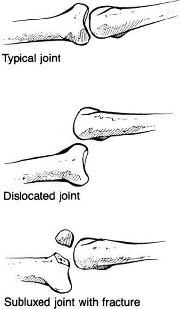
eFIGURE 37.4 Joint subluxation and dislocation.
The nomenclature of subluxations and dislocations is straightforward. Most subluxations and dislocations occur at a joint formed by two bones, and the subluxation or dislocation is named after the affected joint (e.g., posterior dislocation of the distal interphalangeal [DIP] joint of the fourth finger of the right hand). If a joint is formed by three bones, and the two major bones are disarticulated, the subluxation or dislocation is named after the joint involved (e.g., posterior dislocation of the knee). If the joint is formed by three bones, and the minor bone is subluxed or dislocated, the abnormality is named after the minor bone (e.g., lateral dislocation of the patella). As for fractures, direction is assigned with reference to the distal segment.
Specific subluxations and dislocations are frequently seen in the ED and should be skillfully and efficiently reduced, with restoration of anatomic position, by the emergency physician. These include anterior and posterior dislocations of the shoulder joint; anterior and posterior dislocations of the sternoclavicular joint; posterior dislocations of the elbow joint; posterior dislocations of the proximal interphalangeal and DIP joints, often with accompanying avulsion fractures; posterior dislocations of the hip joint; posterior dislocations of the knee joint; lateral dislocations of the patella; and fracture-dislocations of the ankle.
The hallmarks of subluxations and dislocations are pain, deformity, and decreased range of motion. Certain dislocations are associated with specific complications, and these must be ruled out in the evaluation and treatment of the injury. For example, the aorta and trachea are at risk in posterior dislocations of the sternoclavicular joint, and injury to them can lead to rapid respiratory compromise or intrathoracic hemorrhage. The axillary nerve and, less commonly, the musculocutaneous nerve are at risk in anterior shoulder dislocations. Their function must be clearly assessed prior to any attempted reduction and then reassessed after the reduction is complete. The popliteal artery can be stretched, torn, or completely disrupted in a posterior dislocation of the knee. Radiographs are often obtained prior to attempts at reduction and after the reduction has been achieved to document the position of the bones, restoration of anatomic position, and the presence of associated fractures. The need for pre- and postreduction radiographs for all types of dislocations, however, has been challenged in the literature (18,19).
Techniques for the reduction of specific joints are discussed in the chapters dedicated to those joints, but certain principles hold true for all subluxations and dislocations. The length of time between injury and relocation corresponds proportionally to the severity of spasm and swelling that will occur and the difficulty involved in reducing the joint. Adequate pain control and muscle relaxation can facilitate smooth reduction; excess force applied during the reduction may result in fractures, nerve damage, and vascular injury. The technique used to reduce the dislocation should initially replicate the mechanism of injury to release the dislocated bone from the anatomic structure that is keeping it dislocated. In certain dislocations, the incidence of delayed complications is directly proportional to the time from injury to relocation. For example, AVN of the femoral head occurs in 10% to 20% of hip dislocations. The rate is directly proportional to the total time from injury to successful reduction. Similarly, myositis ossificans is a well-recognized complication of posterior dislocation of the elbow. The incidence of myositis ossificans is directly related to three variables: The number of attempts at reduction, the length of time from injury to successful reduction, and whether the joint is adequately immobilized after reduction.
Tendinosis
Tendinosis is a subset of strain since it is an injury to the musculotendinous unit, but it is more appropriately considered a separate entity. In the past, tendinosis was felt to represent inflammation to the musculotendinous unit, and was hence termed “tendinitis.” Recent work has indicated that a more correct term is tendinosis, as there is no significant inflammatory response by the body in these conditions (20). Instead, there is a degeneration of the collagen portion of the tendon in the absence of inflammatory cells. Tendinosis differs from strain in that the pathologic changes are isolated to the tendon and involve the insertion of the tendon into the bone, which occurs via a transitional, calcified fibrocartilage known as Sharpey fibers. Tendinopathy can result from chronic use or from one acute episode of overuse. In acute tendinopathy, the tendon is irritated, and an acute response occurs. Microscopically, there is loss of collagen continuity with an associated increase in ground substance, vascularity, and cellularity. The cellularity results from the presence of fibroblasts and myofibroblasts, not inflammatory cells. If this cycle is not interrupted, permanent changes in the architecture of Sharpey fibers and atrophy of the tendon fibers occur.
Common tendons involved in acute or chronic tendinosis presenting to the ED include patellar, quadriceps, rotator cuff, and the Achilles tendons.
Clinically, tendinosis presents with pain during active range of motion, loss of function, point tenderness over the affected tendon near its insertion into bone and, often, localized edema and erythema. Forced contraction of the affected muscle against resistance elicits pain, which is exacerbated by pressure over the point of insertion. Pain may also be elicited by forced extension of the tendon, stretching the injured fibers.
Calcific tendinitis is an entirely different entity, representing a truly inflammatory response by the body. It is usually associated with chronic inflammation and consists of calcium deposition within the body of the tendon. It is often an indication of tendinous degeneration altering the macrostructure of the tendon. Calcific tendinitis has a characteristic radiographic appearance, revealing the calcium depositions within the tendon. For example, a band of calcification can be seen passing over the bicipital groove in calcific biceps tendinitis at the shoulder.
Muscle Spasms
Muscle spasms are a poorly understood phenomenon in which a voluntary muscle undergoes sustained involuntary contraction, leading to depletion of stored glycogen, localized buildup of lactic acid, and severe pain. The causes of muscle spasm are not known, but hypotheses include localized alterations in blood flow, regional or systemic electrolyte imbalances (especially sodium and calcium; less often, potassium), and postural abnormalities that place a bone in a nonanatomically neutral position, causing attached musculature to fire continuously. For example, overdevelopment of the muscles on one side of the body in a racquet sports athlete can cause latissimus dorsi spasm, as the proprioceptors present in the latissimus sense that the arm is out of the neutral position, resulting in repetitive muscle contraction in attempted compensation.
Muscle spasms typically seen in the ED include spasms of the gastrocnemius, levator scapulae, trapezii, rhomboids, and latissimus dorsi. Rectus abdominus muscle spasm can mimic an acute abdomen.
Treatment may be frustrating for both the patient and the clinician, as complete relief is difficult to achieve. The treatments are not perfect, and many are controversial. Treatment options include freezing the muscle belly with fluoromethane spray, followed by passive, gentle, forced range of motion to “break the spasm”; massage; treatment with muscle relaxants, including diazepam and other benzodiazepines; and pain control with acetaminophen, NSAIDs, or opioid analgesics. Recent research indicates that treatment with muscle relaxants for these conditions may not be as good as basic analgesia (21).
ED EVALUATION
The ED evaluation of any injured patient begins with the basic ABCs: Airway, breathing, and circulation. If the patient is reliable, not intoxicated, and has an isolated injury and the mechanism of injury does not suggest other possible injuries, then evaluation and treatment are limited to the injured joint along with the joints immediately proximal and distal to the injury. The contralateral side of the injured joint or limb should be examined for comparison of range of motion, anatomy, laxity, and neurovascular findings.
In patients with multiple injuries, head trauma, amnesia, intoxication, or medical illnesses resulting in their musculoskeletal injury (e.g., a distal radial fracture after syncope), a complete physical evaluation is mandatory and should follow basic trauma guidelines. This should begin with a primary survey and assessment of the ABCs; evaluation of neurologic disability and Glasgow Coma Scale; then continue with complete exposure of the patient and a search for any accompanying injuries. Once this is complete, the emergency physician should systematically evaluate the underlying medical problem that could have produced the trauma. If a problem is identified, it should be addressed before moving on to the next step in the systematic evaluation. For example, the motorcyclist thrown from the cycle, who presents with an open fracture of the tibia and fibula and flail chest, needs breathing stabilized before the leg is further evaluated. In extremity wounds, hemorrhage can often be rapidly but temporarily controlled with a direct-pressure dressing, ensuring continued circulatory stability until the source of bleeding can be systematically evaluated. After the primary survey, each joint should be palpated and ranged systematically, and the integrity of long bones assessed by inspection, palpation, and range of motion.
As alluded to previously, certain extremity injuries are true orthopedic emergencies, meriting expeditious evaluation and management. These include vascular injuries, compartment syndromes, open fractures and dislocations, and certain dislocated joints (Table 37.1). They should be addressed immediately after the ABCs have been evaluated, stabilized, and recorded on a trauma flow sheet. If vascular compromise is suspected, immediate consultation with an orthopedic or vascular surgeon is advised. When vascular insufficiency is caused by stretching of the vessel across the site of a dislocation or displaced fracture, reduction of the fracture or dislocation may correct the problem. Pulses should be documented after the reduction. If an injury to the vessel itself is suspected, arteriography, color Doppler ultrasound, magnetic resonance angiography, or surgical exploration may be indicated. Color Doppler ultrasound evaluation, followed by close observation with serial neurovascular examinations, is a relatively newer option and is still being validated as a modality. These decisions are usually made in consultation with a surgeon. Blood flow to an ischemic extremity should, ideally, be reestablished within 6 to 8 hours to prevent permanent injuries, including ischemia and necrosis.
Similarly, if compartment syndrome is suspected, it should be ruled out by manometry. This is usually accomplished through emergent consultation with an orthopedic surgeon. If compartment pressures are elevated, emergent fasciotomy is mandatory.
After all life- and limb-threatening injuries have been addressed, attention can be directed toward the identification of individual injuries—fractures, dislocations, sprains, strains, tendinosis, and spasm. This evaluation begins with a thorough history to ascertain the mechanism of injury, timing of the injury, previous injury to the limb, handedness where applicable, and medical problems potentially contributory to current disability. The history should include questions about associated symptoms of pain, paresthesia, weakness, signs of pallor, or deformity.
On physical examination, the emergency physician should note the appearance of the injury, looking for deformity, swelling, erythema, ecchymosis, and exposed bone. Other physical findings include the presence of purulent fluid drainage from the wound, abrasions, or lacerations over the injured area.
The emergency physician should palpate for deformity, local or diffuse tenderness, edema, warmth, and induration. Palpation should include a careful assessment of neurovascular integrity. Patients with vascular compromise will have dusky or mottled skin, diminished distal pulses, and delayed capillary refill. Sensation to light touch may be decreased, and hypesthesia often occurs over an area of acute injury. Active range of motion should be tested. In the majority of acute injuries, the patient is not enthusiastic about actively ranging the joint and passive range of motion, and provocative maneuvers are of limited utility due to pain.
Radiographs are the cornerstone of further evaluation and management of most musculoskeletal complaints. Many fractures can be diagnosed clinically. However, in the vast majority of cases, the diagnosis should be supported radiographically. Radiographs frequently determine the need for operative reduction, the length of time the fracture needs to be immobilized, and potential complications during the rehabilitation process. Criteria for obtaining radiographs of specific injuries are formalized but constantly evolving and are beyond the scope of this chapter. They are addressed in the chapters on injuries to specific joints. The goals of objective criteria for obtaining radiographs include reduction in unnecessary imaging, increased efficiency of patient flow in the ED, decreased exposure to radiation, and containment of healthcare costs. For example, the Ottawa Ankle Rules are objective criteria for obtaining an ankle series after an ankle injury. They state that ankle films are required in patients older than 18 years if pain in the malleolar area is accompanied by inability to bear weight (defined as walking four steps) immediately after the injury and in the ED or by bony tenderness over the posterior aspect of the distal 6 cm of the medial or lateral malleolus. Strict implementation of the Ottawa Ankle Rules has been shown to reduce the number of ankle films ordered by 28%, increase ED efficiency, and decrease ED costs without increasing the percentage of missed fractures (22).
When the decision is made to obtain roentgenograms, the correct views must be obtained, they need to be of sufficient quality to competently evaluate for fracture, and they need to be read thoroughly, in a systematic fashion. Poor-quality films should not be accepted. Many fractures have been missed because of technically inadequate films. In addition, thorough examination of the soft tissue and every bone in the x-ray is essential to avoid missing subtle findings that may indicate fracture or dislocation. For example, the fat pad sign on an elbow x-ray may be the sole indication of a radial head fracture.
Other pitfalls in radiographic interpretation include the following:
• A nutrient foramen, housing a nutrient artery, may be misinterpreted as a fracture. A nutrient foramen will appear less radiolucent than a fracture (very smooth-edged) and will angle obliquely from the edge of the bone through the cortex, and then terminate without involving the cortex on the other side of the marrow.
• Sesamoid bones or accessory ossicles may be confused with fractures. These anatomic variants usually have smooth edges and are well corticated, and no defect in the adjacent bone will be noted. In some instances, a comparison view of the unaffected side can help clarify the structure in question. For example, the patient with pain over the patella following a dashboard injury, who is found to have a two-part patella on x-ray, may have bipartite patella, which is confirmed by its presence on an x-ray of the unaffected knee.
• Fat and clothing folds, casts, or splints may be misinterpreted as fractures, and jewelry or body piercings may be misinterpreted as foreign bodies.
Every x-ray series should include a minimum of two views that are taken at right angles to each other. If the plane of the fracture is at 90 degrees (perpendicular) to the plane of the x-ray beam, the fracture may not be evident on that view but would be readily visible on an x-ray taken in a plane parallel to the plane of the fracture line.
Fractures in the shaft of long bones are often accompanied by additional fractures in that bone proximal or distal to the main fracture site or by dislocations at the joints at either end of the bone. Therefore, all long bone shaft fractures require imaging of the entire length of the bone, including adequate views of its joints. For example, a midshaft femur fracture requires x-rays of the entire shaft, a two-plane knee series, and a two-plane hip series.
Fractures to paired long bones, such as the radius and ulna or the tibia and fibula, may present with different associated fractures. In other words, a fracture to one bone is sometimes accompanied by a distant fracture to the other bone in the pair. For example, medial malleolar ankle fractures resulting from a lateral twisting mechanism sometimes have an associated proximal fibular or fibular head fracture or dislocation. The mechanism of injury explains this association: The twisting force that breaks the medial malleolus is transmitted up the shafts of the tibia and fibula and is dissipated at the weakest point, the proximal fibula, resulting in either fracture or dislocation. Thus, more complex ankle fractures require imaging of the entire shafts of the fibula and tibia and a two-way series of the knee. Certain forearm fractures are commonly associated with a fracture-dislocation of the other bone of the forearm, such as a radial shaft fracture and distal radioulnar joint dislocation, known as a Galeazzi fracture (eFig. 37.5).
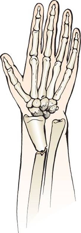
eFIGURE 37.5 Galeazzi fracture dislocation.
Additional views are sometimes helpful. A dedicated scaphoid view can aid in the detection of a subtle scaphoid fracture. An axial view of the calcaneus can reveal a fracture missed on standard ankle and foot series. Many posterior dislocations of the shoulder are not evident, except on an axillary view of the glenohumeral joint, clearly depicting the relationship between the humeral head and the glenoid fossa. In certain instances, comparison views to the patient’s other side are helpful. For example, in distal clavicle osteolysis, characterized by pain and osteolysis of the distal clavicle, bilateral clavicle views help confirm the diagnosis when a larger separation is seen at the affected acromioclavicular joint compared with the unaffected side. Distal clavicle osteolysis occurs in power lifters and in contact with sports who suffer significant blunt trauma to the point (acromion) of the shoulder.
Adequate films, correctly interpreted, are not 100% sensitive for fractures. Growth plate injuries (also known as Salter–Harris injuries) may appear entirely normal on plain radiographs. The diagnosis may not become evident until there has been new bone deposition or resorption at the site of injury. Similarly, nondisplaced, hairline fractures often become visible, or radiolucent, only on radiographs 7 to 10 days after injury, as bone resorption widens the fracture line, hematoma develops, and new bone formation proceeds. If there is potential morbidity associated with a missed fracture on plain x-rays, a clinically fractured bone should be treated as a fracture, or more sophisticated imaging studies should be obtained. A patient with a story and examination consistent with a femoral neck fracture, which has normal plain radiographs and remains nonweight bearing in the ED, should be treated as having a nondisplaced hip fracture. Assessment of a patient’s ability to ambulate is essential prior to discharge, and the inability to ambulate can further confirm the presence of an occult lower extremity fracture. These patients should be kept nonweight bearing, and magnetic resonance imaging should be performed to demonstrate the fracture; a bone scan may also be diagnostic.
Similarly, tenderness in the snuff box (the space on the radial side of the wrist between the extensor pollicis longus tendon and the paired extensor pollicis brevis and adductor pollicis longus tendons) after a fall on an outstretched hand should be treated as a scaphoid fracture, even if the radiographs are normal. If the radiographs show no fracture, immobilization in a thumb spica with follow-up examination and repeat radiographs in 10 days is mandatory (23).
KEY TESTING
Plain radiographs for skeletal injuries must
• Include the appropriate bone/joint injured.
• Have at least two views at right angles to each other.
• For long bone injuries, include the whole length of the bone and the joint above and below
CT Imaging
• Provides greater osseous detail and can do so in multiple planes (axial, coronal, sagittal)
• Consider when the relationship of fracture fragments is complex or will impact down-stream care (e.g., calcaneal fractures, tibial plateau fractures)
• Can be combined with angiographic contrast (CT-A) to identify injuries to major blood vessels
MR Imaging
• Provides greatest soft tissue detail (tendons, ligaments, disc)
• Can also provide imaging in multiple planes (axial, coronal)
• Consider when there is a potential soft tissue injury that will impact care (e.g., spinous ligaments)
• Can be combined with angiographic contrast (MR-A) to identify injuries to major blood vessels
Ultrasound
• An imaging modality being used by some practitioners in the bedside diagnosis of musculoskeletal injuries.
• In trained hands can identify fractures, dislocations, and some soft tissue injuries (e.g., tendon tears).
ED MANAGEMENT
In general, the initial management of musculoskeletal injuries includes immobilization of the affected extremity and administration of pain medication. As previously mentioned, pain related to musculoskeletal injuries is often undertreated in the ED. Once a fracture or dislocation is identified, appropriate analgesia should be provided, and reduction should be performed as needed; this is determined by the location of the injury and the amount of distraction or angulation. The management of specific injuries is discussed in subsequent chapters.
The initial management of open fractures and joints should be directed toward reducing the chances of infection. Gross particulate debris in the wound should be removed. Exposed bone should not be replaced in the wound,because this may further introduce contamination. A sterile dressing should be applied. The extremity is splinted to prevent further tissue and neurovascular injury. Intravenous antibiotic therapy, usually with a first- or second-generation cephalosporin, is instituted (if not contraindicated by drug allergies), and tetanus prophylaxis (sometimes including tetanus immune globulin) is given when indicated. A macrolide antibiotic or trimethoprim–sulfamethoxazole is a good choice for the penicillin-allergic patient. An aminoglycoside such as gentamicin is frequently added to the cephalosporin when there is heavy contamination of the wound. After these initial management steps have been taken, orthopedic consultation should be obtained. Most open fractures and dislocations require formal debridement in the operating room, which should be performed as soon as patient stability will allow (15). The incidence of infection is directly proportional to the length of time from injury until irrigation and formal debridement occurs.
Certain dislocations require expeditious reduction. Dislocated hips are at risk for development of ischemic necrosis, and dislocated elbows are at risk for development of Volkmann ischemic contracture (compartment syndrome, decreased blood flow, necrosis, and contracture after a dislocation). AVN of the hip is thought to be caused by damage to the vessels supplying the femoral head. In general, these vessels are not completely torn in the dislocation; rather, they are stretched, and blood flow through them is compromised. Therefore, the incidence of AVN is directly related to the length of time from injury to adequate relocation of the hip. Similar principles apply to the development of Volkmann ischemic contracture after elbow dislocation. Therefore, reduction should be accomplished as soon as is feasible.
CRITICAL INTERVENTIONS
• Once the patient has stabilized, palpate every bone and put every joint through a range of motion to search for occult injury.
• Relocate or realign any fractures or dislocations that result in neurovascular compromise in an attempt to alleviate the neurovascular deficit as expeditiously as possible.
• Splint fractures as quickly as is possible to minimize pain, prevent injury to adjacent structures, and reduce blood loss associated with these injuries.
• Perform manometry and obtain early consultation for patients with suspected compartment syndrome.
• Administer intravenous antibiotics (first- or second-generation cephalosporin and gentamicin) early for patients with open fractures.
DISPOSITION
ED care is not complete when the diagnosis is made. Appropriate immobilization should be instituted and instructions for immediate care and follow-up given. These should be tailored to the specific injury but, in general, involve rest, ice, and elevation. These simple, cost-effective steps are often overlooked in the rush to discharge patients from the ED and can, in some cases, affect more than the patient’s comfort. For example, the simple application of ice to a lateral ankle sprain during the first 24 hours after injury has been shown to decrease time to return of full function by 50% (24). Conversely, inadequate treatment can lead to decreased mobility, chronic pain, and joint instability.
Depending on the injury, the patient may be instructed to remain nonweight bearing or keep the injured joint immobilized. In other cases, activity as tolerated is appropriate and may speed recovery (e.g., low-back strain). Pain control must be achieved in the ED, and the pain medications given for the outpatient setting should be commensurate with the injury. For example, the patient who requires morphine in the ED for a rib cage contusion should not be discharged with instructions to take acetaminophen as needed for pain. Adequate postinjury pain control improves outcome and decreases repeat visits to the ED.
Patients should be instructed to return to the ED if they experience any signs or symptoms of neurovascular compromise, excessive pain, coldness, erythema, fevers, chills, cough, pleuritic chest pain, or a cast or splint that feels too tight or causes pain. Appropriate follow-up depends on the nature and extent of the injury and can range from a visit to the patient’s primary care doctor in 2 weeks, to orthopedic referral in 10 days’ time, to being seen by a hand surgeon in the next few days for operative repair of a flexor tendon laceration.
Patient with CRPS should be referred early to a physician with expertise in this area, which is usually in the realm of pain management. Frequently, multimodality treatment is required, and a great deal of care coordination must occur.
Common Pitfalls
• Ordering the wrong x-ray because of an inadequate physical examination
• Not recognizing all injuries because the physician’s attention is focused on one specific injury. For example, the patient who falls off a ladder and presents with a grade II lateral ankle sprain may have a calcaneal fracture from the initial impact.
• Relying on poor-quality x-rays or on a single view without obtaining an additional view at 90 degrees to the initial one
• For patients with long bone fractures, failure to include the length of the bone, the joint above and below in adequately exposed radiographs
• Failure to rule out infection or similar condition (olecranon bursitis) in a patient presenting with a swollen joint. It may be a mistake to assume that the swollen joint in the patient with a history of gouty arthritis is another flare of gout if the presentation is atypical or there are other concerning signs.
• Failure to realize that drops of blood or small abrasions on the skin in the area of a fracture are signs of an open fracture
• Failure to access or document a focused neurovascular examination on initial presentation and after reduction of a fracture or dislocation
• Attributing pain to the fracture itself, rather than thoroughly investigating other potential sources of pain, such as a cast problem, neurovascular compromise, or compartment syndrome
• Not evaluating a patient for compartment syndrome with manometry after considering the diagnosis
• Not pursuing additional studies when the plain radiographs are normal in patients with a suspected fracture, especially if they are unable to bear weight
• Undertreating the pain associated with musculoskeletal injuries.
ACKNOWLEDGMENTS
The authors gratefully acknowledge the contributions of David F. Gaieski and Joseph Bernstein to the content of this chapter.
REFERENCES
1. McCaig LF, Burt CW. National hospital ambulatory medical care survey: 2001 emergency department summary. Adv Data. 2003;335:1–36.
2. Henry GL, George JE. Specific high-risk clinical presentations. In: Henry GL, Sullivan DJ, eds. Emergency Medicine Risk Management. Dallas, TX: American College of Emergency Physicians; 1997:475–494.
3. Freedman KB. The adequacy of medical school education in musculoskeletal medicine. J Bone Joint Surg Am. 1998;80:1421–1427.
4. Miner J, Biros MH, Trainor A, et al. Patient and physician perceptions as risk factors for oligoanalgesia: A prospective observational study of the relief of pain in the emergency department. Acad Emerg Med. 2006;13:140–146.
5. Pines JM, Perron AD. Oligoanalgesia in ED patients with isolated extremity injury without documented fracture. Am J Emerg Med. 2005;23:580.
6. Ritsema TS, Kelen GD, Pronovost PJ, et al. The national trend in quality of emergency department pain management for long bone fractures. Acad Emerg Med. 2007;14:163–169.
7. Armfield DR, Kim DH, Towers JD, et al. Sports-related muscle injury in the lower extremity. Clin Sports Med. 2006;25:803–842.
8. McGowan HJ. General principles of fracture management. In: Seidenberg P, Beutler AL, eds. Sports Medicine Resource Manual. 1st ed. Philadelphia, PA: Saunders; 2007.
9. Marcell R, Einhorn TA. The biology of fracture healing. Injury. 2011;42(6):551–555.
10. Swiontkowski MF. The multiply-injured patient with musculoskeletal injuries. In: Bucholtz RW, Heckman JD, eds. Rockwood & Green’s Fractures in Adults. 5th ed. Philadelphia, PA: Lippincott Williams & Wilkins; 2001:47–84.
11. DeLong WG Jr, Born CT, Wei SY, et al. Aggressive treatment of 119 open fracture wounds. J Trauma Injury Infect Crit Care. 1999;46:1049–1054.
12. Perron AD, Brady WJ, Keats TA. Management of common stress fractures: When to apply conservative therapy, when to take an aggressive approach. Postgrade Med. 2002;111:95–106.
13. Klineberg EO, Crites BM, Flinn WR, et al. The role of arteriography in assessing popliteal artery injury in knee dislocations. J Trauma. 2004;56:786–790.
14. Mills WJ, Barei DP, McNair P. The value of the ankle-brachial index for diagnosing arterial injury after knee dislocation. J Trauma. 2004;56:1261–1265.
15. Okike K, Bhattacharyya T. Trends in the management of open fractures. J Bone Joint Surg Am. 2006;88:2739–2748.
16. Wasner G, Schattschneider J, Binder A, et al. Complex regional pain syndrome—diagnosis, mechanisms, CNS involvement and therapy. Spinal Cord. 2003;41:
61–75.
17. Gibbons JJ, Wilson PR. RSD score: A criteria for the diagnosis of reflex sympathetic dystrophy and causalgia. Clin J Pain. 1992;8:260–263.
18. Hendey GW. Necessity of radiographs in the emergency department management of shoulder dislocations. Ann Emerg Med. 2000;36:108–113.
19. Hendey GW, Chally MK, Stewart VB. Selective radiography in 100 patients with suspected shoulder dislocation. J Emerg Med. 2006;31(1):23–28.
20. Khan KM, Cook JL, Bonnar F, et al. Histopathology of common tendinopathies: Update and implications for clinical management. Sports Med. 1999;27:
393–408.
21. Turturro MA, Frater CR, D’Amico FJ. Cyclobenzaprine with ibuprofen vs. ibuprofen alone in acute myofascial strain: A randomized double-blind clinical trial. Ann Emerg Med. 2003;41:818–826.
22. Stiell IG, Greenberg GH, McKnight RD, et al. Decision rules for the use of radiography in acute ankle injuries: Refinement and prospective validation. JAMA. 1993;269:1127–1132.
23. Perron AD, Brady WJ, Keats TE, et al. Orthopaedic pitfalls in the emergency department: Scaphoid fracture. Am J Emerg Med. 2001;19:310–316.
24. Tiemstra JD. Update on ankle sprains. Am Fam Phys. 2012;85(12):1170–1176.