
Carl A. Germann and Megan L. Fix
Hip injuries are common and associated with notable morbidity and mortality, especially in older patient populations. It is predicted that by the year 2050, 6.3 million hip fractures will occur yearly worldwide (1). Causes of hip fracture are primarily pathologic (from osteopenia) and falls, whereas motor vehicle collisions account for the majority of hip dislocations.
Anatomically, the hip is an articulation between the femoral head and the acetabulum. The femoral head is firmly seated in the acetabulum, which is reinforced by labral cartilage, capsule, overlying ligaments, and proximal musculature of the lower extremity (Fig. 43.1). It is a very stable articulation, requiring violent injury to fracture or dislocate. The femur is the longest and strongest bone in the human body and is often subjected to substantial forces produced during powerful muscle contraction, weight transmission, or trauma. The length, angle, and narrow circumference of the femoral neck allow substantial range of motion at the hip, but these same characteristics subject the neck to incredible shearing forces.

FIGURE 43.1 Anatomy of the hip. Anterior and posterior views of the femur (top) and division of the proximal femur (bottom).
Primary sources of blood flow for the femoral head are the retinacular branches of the circumflex femoral artery. These arteries enter the femur in the region of the femoral neck; fractures proximal to their entry can result in disruption of blood flow and cause avascular necrosis (AVN) of the femoral head.
ED EVALUATION
During the initial evaluation, details of the mechanism of injury as well as a thorough physical examination and search for associated neurovascular injuries are imperative. The initial radiographic studies in patients with suspected hip and femur injuries are the anteroposterior (AP) and lateral views. When analyzing the AP view of the hip, there are two important radiographic lines that help identify femur fractures and slipped capital femoral epiphysis (SCFE): Shenton and Klein lines. The Shenton line is a gentle arc seen on the AP view that runs along the inferior border of the femoral neck and extends to the inferior border of the superior pubic ramus (eFig. 43.1). The Shenton line will have a step or discontinuity in subtle cases of femoral neck and head fractures. The line may also appear disrupted in the radiograph of a normal hip, if it is not a true AP view. Klein line runs along the superior border of the femoral neck and intersects the superior portion of the femoral head (eFig. 43.2). The Klein line will project superior to a subtle fracture of the femoral head. The Klein line will also project superior to the epiphysis in a subtle case of SCFE.

FIGURE 43.2 Posterior hip dislocation.

eFIGURE 43.1 The Shenton line. (From Gruber JE, Gibbs MA, Ferrera PC, et al. Trauma Management: An Emergency Medicine Approach. St. Louis, MO: Mosby; 2001:398 (Figure 26–2), with permission.)

eFIGURE 43.2 The Klein line. (From Gruber JE, Gibbs MA, Ferrara PC, et al. Trauma Management: An Emergency Medicine Approach. St. Louis, MO: Mosby; 2001:398 (Figure 26–3), with permission.)
Approximately 2% to 10% of all hip fractures are radiographically “occult” on plain films (2). Therefore, if clinical suspicion remains and plain radiographs are negative, magnetic resonance imaging (MRI) is indicated. Although computed tomography (CT) scan can be used, small studies suggest that it is inferior to MRI (2,3). CT does seem to have merit when the patient does not have significant osteoporosis or in the setting of significant trauma (3). MRI is the imaging modality of choice with a sensitivity and specificity approaching 100%. In studies of patients with suspicion of hip fracture and normal plain radiographs, MRI showed occult fracture in 37% to 55% of patients (4).
KEY TESTING
• The initial radiographic studies in patients with suspected hip and femur injuries are the AP and lateral views.
• If clinical suspicion remains high and plain radiographs are negative, MRI is recommended to evaluate for occult fracture.
• Failure to detect these injuries results in increased mortality, risk of subsequent displacement, and a higher incidence of complications such as AVN.
ED MANAGEMENT
Hip Dislocations
The relationship of the femoral head to the acetabulum is used to classify dislocations into posterior, anterior, inferior, and central. Posterior dislocations account for 80% to 90% of cases. Anterior dislocations are seen in 10% to 15% of patients. Inferior dislocation (luxatio erecta) of the hip is very rare and occurs almost exclusively in children younger than 7 years of age. Central dislocation is often used to describe the translocation of the hip joint causing a comminuted fracture of the acetabulum.
The neurovascular examination after a hip dislocation should focus on the sciatic nerve and femoral nerve and vessels. Sciatic nerve palsy is more likely to occur in conditions such as traumatic dislocation of a nonprosthetic hip or during the total hip arthroplasty (THA) procedure itself. Sciatic palsies may be present in approximately 10% of patients who experience traumatic hip dislocation. The sciatic neuropathy may produce paralysis of the peroneal muscles. The femoral artery and its branches are the most likely vessels to be injured, particularly after an anterior dislocation. The development of AVN of the femoral head caused by arterial injury has been reported in 1% to 17% of dislocations (5). Risk factors for the development of AVN include the total dislocation time, severity of the injury, number of reduction attempts, and presence of comorbid conditions. The incidence of AVN, traumatic arthritis, permanent sciatic nerve palsy, and joint instability logarithmically increases with the length of time for which the hip remains dislocated (6). Of course, prosthetic femoral heads are not at risk for developing AVN.
Posterior Hip Dislocations
Posterior hip dislocations are the most common type of hip dislocation and result from force applied to a flexed knee and hip. These injuries usually occur as a result of motor vehicle crashes where a seated occupant typically has the hip adducted, flexed, and internally rotated at the time of impact. This injury may be prevented by the use of a lap belt.
In posterior hip dislocations, the limb is often found flexed, adducted, internally rotated, and shortened. The knee of the affected extremity often rests on the opposite thigh. The greater trochanter may be unusually prominent. Likewise, the femoral head can often be palpated in the buttocks region (Fig. 43.2 and eFig. 43.3). Examination of the leg should ensure vascular function (rarely compromised) and include neurologic evaluation for possible sciatic nerve injury. Up to 50% of dislocations are associated with acetabular or femoral head fractures.
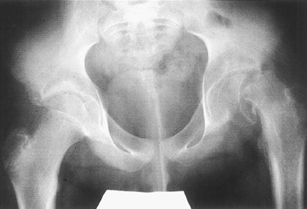
FIGURE 43.3 Slipped capital femoral epiphysis. A 13-year-old boy with a severe left-sided SCFE. (From Swiontkowski MF, Stovitz SD. Manual of Orthopaedics. 6th ed. Philadelphia, PA: Lippincott Williams & Wilkins; 2006.)

eFIGURE 43.3 Posterior dislocation of the hip. A: Mechanism of injury: The knee (1) strikes the dashboard. The thigh (2) is forced into flexion and adduction, and the femoral head is driven backward out of the acetabulum. B: Ischial dislocation: The hip (1) is flexed. The hip is markedly adducted, so that the knee (2) of the affected limb lies on the opposite thigh. The limb (3) is in extreme internal rotation. The greater trochanter and buttock (4) on the affected side are unusually prominent. C: Iliac dislocation: As in (B), the hip is flexed and is adducted (2).
Optimally, posterior dislocations should be reduced within 6 hours to minimize the risk for AVN (7). Therefore, this procedure should be attempted in the emergency department (ED) (eFig. 43.4). Adequate analgesia and muscle relaxation must be obtained using appropriate agents (e.g., a short-acting opioid and a short-acting benzodiazepine or propofol) and monitoring.
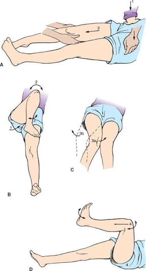
eFIGURE 43.4 Posterior dislocation of the hip: method of manipulative reduction. A: The assistant holds downward pressure (1) on the anterior superior iliac spine. With the knee flexed (2), the operator pulls on the limb in the line of deformity. B: The assistant slowly brings the thigh (1) to 90 degrees of flexion. The assistant gently rotates the thigh internally and externally and rocks the thigh (2) backward and forward to disengage the head from the external rotator muscles and the posterior capsule. C: The assistant relocates the femoral head by further internal rotation and extension of the thigh (3a) or external rotation and extension of the thigh (3b). D: The assistant pushes firmly on the trochanter to direct the femoral head into the acetabulum while the limb is rotated and extended (4). Forceful rotation should be avoided, because it can fracture the femoral neck. If the reduction is not accomplished with two adequate attempts, open reduction is indicated.
Among the several techniques described in the literature, the Allis technique is commonly used to relocate posterior hip dislocations. After the patient is adequately sedated, the hip is flexed and an upward traction is applied to the supine patient. An assistant may apply pressure to the iliac crests, stabilizing the pelvis from lifting off the bed. To apply constant traction to the leg, it is often necessary to stand on the bed and straddle the patient. Upward traction and continued longitudinal force is applied with gentle internal and external rotation. Successful reduction is often felt by a dull click. After reduction, the extremity should be placed in extension and neurovascular function of the extremity should be reassessed. Postreduction radiographs should be obtained to identify the proper location of the femoral head. General anesthesia is occasionally necessary if procedural sedation is unsuccessful.
Anterior Hip Dislocations
Anterior hip dislocations occur most commonly from a direct blow to the abducted hip. A distally applied force causes the femoral head to tear through the anterior capsule and lever out of the acetabular cup. The mechanism of injury is most often an MVC during which the occupant has the hip abducted and externally rotated at the time of impact or a fall or sports injury causing forced hyperextension. Anterior dislocations are classified as obturator, iliac, and pubic (eFig. 43.5).
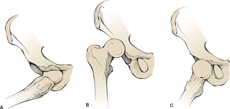
eFIGURE 43.5 Anterior hip dislocations. Anterior obturator hip dislocation (A), anterior iliac hip dislocation (B), and anterior pubic hip dislocation (C).
On physical examination, patients with anterior obturator dislocations present with abduction, external rotation, and flexion of the involved extremity. Patients with anterior iliac and pubic dislocations present with slight abduction, external rotation, and extension of the involved extremity (eFig. 43.6). Following anterior dislocation, a careful neurovascular examination is mandatory as the femoral artery, vein, or nerve are prone to injury.

eFIGURE 43.6 Anterior dislocations of the hip. A: Obturator dislocation. The hip (1) is slightly flexed. The limb (2) is externally rotated. The thigh (3) is abducted. B: Pubic dislocation. The extremity (1) is in severe external rotation (90 degrees). The extremity (2) is abducted only slightly (15 to 20 degrees), and it is slightly flexed. The femoral head (3) can readily be palpated in the inguinal region.
Early reduction is recommended to minimize neurovascular complications, such as AVN. A modified Allis maneuver is a commonly used reduction technique for anterior dislocations. Like the Allis technique, the patient is placed supine and an assistant stabilizes the pelvis. Traction is applied along the long axis of the femur with slight hip flexion. Gentle adduction and internal rotation may encourage reduction. If reduction is unsuccessful using common procedural sedation techniques, closed reduction under general anesthesia may be required.
Prosthetic Hip Dislocations
More than one million hip arthroplasties are done every year worldwide, and the number is projected to double within the next two decades given the ageing population and obesity epidemic (8). After THA, instability in the form of dislocation is second in frequency to joint loosening as a major complication of this procedure. Dislocation of the prosthesis occurs in 1% to 3% of patients with primary THA and in 5% to 20% of patients with a revised THA (9). Dislocations after THA can occur in a posterior or anterior direction. Posterior dislocation is the most common and accounts for 75% to 90% of THA dislocations (10). Dislocations often occur in the first 3 months postoperatively and are generally caused by relaxed soft tissues and immature scar formation (11). Over time, stretching of the pseudocapsule caused by extremes of motion may lessen soft tissue constraints and allow for dislocation.
Reduction techniques for prosthetic hip dislocations are identical to those described earlier. While a dislocated prosthetic hip carries no risk of AVN, injury to the sciatic nerve can occur.
Hip Fractures
Given the increasing elderly population and number of patients with osteoarthritis of the hip, the number of hip fractures may double over the next 20 years, reaching half a million a year by 2050 (12). A third of those over age 65 who live at home and half of institutionalized individuals are estimated to fall yearly. Morbidity in this age group is staggering, with a third dying within 1 year (13). Thirty percent of those who were independently ambulatory prefracture require permanent postinjury crutches or a walker, and 7% of them become permanently bedridden (14).
The term hip fracture refers to a fracture of the femur proximal to the subtrochanteric region. Hip fractures are generally classified into six anatomic groups: femoral head and neck fractures, intertrochanteric fractures, subtrochanteric fractures, and isolated fractures of greater or lesser trochanters. The terms intracapsular (femoral head and neck) and extracapsular (intertrochanteric, subtrochanteric, and greater and lesser trochanter) are also commonly used.
Currently, treatment of these fractures is hemiarthroplasty or open reduction and internal fixation (ORIF) for femoral neck fractures.
Femoral Head Fractures
Isolated fractures of the femoral head are relatively uncommon and most often due to motor vehicle collisions. They are more common in younger patients as the force required often causes femoral neckfractures in older patients. As with dislocations of the hip, acetabular fractures may also be seen. These fractures usually occur in conjunction with hip dislocations caused by severe shearing forces. Femoral head fractures occur in 6% to 16% of hip dislocations (15).
Femoral head fractures may not be visualized on routine radiographs and CT or MRI may be required to confirm the diagnosis. Early recognition and reduction is important to maximize future joint function and minimize complications such as arthritis, AVN of the femoral head, or myositis ossificans. Urgent orthopedic consultation is mandatory. In most cases, satisfactory results can be obtained with closed reduction. However, if the hip cannot be reduced by manipulation or if reduction of the femoral head fragment is unsatisfactory, open reduction will be required. Unfortunately, complications may occur regardless of proper intervention and result in the need for prosthetic replacement.
Femoral Neck Fractures
It is estimated that 5% to 10% of people reaching their 80s will have a femoral neck fracture. These fractures occur below the femoral head and above the greater trochanter and are much more common than femoral head fractures. In the elderly, these fractures may be pathologic, occurring with a fall or minimal trauma. In younger patients, they are usually associated with high-energy mechanisms such as motor vehicle trauma. Osteoporosis is a major risk factor, making the injury four times more common in women than in men.
Several classifications systems have been described but are not consistently used because of poor interrater reliability and limited clinical utility. More commonly, femoral neck fractures are classified as either nondisplaced or displaced. Most femoral neck fractures are displaced and present with the limb externally rotated, abducted, and slightly shortened. Fifteen percent to 20% of all femoral neck fractures are nondisplaced. The prognosis for nondisplaced fractures is excellent; functional outcome is favorable and the majority of patients heal without complication. However, a nondisplaced femoral neck fracture possesses no inherent stability and may become displaced without surgical fixation.
Stress fractures represent a unique type of femoral neck fracture. These fractures most commonly occur in young, athletic individuals, but they also occur in those with pathologic bones, (rheumatoid arthritis, renal osteodystrophy, and chronic steroid use). Stress fractures may be bilateral and are usually the result of repetitive abnormal stress on normal bone or repetitive normal stress on abnormal bone. The pain is often gradual in onset. Ambulation is common, and patients complain of pain with weight bearing. The usual signs of fracture are missing, both clinically and radiographically. Bone scan or MRI is indicated in any high-risk individual who cannot bear weight. Treatment is based upon the degree of injury and the state of the underlying bone. Treatment ranges from resting the affected hip to prosthetic hip replacement.
AVN and nonunion are the two major complications of femoral neck fractures. AVN of the femoral head is most common and may occur despite optimal treatment. It is identified on x-ray as an increase in density in the region of the femoral head thought to be new bone deposition.
Early and precise anatomic reduction reduces the risk of AVN. Prompt recognition and orthopedic consultation in the ED may help minimize AVN and other sequelae. Treatment of displaced fractures often consists of ORIF, hemiarthroplasty, or THA.
Intertrochanteric Fractures
These are the most common types of hip fractures with the majority being caused by falls in the elderly. Intertrochanteric fractures are often considered pathologic, most commonly secondary to osteoporosis. Other causes include cancer and, less commonly, Paget disease. X-ray findings usually show comminution, the majority with three, four, or more fragments (16).
Emergency diagnosis is usually straightforward. Any movement causes extreme pain, and the hip may be tender to palpation. The affected leg is shortened and externally rotated, and weight bearing is not possible. The diagnosis is usually confirmed with a single AP view of the hip. A lateral or groin view may be necessary to evaluate the degree of comminution.
The intertrochanteric fracture is extra-articular and typically extends in a relatively straight line between the greater and lesser trochanters (eFig. 43.7). A variety of classifications are used to describe these fractures with no universal agreement. The Boyd and Griffin classification uses four categories (eFig. 43.7). When obtaining orthopedic consultation, precise anatomic description may be more useful than a specific classification type.

eFIGURE 43.7 Boyd and Griffin classification of intertrochanteric hip fractures. (Adapted from Rockwood CA Jr, Green DP, Bucholz RW. Rockwood and Green’s Fractures in Adults. 3rd ed. Philadelphia, PA: Lippincott Co.; 1991.)
Initial management consists of immobilization with sandbags or Buck traction (2 to 4 kg) which helps decrease blood loss. Intravenous volume replacement, with crystalloid or blood, may be necessary because patients can lose 1 to 2 L of blood with these fractures. Many of these patients have chronic medical problems that should be addressed (including the cause of the fall), and this history will help to establish whether the patient is an operative candidate. Laboratory evaluation should include the standard preoperative labs, as well as analysis necessary to ascertain cause of the fall (may include neurologic and cardiovascular evaluations).
Operative repair has historically been considered safer than conservative therapy. This opinion is a result of the morbidity and mortality associated with extended immobilization. Surgical reduction and fixation should be considered urgently rather than emergently, and all patients should have a thorough preoperative evaluation. Selected fractures may be managed by splinting, traction, or spica immobilization as determined by the consulting orthopedist and the patient’s underlying medical conditions. Complications are common and reflect the frail status of most of these patients. Mortality rates are up to 15% (17).
Greater and Lesser Trochanteric Fractures
Greater trochanteric fractures are rare and occur as a result of direct trauma in adults or secondary to apophyseal avulsion as a result of forceful muscular contraction in young patients (ages 7 to 17). Tenderness to palpation is usually present over the greater trochanter. Patients are usually ambulatory with a limp. When caused by direct trauma, these fractures are rarely displaced and can be difficult to see on routine radiographs, so comparison views of the other leg are recommended. The direct blow (adult) fracture is usually comminuted without significant displacement whereas the epiphyseal avulsion (youth) type is usually displaced but not comminuted. Management varies from adduction splint and bed rest, to ORIF. In most instances, the prognosis is good, regardless of the method used.
Lesser trochanteric fractures usually occur in the young: 85% occur before age 20. These fractures usually occur from forceful contraction of the iliopsoas muscle during strenuous activities. The lesser trochanter can be displaced up to 1 cm. This injury in an adult mandates a search for pathologic bone. Patients have pain and tenderness in the femoral triangle. In a complete avulsion, they are unable to lift the affected extremity in the sitting position (Ludloff sign). Radiographic imaging frequently requires comparison views. Treatment is usually conservative, unless there is significant displacement and outcomes are uniformly good (18).
Subtrochanteric Fractures
Subtrochanteric fractures are defined as fractures occurring between the lesser trochanter and a point 5 cm distally. These fractures account for 10% of hip fractures and are associated with more severe trauma or pathologic bone (such as osteoporosis, Paget disease, or bony metastasis).
Clinical presentation is similar to that of intertrochanteric fractures. The forces of the muscles extending across this region frequently result in abduction, flexion, and external rotation. Young patients with these fractures are subject to violent forces, and associated injuries or other associated hip fractures are common. Blood loss can be substantial in fractures of the midshaft femur. Classification is varied and complex. Anatomic description is best used when describing these fractures.
Treatment involves immobilization, analgesia, volume replacement (if indicated), and timely orthopedic intervention. Examination for associated neurovascular injury should be performed. The presence of a tense, swollen upper thigh should be evaluated with angiography or Doppler for an acute vascular injury. Prompt vascular surgery consultation should be obtained if this is suspected. Operative reduction and fixation (usually intramedullary nail) is used based on specific patient and fracture characteristics.
Acetabular Fractures
Acetabular fractures should be considered in patients with femoral head dislocations and, more commonly, pelvic fractures. The force of injury applied to the femur from impact onto the dashboard is transmitted to the acetabulum.
There are four types of acetabular fractures: posterior lip fracture (most commonly associated with posterior hip dislocations), transverse acetabulum fracture, anterior fracture, and ilioischial (or posterior) fracture. In the ilioischial fracture, the entire posterior column (the bone extending from the ilium to the ischium) becomes separated from the pelvis. An occult acetabular fracture should be suspected when the patient gives a history of a recent fall, but the pelvic radiograph is normal. Oblique views, CT scan, MRI, and bone scan are often necessary for diagnosis, as initial films generally do not visualize the injury. MRI is the gold standard for diagnosis of clinically suspected occult injury (3).
Acetabular fractures are managed in consultation with an orthopedic surgeon. ED management involves immobilization, analgesia, volume replacement (if necessary), and identification of associated injuries.
Slipped Capital Femoral Epiphysis
SCFE is the most common hip disorder in adolescents. If this injury is untreated, it can result in profound lifelong morbidity due to AVN. The diagnosis is initially frequently missed. SCFE is a Salter–Harris type 1 fracture of the femoral capital epiphysis where the head of the femur slips posteriorly and inferiorly. This fracture occurs in boys twice as often as in girls. SCFE prior to age 8 mandates a metabolic workup for endocrine disorders, although ED treatment remains the same. Most affected children are obese, suggesting a primary mechanical problem. However, the disease is considered multifactorial. Up to half of SCFEs present bilaterally and hips are usually affected within 18 months of each other (19).
The most common presentation is a limping obese adolescent, complaining of hip pain. Because of referred pain from the hip, a common presentation is knee pain (15%). The average onset is 13.5 years for boys and 12 years for girls. The hip is externally rotated with an antalgic gait. Bilateral hip radiographs are mandatory and need to include AP and frog-leg lateral views. Klein line will not project through the femoral head if SCFE is present (Fig. 43.3).
All patients with SCFE should be nonweight bearing, and need operative percutaneous fixation of the femoral head. There is controversy as to specifics including timing or surgery and value of reduction, thus immediate consultation with orthopedics is indicated. The most common complication of SCFE is AVN, which ultimately requires hip replacement. Chondrolysis is another potential complication that has decreased with improved surgical techniques, and now occurs in about 1% to 2% of SCFE cases (19).
CRITICAL INTERVENTIONS
• Evaluate patients with hip dislocations for associated acetabular, femur, or knee injuries.
• Perform early closed reduction of hip dislocations (<6 hours is ideal). This treatment lowers the risk of AVN and posttraumatic osteoarthritis.
• Perform a CT or MRI in patients with persistent hip pain in the absence of radiographic findings to identify an occult hip fracture.
DISPOSITION
Orthopedic consultation is recommended for all hip dislocations and fractures. Hip dislocations require careful observation with frequent neurovascular checks. Young healthy patients with nondisplaced femoral neck fractures are typically managed by keeping weight off the joint. Occasionally they will require surgical fixation. Both elderly and young patients with intertrochanteric and subtrochanteric fractures require surgical fixation. Hospital admission for all elderly patients with acute hip fractures is recommended. These patients generally require evaluation and consultation for preoperative medical clearance, as well as frequent neurovascular checks. If there is a strong clinical suspicion of occult disease, then further imaging (bone scanning or MRI) is appropriate, either in the ED or as an inpatient.
Common Pitfalls
• Failure to consider SCFE in adolescent patients with knee or hip pain.
• Failure to image the hip in an elderly patient with a history of a recent fall and a complaint of knee pain.
• Failure to realize that hip pain and inability to walk, even in the absence of trauma, may be due to a hip fracture and advanced imaging should be considered (MRI).
• Failure to look for acetabular fractures in patients with hip dislocations and inferior pubic ramus fractures.
REFERENCES
1. Dennison E, Cole Z, Cooper C. Diagnosis and epidemiology of osteoporosis. Curr Opin Rheumatol. 2005;17:456–461.
2. Lubovsky O, Liebergall M, Mattan Y, et al. Early diagnosis of occult hip fractures MRI versus CT scan. Injury. 2005;36:788–792.
3. Cannon J, Salvatore S, Munro M. Imaging choices in occult hip fracture. J Emerg Med. 2009;37(2):144–152.
4. Pandey R, McNally E, Ali A, et al. The role of MRI in the diagnosis of occult hip fractures. Injury. 1998;29:61–63.
5. Yang EC, Cornwall R. Initial treatment of traumatic hip dislocation in the adult. Clin Orthop Relat Res. 2000;(377):24–31.
6. Brooks RA, Ribbans WJ. Diagnosis and imaging studies of traumatic hip dislocation in the adult. Clin Orthop Relat Res. 2000;(377):15–23.
7. Vecsei V, Schwendenwein E, Berger G. Hip dislocation without bone injuries. Orthopade. 1997;26:317–326.
8. Pivec R, Johnson AJ, Mears SC, et al. Hip arthroplasty. Lancet. 2012;380:1768–1777.
9. Yuan L, Shih C. Dislocation after total hip arthroplasty. Arch Orthop Trauma Surg. 1999;119:263–266.
10. Morey BF. Instability after total hip arthroplasty. Orthop Clin North Am. 1992;23:237–248.
11. DeWal H, Su E, DiCesare PE. Instability following total hip arthroplasty. Am J Orthop. 2003;32(8):377–382.
12. AAOS Bulletin. Femoral Neck Fracture (Adult). Chicago, IL: American Academy of Orthopaedic Surgeons; 1989.
13. Parker MJ, Anad JK. What is the true mortality of hip fractures? Public Health. 1991;105:443–446.
14. Finsen V, Borset M, Rossvoll I. Mobility, survival and nursing-home requirements after hip fracture. Ann Chir Gynaecol. 1995;84:291–294.
15. Asghar FA, Karunakar MA. Femoral head fractures: Diagnosis, management, and complications. Orthop Clin North Am. 2004;35:463–472.
16. Rogers LF. The Radiology of Skeletal Trauma. 2nd ed. New York, NY: Churchill Livingstone; 1992.
17. Rudman N, McIlmail D. Emergency department evaluation and treatment of hip and thigh injuries. Emerg Med Clin North Am. 2000;18(1):29–67.
18. Williams-Johnson J, Williams E, Watson H. Management and treatment of pelvic and hip injuries. Emerg Med Clin North Am. 2010;28(4):841–859.
19. Peck D. Slipped capital femoral epiphysis: Diagnosis and management. Am Fam Physician. 2010;82(3):258–262.