
Christine B. Irish and Christopher T. Bowe
Knee trauma accounts for more than one million emergency department (ED) visits each year (1). The knee joint is made up of bones, ligaments, muscles, and cartilage (Fig. 44.1). The three bones that make up the knee are the distal femur, patella, and proximal tibia. The femoral condyles articulate with the proximal tibia (tibial plateau) to form the weight-bearing portion of the knee joint. The patella does not function in weight bearing, but rather provides protection to the distal femur, in addition to improving stability and strength of the extensor mechanism.

FIGURE 44.1 Anatomy of the knee.
The knee relies on four major ligaments: the medial collateral ligament (MCL), the lateral collateral ligament (LCL), the anterior cruciate ligament (ACL), and the posterior cruciate ligament (PCL) for stability and proper function (2). The MCL provides most of the resistance to valgus stress, while the LCL protects from varus stress. The cruciate ligaments are located within the intercondylar notch. The ACL prevents anterior displacement of the tibia relative to the femur while the PCL prevents posterior displacement (2). The anatomy of the posterior lateral corner of the knee is complex; its major structures consist of the LCL, the arcuate complex, the popliteal tendon, and the popliteal-fibular ligament (3). The posterolateral corner primarily resists posterior lateral rotation of the tibia relative to the femur but also contributes to resisting posterior tibial translation. The posteromedial corner of the knee consists primarily of the posterior oblique portion of the MCL and the associated joint capsule. These structures provide resistance to valgus stress and posterior medial tibial translation. The menisci are cartilage positioned on the articulating surface of the tibia. The menisci function to provide a contact surface between the tibia and femur and distribute and dissipate forces within the knee (4).
The muscles involved with normal knee function are divided into extensors and flexors. Extension is primarily provided by the quadriceps muscles, composed of the vastus medialis, vastus intermedius, vastus lateralis, and rectus femoris. The tendons of these muscles combine to form the quadriceps tendon, which inserts into the superior patella and the medial and lateral retinaculum of the joint capsule. This tendon then becomes the patellar tendon, which connects to the tibial tubercle (5). The primary flexors of the knee are the hamstring muscles, consisting of the semimembranosus, semitendinosus, and the two heads of the biceps femoris of the posterior thigh.
The major vascular structures around the knee are located posteriorly. The popliteal artery is fixed proximally. This tethering makes the popliteal artery susceptible to injury by traction forces, as seen with dislocations. The popliteal artery is also at risk of disruption by fractures or penetrating wounds (6). There is limited collateral circulation, so damage to the popliteal artery places the viability of the lower limb in jeopardy. The sciatic nerve separates into the tibial and peroneal nerves proximal to the knee. The tibial nerve passes posteriorly and is therefore susceptible to the same mechanisms of injury as the popliteal artery, while the peroneal nerve courses superficially around the lateral aspect of the knee to the proximal fibula (6).
CLINICAL PRESENTATION
Fractures
Distal femoral fractures are typically described relative to the femoral condyles and are separated into supracondylar or intercondylar (eFig. 44.1) (7). The proximity of the femoral artery and peroneal nerves to the bone requires that the neurovascular status of the extremity be evaluated and documented in the setting of a fracture.

eFIGURE 44.1 Distal femur fractures. A: Y-shaped intercondylar and B: T-shaped intercondylar.
Tibial plateau fractures occur as a result of varus–valgus forces, axial loads, or both. Fractures of the tibial plateau may cause a major impairment in stability and function of the knee (8). The potential for neurovascular injuries is much higher in patients with proximal tibial fractures than in distal femoral fractures. The potential for the development of a compartment syndrome is also significant. Paresthesias and paralysis from compartment syndromes may be mistaken for a primary nerve injury. Injuries to the menisci, as well as to the collateral and cruciate ligaments, should be suspected as well.
A fracture of the tibial tuberosity often represents an avulsion injury caused by the patellar tendon. This is an uncommon fracture. It occurs with either strong flexion or extension against resistance. Fractures of the tibial tuberosity most commonly occur before closure of the epiphysis. The differential diagnosis of pain over the tibial tuberosity should also include Osgood–Schlatter disease, which is a traction injury to the apophysis of the tibial tuberosity. Although it is most common in boys aged 10 to 13 years, it may be seen in girls, usually aged 8 to 13 years. It is thought that repetitive trauma to the epiphysis results in incomplete separation of parts of the cartilaginous and osseous portions of the tibial tuberosity. Diagnosis is based on the history and physical examination. The tubercle is painful, tender, and usually enlarged. Pain is exacerbated by extension of the knee, particularly against resistance.
Patellar fractures result most commonly from direct forces. Less common are fractures resulting from severe tension placed on the patella by strong contraction of the quadriceps; the usual result is a transverse fracture, which may disrupt the extensor mechanism (5).
Knee Dislocation
Knee dislocations are orthopedic emergencies and require immediate attention. Knee dislocations are generally associated with high-energy trauma. The dislocation is described as anterior, posterior, medial, lateral, or rotary based on the position of the tibia relative to the femur. For a dislocation to occur, significant trauma to the surrounding soft tissue is generally seen, including the collateral ligaments, cruciate ligaments, menisci, joint capsule, and neurovascular structures. Physical examination may be difficult, and a dislocation may spontaneously reduce before the patient arrives at the ED. The patient may describe a history of abnormal position of the joint before reduction. Because of the concern for peroneal nerve and popliteal artery injury, specific attention should be devoted to this aspect of the physical examination. The absence of distal pulses is highly correlated with significant vascular injury; the presence of pulses, however, does not eliminate the possibility of vascular damage (9). Considering the significant mechanisms of injury and the potential for vascular damage, compartment syndrome is also a potential complication.
Patellar Dislocation
The patella may be completely dislocated or subluxed from the intercondylar groove as a result of a direct blow or by hyperflexion of the knee joint. The patella almost always displaces laterally. Diagnosis is usually made by careful physical examination. If the patella has spontaneously reduced, plain films will confirm proper placement and identify any bony injury (9).
Ligamentous Injuries
LCL injuries are more commonly seen after falls and motor vehicle collisions (MVCs). MCL and LCL tears are associated with cruciate ligament injuries, with the LCL injury less likely to occur as an isolated event (2). The patient generally presents with tenderness along the distribution of the ligament. There may be a history of “popping” and abnormal bending of the knee. The physical examination should include the diagnostic tests discussed below, with varus and valgus forces applied to the knee at extension and 30 degrees of flexion.
Injuries to the cruciate ligaments are common with low-velocity trauma as well as with high-energy forces. The ACL is weaker than the PCL and is more commonly injured. Tests for ACL injuries include the Lachman, anterior drawer, and pivot shift tests. The Lachman test is the most sensitive. PCL stability is evaluated by the posterior drawer and posterior sag tests (2).
Meniscal Tears
Meniscal injuries generally occur with a twisting force applied to the knee. Patients frequently complain of hearing a “pop,” which is common but not specific to meniscal injuries. Often, the patient reports locking of the knee, usually a result of a “bucket-handle” tear, with a free central portion of the cartilage becoming lodged in the intercondylar notch. The medial meniscus is most commonly affected. The patient usually presents with a joint effusion.
Tendon Injuries
The extensor mechanism includes the quadriceps tendon, the patella, and the patellar tendon. Injuries to these structures, or avulsion of the tibial tubercle, can disrupt the extension of the knee. Disruption may result from high-energy or penetrating trauma but is also seen with low-energy forces (9). The quadriceps tendon is more commonly ruptured and usually ruptures just proximal to the patella (10). The patellar tendon frequently ruptures at the insertion into the patella. The physical examination generally reveals a swollen, tender knee, and a palpable deficit may be present. Tenderness is usually identified at the site of injury. To evaluate the tendons properly, the patient’s ability to extend the knee fully against resistance must be tested. The diagnosis is usually suspected clinically. Partial injuries may be difficult, if not impossible, to detect in the ED.
DIFFERENTIAL DIAGNOSIS
Common important bony injuries to the knee include fractures, dislocations, and subluxations. Of further concern are tendon and cartilage injuries, as well as injury to the vasculature and nerves surrounding the knee.
ED EVALUATION
The history is extremely useful in guiding the evaluation of the injured knee. The chief complaint, mechanism of injury, and the exact nature and timing of symptoms will help to differentiate acute trauma from chronic injuries. The traumatic mechanism should be fully explored, as well as the position of the knee at the time of injury. The history should also assess for additional injuries, particularly to the hip and ankle.
The physical examination begins with visual inspection of the knee, followed by the remainder of the lower extremity. The examiner should note any obvious deformity, ecchymosis, erythema, edema, or cutaneous lesions. Often, comparison with the uninjured knee will be helpful in assessing for joint effusion and local edema. Following gross inspection, palpation will elicit specific areas of tenderness, crepitus, abnormal skin temperature, or the presence of an effusion. Finally, the examiner should assess active and passive range of motion, alignment, and neurovascular status (2). The comprehensive examination will include evaluation of the knee in the standing, sitting, and supine positions, when not limited by pain or swelling.
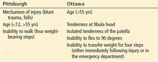
After completion of a thorough physical examination, appropriate radiographic studies may be ordered when clinically indicated. Several clinical decision rules regarding the use of radiographs in the acutely injured knee have been proposed (Table 44.1). Prospective validation has shown excellent sensitivity of these rules (1). Adherence to these clinical decision rules can substantially reduce the incidence of unnecessary radiographs (11). Standard imaging includes the anteroposterior and lateral views and can be supplemented with sunrise (skyline) and tunnel views to evaluate the patellofemoral joint and intercondylar notch.
TABLE 44.1
Decision Rules for the Use of Radiographs in the Acutely Injured Knee
If radiographs are not indicated or are negative, the assessment of potential injuries should continue with a systematic examination of the soft tissues, as well as testing of weight bearing and gait. As previously noted, the unaffected knee should also be examined for comparison. Any obvious discrepancies between the patient’s joints should be noted and are more important than absolute laxity. During examination of the uninjured knee, the physician should demonstrate the specific maneuvers that will be performed on the affected joint. This can diminish the patient’s fears and improve cooperation during examination of the affected knee. Often, the presence of excessive tenderness or large joint effusions will make the examination difficult. In these cases, tensing of the musculature secondary to pain may give the impression of more joint stability than is actually present (2). To facilitate a more accurate examination and appropriate treatment recommendations, it is often necessary to immobilize the injured knee in the ED and arrange for a follow-up examination within 5 to 7 days (12).
Specific Tests for Soft Tissue Injuries
Collateral Ligaments
To assess for integrity of the LCLs and MCLs, the knee should be examined in both full extension (0 degree) and at 30 degrees of flexion. The tibia and femur should be stabilized by the examiner’s hands while applying varus and valgus force across the joint line. Varus force tests for LCL laxity and is noted by lateral joint line opening, whereas valgus force assesses MCL stability and is noted by medial joint line opening. In both tests, the endpoint should be compared to the uninjured knee (Fig. 44.2A,B) (2).

FIGURE 44.2 Examination of the knee. A: valgus stress at 0- and 30-degree flexion, as viewed from the side; B: valgus stress at 0-degree flexion, as viewed from above; C: anterior drawer test; D: McMurray test; and E: Apley test.
Cruciate Ligaments
Given that the primary function of the ACL is to prevent excessive anterior movement of the tibia relative to the femur, the focused physical examination should assess for abnormal anterior tibial translation. The Lachman test is the test of choice to detect ACL injury (2). The patient should be supine with the knee flexed at 30 degrees. The examiner’s hands are placed on the anterolateral femur and posterior proximal tibia. Anterior force is then directed on the posterior tibia, with a positive result noted when there is excessive anterior displacement of the tibia relative to the femur and lack of a distinct endpoint. A positive result indicates an ACL laxity or rupture (13). The Lachman test has a high sensitivity and specificity for ACL injury (approaching 85% and 95%, respectively) (12); False negatives most often result from surrounding muscular contraction in the presence of hemarthrosis, pain, and guarding. False positives may be noted in cases of PCL rupture where the knee is initially subluxed posteriorly (2).
The anterior drawer test may also be utilized to evaluate for ACL injury but it is noted that the accuracy is limited due to hamstring spasm and difficulty achieving appropriate positioning in the acutely injured patient. The knee should be flexed to 90 degrees and stabilized, with the foot flat on the stretcher in a neutral position (Fig. 44.2C). The examiner holds the proximal tibia with both hands and exerts anterior force. Significant displacement of the tibia anteriorly (compared with the unaffected knee) suggests rupture (2).
A third test for ACL injury is the pivot shift, which has a good positive predictive value but poor specificity (12). The examiner should lift the leg by the distal tibia with the knee fully extended. With an ACL rupture, the tibia is subluxed anteriorly in this position. The examiner then applies a mild valgus force while the knee is carefully flexed through 20 to 40 degrees. At this point, the tibia jumps into a reduced position and the examiner and the patient may appreciate a sudden clunk (2). Pain may hinder the performance of this test in the acutely injured knee.
The posterior drawer test is the reverse of the anterior drawer test and is the most accurate test of isolated PCL injury, with sensitivity and specificity of 90% and 99% respectively (2). The posterior Lachman test may be selected as an alternative to the posterior drawer test if flexion to 90 degrees is limited, as the posterior Lachman test is performed in only 30 degrees of flexion. A third technique for detection of PCL injury is the posterior sag test. The examiner holds the knee in 90 degrees of flexion with the thigh supported by a pillow and the foot secured. In this position, the tibia will displace posteriorly or “sag” if the PCL is ruptured (2).
Meniscus
The McMurray test is commonly used to identify meniscal injury (Fig. 44.2D). While the patient is supine, the examiner holds the knee (flexed at 90 degrees) with one hand and holds the lower leg or foot in the other hand. Then the examiner applies internal and external rotation to the lower leg while flexing and extending the knee which is palpated for catching or clicking sensations. A medial meniscal tear should be suspected if clicking occurs as the leg is extended and externally rotated after being completely flexed. Clicking with internal rotation during extension is used to help identify a lateral meniscal injury. A positive test does not guarantee a meniscal injury but indicates the need for further evaluation (14).
Another test used to identify meniscal injury is the Apley test (Fig. 44.2E). The patient is placed in the prone position, and the knee is flexed to 90 degrees. The examiner applies upward force to the tibia while it is rotated, and the knee is extended (14). A positive test is noted with pain and clicking in the joint and suggests a meniscal injury.
Patellar Subluxation
The apprehension sign is seen with patellar subluxation during manual translation maneuvers. The knee is positioned in extension and the examiner exerts lateral force on the patella. In patients with patellar subluxation, this manipulation is painful and induces apprehension. A positive test is noted when the patient contracts the quadriceps and attempts to flex the knee to resist subluxation (2).
Extensor Mechanism
The extensor mechanism of the knee includes the quadriceps muscle, quadriceps tendon, patella, patellar tendon, and surrounding retinacula. As expected, injuries to the extensor mechanism may lead to the inability to extend the leg. Physical examination of the knee should include an evaluation of active leg extension; decreased ability to extend indicates an injury to the extensor mechanism (15).
Diagnostic Studies
As previously mentioned, several excellent clinical decision rules exist to help in the decision to order plain films (Table 44.1). Radiographs help diagnose fractures, effusions, dislocations, foreign bodies, and calcified loose bodies. Additional findings include fluid–fluid levels that may represent lipohemarthrosis (fat floating on synovial fluid and an indication of an occult fracture) in the appropriate clinical setting. The presence of such a finding warrants further evaluation utilizing computed tomography (CT) scanning, or magnetic resonance imaging (MRI).
CT scanning may be used to better define bony injuries, particularly tibial plateau fractures. Ultrasound and MRI are indicated to evaluate the soft tissues of the knee. Angiography can be used after knee dislocation to rule out an occult injury to the popliteal artery, and should be considered even in the presence of symmetric pedal pulses (1). Angiography is also indicated in the presence of penetrating trauma in proximity to vascular structures and (16).
Occasionally, it may be difficult to clinically distinguish hemarthrosis from an infective or inflammatory joint effusion. Arthrocentesis and joint-fluid analysis may be helpful in these cases to determine the etiology of an effusion, particularly when the traumatic mechanism is unclear. An acute hemarthrosis may suggest a significant injury, such as an ACL or PCL tear, and the presence of lipid droplets in a bloody effusion can signify an occult fracture. Arthrocentesis of acute hemarthrosis can be quite therapeutic in providing pain relief, however this practice is controversial given the rate of reaccumulation and the small risk of introducing infection associated with the procedure.
For patients with deep lacerations of the knee, it is important to evaluate the integrity of the joint capsule. Methylene blue should be injected into the joint, and simultaneous inspection of the wound will reveal dye extravasation in cases of joint capsule disruption.
KEY TESTING
• Use of the Ottawa Knee Rules is recommended to reduce unnecessary radiographs in acute trauma setting.
• Remember to examine the ipsilateral hip and ankle for associated injury.
• CT scanning may be particularly useful in patients with tibial plateau fractures.
• Methylene blue injection is useful to assess joint space involvement in deep lacerations.
ED MANAGEMENT
Fractures
The initial management of patients with distal femur fractures includes immobilization, pain management, and evaluation for associated injuries. Early orthopedic consultation is indicated for development of definitive care plans (skeletal traction or surgical repair).
For tibial plateau fractures, diagnostic tests include plain radiographs and CT scans. CT scanning has demonstrated improved accuracy over plain radiography as it is more sensitive for detecting subtle nondisplaced fractures as well as occult lesions. With severely comminuted fractures, CT scanning can quantify the extent of the articular surface involved and the amount of displacement or depression of the fracture line. The additional information gained by CT scanning is useful for planning operative repair, and will often modify the surgical plan (1).
Complications of tibial plateau fractures include vascular injury, deep venous thrombosis (DVT), and compartment syndrome. Any indication of distal ischemia, including paresthesias, diminished distal pulses, or a diminished ankle-brachial index should immediately alert the physician to the possibility of a vascular injury, and an arteriogram should be promptly obtained. Although not generally a concern in the acute setting, DVT is a recognized sequelae of this injury and is most easily diagnosed by lower extremity ultrasound. If significant swelling and tenderness are present, the physician should suspect a developing compartment syndrome. The diagnosis of compartment syndrome is suggested by the presence of the five Ps (pain, pallor, paresthesias, paralysis, pulselessness); however, these findings are not universally present initially and early diagnosis is critical. Compartment pressures should be measured to definitively rule out the diagnosis.
Standard management of tibial plateau fractures includes immobilization, evaluation for other injuries, and timely orthopedic consultation. Depressed fractures require surgical reduction. Because of the risk of compartment syndrome and the possibility of delayed onset, patients with tibial plateau fractures are often admitted for observation.
Tibial tuberosity fractures are generally diagnosed on plain radiographs. In cases of suspected Osgood–Schlatter disease, plain radiographs may be difficult to interpret, as fragments of the tuberosity may be a normal variant. Tibial tuberosity fractures can require open reduction but are usually treated with immobilization. Osgood–Schlatter disease is treated with rest, occasionally plaster cast immobilization, and rarely surgical excision of loose ossicles to relieve symptoms.
Diagnosis of patellar fractures is generally made with plain radiographs. A bipartite patella may be confused with a fracture. Often, the margins of a bipartite patella are rounded and well corticated but, in the setting of acute trauma, it may be difficult to differentiate from an acute fracture. Management is based on the type of fracture, degree of separation, and integrity of the extensor mechanism. Orthopedic referral is indicated. Simple nondisplaced fractures are treated with immobilization and outpatient orthopedic referral. Fractures that disrupt the extensor mechanism, typically displaced transverse or severely comminuted fractures, will require open reduction and internal fixation for optimal results.
Knee Dislocation
Arteriography has been used in the evaluation of knee dislocations to identify injuries to the popliteal artery. If there are signs of vascular injury (absent/decreased pulses, a diminished ankle-brachial index [<0.90], expanding hematoma, hemorrhage, ischemia, or bruit), the patient requires an arteriogram (6). Patients without hard signs may be admitted for serial examinations although some authors have advocated the use of routine arteriography in all patients with posterior knee dislocations, even in patients with intact distal pulses. There is disagreement about mandatory arteriography in patients with intact distal pulses, as the majority of these patients will have nonsurgical lesions, such as intimal tears. Therefore, others advise a more selective approach to arterial imaging, combined with admission and frequent neurovascular examinations (17). CT angiography (CTA) is being used with increasing frequency in place of traditional arteriography to assess for vascular injury, given the reduced radiation dose and less invasive nature of CTA (6).
Emergency management involves rapid reduction of the dislocation, particularly in cases with vascular compromise. Reduction can be accomplished by longitudinal traction (eFig. 44.2). If the clinical status permits, intravenous sedation and analgesia may facilitate the reduction. The limb’s neurovascular status must be reassessed periodically, particularly after manipulation. Orthopedic and vascular surgery consultations should be obtained (6).

eFIGURE 44.2 Technique for reduction of knee dislocation.
Patellar Dislocation
Emergency management of patellar subluxations involves acute reduction. Reduction is typically performed by applying medial force to the patella while the knee is passively extended. Once reduced, the knee is immobilized, and the patient is treated with elevation, ice, crutches, and outpatient orthopedic follow-up.
Ligamentous Injuries
Treatment of patients with collateral ligament injuries is based on the severity of the injury. These patients should be initially treated with immobilization, ice, and crutches. The patient should be referred for timely orthopedic follow-up.
For patients with cruciate ligament injuries, plain radiographs are often nonspecific. Sonography and MRI scanning are used to evaluate the cruciate ligaments and arthroscopy is useful for both diagnosis and treatment (13). Initially, immobilization and orthopedic consultation are necessary.
Meniscal Tears
MRI scanning is very helpful in identifying meniscal injuries. Arthroscopy is generally reserved for treatment, as the accuracy of the MRI is well documented (1). Initial treatment consists of immobilization, analgesia, and outpatient orthopedic referral within 1 week.
Tendon Injuries
X-ray findings may include a high-riding patella (patella alta) with a patellar tendon rupture or low-riding patella (patella baja) with quadriceps tendon rupture (15). CT or MRI may be necessary but can be deferred to outpatient follow-up. A complete rupture requires surgical repair. Incomplete ruptures usually will heal in 6 to 8 weeks with immobilization. Complete ruptures necessitate outpatient orthopedic referral within 5 to 7 days for further evaluation and definitive management.
Bursitis
If bursitis is suspected, rest, ice, and nonsteroidal anti-inflammatory medications are prescribed. The patient may be referred to an orthopedist for follow-up.
CRITICAL INTERVENTIONS
• Rapidly reduce knee dislocations utilizing longitudinal traction after intravenous sedation and analgesia, especially when vascular compromise is present.
• Perform arteriography in patients with knee dislocations, if there are hard signs of vascular injury (absent pulses, expanding hematoma, hemorrhage, ischemia, or bruit).
• Administer appropriate intravenous antibiotics in patients with open fractures.
DISPOSITION
Many of the knee injuries described may be evaluated in the ED and safely referred for orthopedic follow-up as an outpatient. The emergency physician should ensure that the knee is properly immobilized and provide the patient with appropriate analgesia and clear activity restrictions (weight bearing, crutches, etc). Follow-up appointments should generally take place within 1 week.
More severe injuries often require hospital admission. Significantly displaced fractures, open fractures, fractures with associated vascular or nerve damage, and injuries with the potential to develop compartment syndrome should all be admitted for further evaluation and treatment. Such injuries include distal femur fractures, tibial plateau fractures, patellar fractures (with disruption of the extensor mechanism), and knee dislocations.
Common Pitfalls
• Performing a physical examination while the patient remains in the wheelchair.
• Failure to examine the hip carefully. Knee pain is often referred from the hip, and knee trauma may precipitate hip injuries.
• Misinterpreting a bipartite patella as a patellar fracture on plain films.
• Failure to examine the knee in full extension against resistance when evaluating the extensor mechanism.
• Failure to diagnose a proximal fibula fracture or dislocation resulting from an ankle injury.
REFERENCES
1. Tuite MJ, Daffner RH, Weissman BN, et al. ACR appropriateness criteria acute trauma to the knee. J Am Coll Radiol. 2012;9:96–103.
2. Lubowitz JH, Bernardini BJ, Reid JB. Current concepts review. Comprehensive physical examination for instability of the knee. Am J Sports Med. 2008; 36(3):577–594.
3. Vinson EN, Major NM, Helms CA. The posterolateral corner of the knee. AJR Am J Roentgenol. 2008;190(2):449–458.
4. Roulsen MR, Johnson DL. Meniscal injuries in the young, athletically active patient. Phys Sportsmed. 2011;39(1):123–130.
5. Scolaro J, Bernstein J, Ahn J. Patellar fractures. Clin Orthop Relat Res. 2011; 469(4):1213–1215.
6. Howells NR, Brunton LR, Robinson J, et al. Acute knee dislocation: An evidence based approach to the management of the multiligament injured knee. Injury. 2011;42:1198–1204.
7. Crist BD, Della Rocca GJ, Murtha YM. Treatment of acute distal femur fractures. Orthopedics. 2008;31(7):681–690.
8. Manidakis N, Dosani A, Dimitriou R, et al. Tibial plateau fractures: Functional outcome and incidence of osteoarthritis in 125 cases. Int Orthop. 2010; 34(4):565–570.
9. Ramseier LE, Werner CL, Heinzelmann M. Quadriceps and patellar tendon rupture. Injury. 2006;37:516–519.
10. Hak DJ, Sanchez A, Trobisch P, et al. Quadriceps tendon injuries. Orthopedics. 2010;33(1):40–48.
11. Jenny J, Boeri C, El Amrani H. Should plain X-rays be routinely performed after blunt knee trauma? A prospective analysis. J Trauma. 2005;58(6):1179–1182.
12. Spindler KP, Wright RW. Anterior cruciate ligament tear. N Engl J Med. 2008; 359(20):2135–2142.
13. Siegel L, Vandenakker-Albanese C, Siegel D. Anterior cruciate ligament injuries: Anatomy, physiology, biomechanics, and management. Clin J Sport Med. 2012; 22:349–355.
14. Hegedus EJ, Cook C, Hasselblad V, et al. Physical examination tests for assessing a torn meniscus in the knee: A systematic review with meta-analysis. J Orthop Sports Phys Ther. 2007;37(9):541–550.
15. Tuong B, White J, Louis L, et al. Get a kick out of this: The spectrum of knee extensor mechanism injuries. Br J Sports Med. 2011;45:140–146.
16. Medvecky MJ, Zazulak1 BT, Hewett TE. A multidisciplinary approach to the evaluation, reconstruction and rehabilitation of the multi-ligament injured athlete. Sports Med. 2007;37(2):169–187.
17. Miranda FE, Dennis JW, Veldenz HC, et al. Confirmation of the safety and accuracy of physical examination in the evaluation of knee dislocation for injury of the popliteal artery: A prospective study. J Trauma. 2002;52(2):247–252.