
Trevor J. Lewis
Epistaxis is a frequent problem encountered by the emergency physician, accounting for approximately 1 in 200 emergency department (ED) visits in the United States (1). The incidence is bimodal, with peaks at ages younger than 10 and between 70 and 79 years of age (2). The higher incidence in children is attributed to local digital trauma in combination with reduced humidity during winter months (1,3). Pediatric epistaxis is usually benign and treated with local measures (4). Elderly patients are more likely to require invasive therapy and admission, because of both systemic causes of epistaxis and increased endothelial degeneration (3). Epistaxis is rarely life-threatening, and a majority of patients can be managed definitively by the emergency physician. When bleeding is not controlled by local measures, the source is usually posterior, and urgent otolaryngology consultation is required. Most posterior bleeds occur in older patients with the mean age of occurrence reported as 64 years of age (5).
The vascular supply to the nose is extensive, consistent with its function of warming and humidifying air. The blood supply to the nasal mucosa arises from the internal maxillary and facial arteries via the external carotid as well as from the posterior and anterior ethmoidal arteries via the internal carotid (6). The sphenopalatine artery, the terminal branch of the maxillary artery, supplies the lateral nasal wall below the middle turbinates and is the vessel responsible for most posterior epistaxis (7). The superior labial artery and greater palatine artery, terminal braches of the facial and maxillary arteries, respectively, supply the anterior portions of the septum and floor of the nasal cavity. The anterior and posterior ethmoidal arteries supply both the septum and the lateral aspect of the nose superiorly. The confluence of these terminal arteries is referred to as Kiesselbach plexus or Little area. The majority of anterior nosebleeds occur in this area (Fig. 69.1).
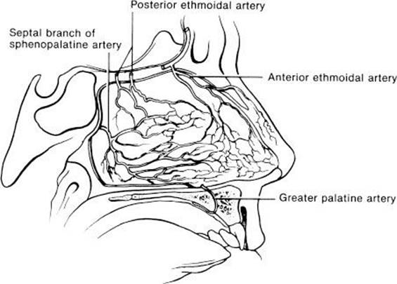
FIGURE 69.1 Blood supply to Kiesselbach plexus.
CLINICAL PRESENTATION
Epistaxis usually presents as spontaneous unilateral bleeding from a discrete lesion. Bilateral epistaxis is more commonly due to trauma or a posterior source of bleeding. Approximately 90% of nosebleeds are anterior, which means the bleeding source can be visualized in the anterior portion of the nasopharynx. The remaining 10% are posterior hemorrhages, in which a bleeding source is not directly visible. Posterior bleeds may present with nausea, hematemesis, anemia, hemoptysis, or melena (7) or may present with sudden copious bleeding due to involvement of larger vessels.
Patients with an underlying coagulopathy may present with other manifestations of bleeding such as bruising, petechiae, bleeding gums, hematuria, or gastrointestinal bleeding. Patients with a significant amount of blood loss may present with signs and symptoms of hypovolemia such as lightheadedness, fatigue, tachycardia, or hypotension.
DIFFERENTIAL DIAGNOSIS
Epistaxis usually occurs from an interaction of factors that damage the superficial blood vessels of the nasal mucosa. The differential diagnosis includes both local and systemic causes, although the majority of cases are idiopathic (8). Local causes include direct facial trauma, nose picking (epistaxis digitorum), and chemical nasal irritants such as cigarette smoke, cocaine, and steroid nasal sprays. Epistaxis may be due to inflammatory disorders such as upper-respiratory infection, sinusitis, and allergic rhinitis. Environmental causes such as dry air during the winter months have been correlated with higher rates of admission for epistaxis (9). Other causes include foreign bodies (especially in children, the developmentally delayed, and elderly institutionalized patients), iatrogenic causes such as nasogastric and nasotracheal intubation, surgical complications, and overzealous treatment of self-limited hemorrhage.
Hypertension has been cited as a cause of epistaxis, but the relationship is controversial. Although studies have shown a higher prevalence of hypertension in epistaxis it is postulated that the sight of blood may cause a hypertensive or “white coat” response (8). Many hereditary bleeding disorders can present with epistaxis, including hemophilia, von Willebrand disease, and thrombocytopenia. Heavy alcohol use and a history of renal failure also increase the risk of epistaxis.
Numerous medications increase the risk of epistaxis via inhibition of normal clotting mechanisms. The most common class is nonsteroidal agents (including aspirin), which interfere with the cyclooxygenase pathway and inhibit platelet aggregation (6). Other commonly implicated medications include warfarin and other antithrombotic agents as well as antiplatelet medications. Many herbal supplements have anticoagulant properties including those that contain garlic, gingko, ginseng, and vitamin E (9).
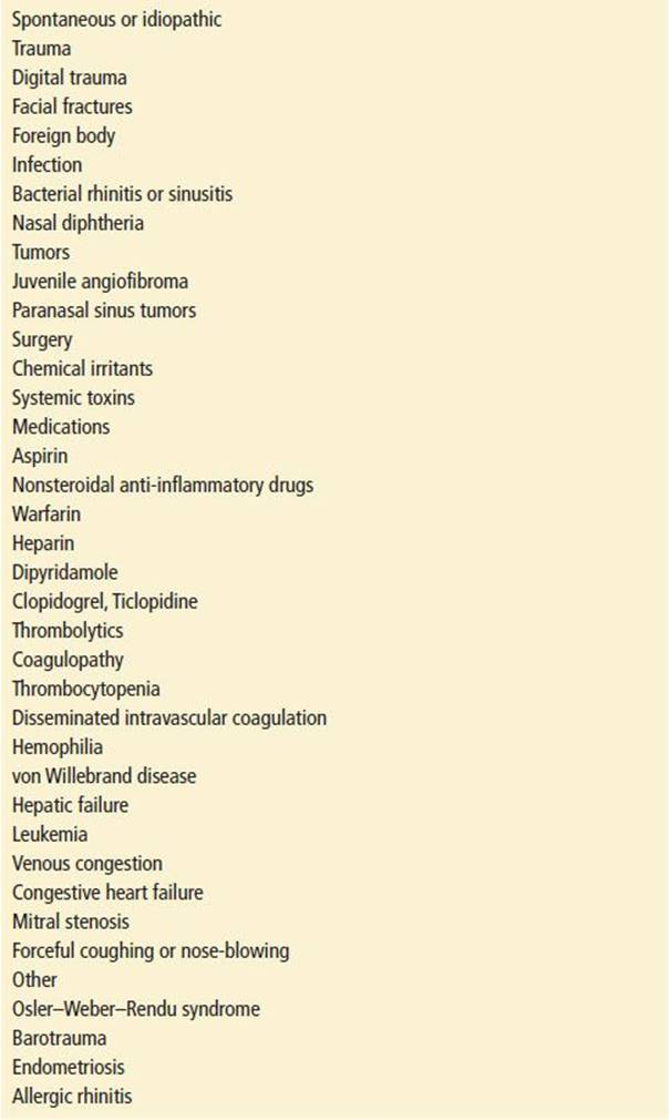
Occasionally, patients present with massive epistaxis that is initially confused with hemoptysis or hematemesis. Blood from an episode of epistaxis that is swallowed or aspirated may further confuse the issue. If blood is coming from the nose and mouth and the source is unclear, the patient should be asked to sit upright, blow the nose, and rinse the mouth with water, and the oropharynx should be examined under suitable light. Blood dripping from the posterior nasopharynx confirms nasal hemorrhage. Table 69.1 lists some causes of epistaxis.
TABLE 69.1
Causes of Epistaxis
ED EVALUATION
Initial evaluation of the patient with epistaxis should focus on control of the bleeding to facilitate a directed history and physical examination and to alleviate the patient’s anxiety. These measures should be initiated in triage and include the direct application of pressure by the thumb and index finger to the nasal alar area and the anterior septal area. The patient should be instructed to bend forward at the waist and spit out any blood that may pool in the throat. Swimmer’s clips may be applied to the nasal area to control hemorrhage (10). Patients presenting with massive epistaxis or unstable vital signs require immediate resuscitation and airway management as well as direct control of the bleeding site. This precludes a lengthy history and physical examination. Once the patient has been stabilized, a more detailed history and examination can be obtained.
The history should focus on the initial site, duration, and amount of bleeding. One should inquire about precipitating events, including trauma and intranasal medications (illicit or otherwise) (3) as well as previous episodes of bleeding and whether control was achieved at home or in the hospital. Episodes of hematemesis may suggest a posterior source of epistaxis. Easy bruising or prolonged bleeding after minor surgical procedures suggests the possibility of coagulopathy, and recurrent episodes of epistaxis, even if self-limited, should raise suspicion for intranasal tumors. Use of alcohol and medications, especially aspirin, nonsteroidal anti-inflammatory agents, warfarin, or other antithrombotic or antiplatelet agents, should be noted, as these not only predispose to epistaxis but make treatment more difficult.
KEY TESTING
• Clotting studies (PT [INR], PTT): only for suspected bleeding diathesis, liver disease, or the anticoagulated patient (11,12).
• CBC, Platelets: if the patient relates a history of easy bruising, platelet disorder, neoplasia, or recent chemotherapy.
ED MANAGEMENT
The first priority in the management of epistaxis is to address the airway, breathing, and circulation (ABCs). Patients with unstable vital signs or severe hemorrhage should have intravenous access, as well as continuous cardiac monitoring and pulse oximetry. Massive hemorrhage may require endotracheal intubation to protect the airway and facilitate nasal packing. Elderly patients with massive bleeding may deteriorate rapidly, so aggressive resuscitation is vital (8). Rapid control of epistaxis that is associated with multiple trauma or significant facial injuries is best secured immediately with epistaxis balloons (13). This allows other life-threatening injuries to be addressed expeditiously. In patients with nontraumatic massive epistaxis, rather than making lengthy attempts at locating a bleeding source, the physician should proceed directly to packing with nasal balloons or tampons.
Most cases of epistaxis, however, are minor, and the episode frequently terminates with the proper application of pressure. When simple compression fails to stop the bleeding, the source of epistaxis must be located and then treated appropriately. Available treatments include topical vasoconstrictors, chemical or electrical cautery, nasal packing using prefabricated nasal tampons or epistaxis balloons, or traditional nasal packing. Every ED should have a prepackaged epistaxis tray or have key materials readily available (Table 69.2).
TABLE 69.2
Equipment Used for Management of Epistaxis

Preparation is the key to successful management. The physician should wear gown, gloves, and protective eyewear in anticipation of droplet spread of blood. The patient should be seated in an adjustable examination chair and draped from the neck down. A headlamp or mirror provides an excellent light source for optimal visualization.
The patient should be sitting upright or leaning slightly forward to prevent blood from tracking into the pharynx. The chair should be adjusted so that the patient is just below eye level and the patient’s head comfortably positioned against the headrest to prevent sudden movements during treatment. The patient should hold an emesis basin under the chin to catch blood running from the nose or mouth.
The patient should be instructed to blow the nose to clear the nasopharynx of blood and clots, even if bleeding has ceased, as local fibrinolysis of clots can result in persistent oozing or recurrence of epistaxis. This also allows for improved access for topical anesthesia (8). An initial attempt to locate the bleeding site should then be undertaken. The nasal speculum is used to spread the nares vertically, with the instrument held in such a manner that the index finger rests on the bridge of the nose for stabilization. The examination begins at Kiesselbach plexus, moving superiorly and posteriorly along the nasal septum, and concludes with the turbinates and lateral wall. Particular attention should be directed to common sites of bleeding: Kiesselbach plexus, the posterior floor of the nasal cavity, the junction of the anterior third and posterior two-thirds of the nasal septum, and the high anterior septum (11). With a well-illuminated and thorough examination, a bleeding point can almost always be identified.
Control of Anterior Epistaxis
If the bleeding point is identified and bleeding is not too brisk, application of oxymetazoline (Afrin) alone, or in combination with chemical cautery (silver nitrate), has been shown to be effective in the management of anterior epistaxis. Studies have indicated Afrin alone controlled 65% of nosebleeds in consecutive patients presenting to the ED (14). Other topical vasoconstrictors include phenylephrine (Neo-Synephrine) or epinephrine (1:1,000) applied to the area via cotton pledgets.
Electrical cautery has little advantage over chemical cautery with silver nitrate sticks (15). The nasal mucosa should be preanesthetized with 4% lidocaine solution on a cotton pledget to reduce discomfort. The principle of cautery is to injure the nasal mucosa and underlying vasculature, resulting in immediate coagulation and vasoconstriction and immediate cessation of bleeding (3). The proper technique is to gently roll the applicator tip over the mucosa for no more than 5 seconds (16) until a grey eschar forms. Brisk bleeding may be slowed by cauterizing the four quadrants around the bleeding site or any identifiable vessel just proximal to the area of bleeding. Brisk bleeding may also decrease the effectiveness of the cautery by rapidly washing away the silver nitrate. Diffuse or extensive cautery should be avoided, and only one side of the septum should be cauterized at one time, so as to avoid septal necrosis or perforation.
For bleeding that is more brisk, pledgets soaked with an anesthetic-vasoconstrictor solution should be inserted into the nose on the side of the bleeding to provide hemostasis and anesthesia (17). An acceptable pledget can be fashioned by winding a piece of cotton tightly around the bayonet forceps. The cotton is pulled off the forceps and soaked in 4% cocaine or a 1:1 solution of 4% topical lidocaine and epinephrine (1:1,000), phenylephrine, or oxymetazoline. Oxymetazoline is suggested for increased safety in the elderly and hypertensive patients (18). Multiple pledgets may be placed, one above the other, onto the floor of the nasal cavity and allowed to remain in place for 10 to 15 minutes. If this fails to stop the bleeding, then cautery should still be attempted over the now well-anesthetized area.
If direct attempts to cauterize a bleeding point fail, anterior nasal packing should be placed. At this point, it may be a good idea to reconsider whether bleeding is posterior, because placing a posterior pack will necessitate first removing any anterior packing. Furthermore, if the site of bleeding is from the meatus, or from the medial surface of a turbinate, packing may prove to be of little value, and attempts to arrest the bleeding with other methods will be needed.
Traditional anterior nasal packing with Vaseline gauze has been supplanted by preformed nasal tampons or epistaxis balloons. Several nasal tampons are currently available, including Merocel, Rhino Rocket, and Rapid Rhino Nasal Pac with Gel Knit. The 85% success rate of Merocel anterior packing is similar to that of traditional Vaseline gauze (8). After sufficient anesthesia is obtained, the Merocel tampon is lubricated with antibiotic ointment and inserted at a 45-degree angle into the nasal cavity for a distance of 1 to 2 cm. Antibiotic ointment is used to minimize infection as well as to lubricate the packing during insertion. The length of the Merocel is grasped with bayonet forceps and the long axis of the Merocel is rotated into the horizontal plane and then pushed straight backward into the nasal cavity. Merocel is supplied in different lengths that may be trimmed with scissors for both anterior and anterior–posterior hemorrhage. If an anterior–posterior sponge is employed, it should be inserted until it touches the back wall of the posterior nasal space. If the pack does not fully rehydrate with blood, then saline should be applied. The drawstring is secured to the patient’s cheek.
The Rhino Rocket is a nasal tampon that is inserted using a supplied applicator. The nose should first be anesthetized with 4% lidocaine. The tampon is lubricated with antibiotic ointment and inserted along the floor of the nose. The Rapid Rhino Nasal Pac with Gel Knit is the newest nasal tampon. This is an inflatable balloon coated with a carboxymethylcellulose hydrocolloid compound. The compound acts as a platelet aggregator and lubricant when soaked in water (7). To insert the Rapid Rhino, the tampon is soaked in sterile water for 30 seconds and then completely inserted into the patient’s nostril. The attached balloon is then inflated with 10 to 15 cc of air. Compared to the Rhino Rocket, the Rapid Rhino appears to be less painful to insert and easier to remove and to have similar effectiveness in stopping epistaxis (17).
Traditional anterior nasal packing is more difficult but may be considered if the foregoing measures are unavailable or unsuccessful. The nose should be packed with sterile petrolatum ribbon gauze (0.5 to 1 in wide), to which is added an antibacterial ointment to prevent toxic shock syndrome. Packing begins by grasping the ribbon with the bayonet forceps about 6 in from its end. The ribbon is then placed in the nasal cavity as far back as possible, while making sure that the free end still protrudes from the nose. This first pass is pressed onto the floor of the nasopharynx with the closed forceps. The ribbon is then grasped about 4 to 5 in from the nasal alae, the nasal speculum is repositioned so that the lower blade holds the ribbon against the lower border of the nasal alae, and a second strip is brought into the nose and pressed downward. This process is continued superiorly in a stair-step fashion until there is no room left in the nose. Both ends of the ribbon must protrude from the anterior end of the nose, and the nostril is covered with a piece of gauze and secured with tape (19).
Other alternatives to the traditional anterior nasal pack include calcium sodium alginate (Kaltostat), Oxycel, Avitene, and balloon tamponade. Topical thrombogenic agents such as fibrin glue, gelatin sponge (Gelfoam), and microfibrillar collagen powder (Avitene) are useful in cases of epistaxis associated with coagulopathy when applied directly to the bleeding area. Floseal, a hemostatic sealant composed of collagen-derived particles and topical-derived thrombin, is applied as a gel to control epistaxis. When compared to traditional nasal tampons Floseal was found to be better tolerated and to provide superior epistaxis control. It is more costly, however (2). Thrombin-JMI epistaxis kit is a bovine derived topical thrombin that is reconstituted with saline and applied via a nasal mist device.
Control of Posterior Epistaxis
Bleeding that begins with the sensation of blood in the posterior pharynx, bleeding that cannot be localized anteriorly, or bleeding that fails to arrest with anterior packing is considered posterior and needs to be managed in consultation with an otolaryngologist. However, bleeding should be initially controlled by the emergency physician using one of a number of available techniques.
The traditional method of posterior packing with gauze rolls attached to strings is cumbersome and should be replaced with the use of an epistaxis balloon or inflatable Foley catheter. Balloon catheters are easier to install and just as effective as traditional posterior nasal packs (20). Posterior nasal packing should always be accompanied by anterior nasal packing. When the posterior balloon is inflated and pulled forward, it acts to seal the posterior choana. The anterior naris is then packed with a nasal tampon or by inflation of an anterior balloon, effectively tamponading the entire cavity. Hence, it must be kept in mind that if the posterior pack dislodges, a large amount of blood can be aspirated (20). Posterior packs are uncomfortable when placed and usually require opioids for analgesia. Continuous pulse oximetry and cardiac monitoring are mandatory as posterior packing can cause hypoxemia, apnea, and cardiac dysrhythmia because of stimulation of the nasopulmonary reflex (3).
The commonly used balloons for control of posterior epistaxis include the Epistat nasal catheter, the Brighton balloon, and the Simpson balloon. The Epistat catheter is placed by inserting the catheter’s entire length along the floor of the nose. The posterior balloon (white valve) is inflated with 10 cc of saline and pulled forward to occlude the posterior choana. The anterior balloon (clear valve) is then inflated with up to 30 cc of saline, unless the bleeding stops or the septum starts to bow. Rapid Rhino has a similar 9 cm dual port posterior pack.
If a Foley catheter is to be used, a 12 to 16Fr catheter with 30-cc balloon is placed into the nose along the floor of the nasopharynx until the tip is visible through the mouth in the posterior pharynx. It is then slowly inflated with 15 mL of saline, pulled anteriorly until it is firmly set against the posterior choana, and secured into place with an umbilical clamp wrapped around a dental roll or matted 4- × 4-in gauze, so as to avoid direct pressure necrosis of the ala or columella.
A traditional posterior pack is fashioned from a 4- × 4-in gauze rolled into a 1-in roll and secured with 3-silk sutures or umbilical tapes. This pack is brought into position in the posterior choana by passing a small red rubber catheter through each nostril and back out of the mouth, securing each of the two outer sutures to the catheters and pulling the catheters out of the nose again, drawing the pack through the mouth and into position. Each of the sutures is tied together over a gauze buttress to prevent pressure necrosis of the columella. The third suture is brought out through the mouth and taped to the cheek to facilitate removal. Posterior packing is successful in stopping posterior bleed 70% of the time (21).
If unable to control posterior bleeding with traditional packing, emergent otolaryngology evaluation is necessary for possible endoscopic evaluation and cautery/ligation. Many otolaryngologists opt for early management of posterior bleeds with endoscopic cautery under direct visualization in the operating suite, early surgical control using arterial ligation, or referral to interventional radiology for selective arterial embolization (22). If time permits and bleeding is not severe, early consultation may spare patients with posterior nasal hemorrhage the discomfort of posterior packing altogether.
CRITICAL INTERVENTIONS
• Aggressively resuscitate patients with significant hemorrhage and signs of hypovolemia.
• Instruct patients who present to triage with epistaxis to apply squeezing pressure to the nose, as most nosebleeds are anterior and will terminate with this maneuver.
• For posterior epistaxis, control the hemorrhage with a balloon catheter, consult an otolaryngologist, and admit to the hospital.
DISPOSITION
Patients with a history of severe epistaxis or recurrent episodes of bleeding should be referred to an otolaryngologist for an outpatient examination to rule out a neoplastic process. Children with recurrent events should be evaluated for bleeding disorders (23). Patients with anterior packing should have follow-up for packing removal in 48 to 72 hours.
All patients with posterior packing should to be admitted to a high-acuity setting. These patients are at risk for cardiac and pulmonary complications and necessitate close monitoring. Clinicians should also strongly consider admitting elderly patients or those with cardiac or pulmonary disease who require anterior packing. Patients who are coagulopathic or anemic often require admission for transfusion.
Patients who are discharged from the ED should be instructed to avoid blowing or picking the nose, straining, bending over, or participating in sports and strenuous activities and to sneeze with their mouth open. First aid instructions should be given on how to control epistaxis at home and when to seek medical help. Home humidifiers and saline nasal sprays may be helpful in drier, colder months, and patients should avoid any manipulation of the nose. Topical Naseptin cream or petroleum jelly may limit recurrent bleeds (8). Patients discharged with nasal packing should be prescribed an antibiotic, such as ampicillin/sulbactam or a second-generation cephalosporin, to prevent toxic shock syndrome or sinusitis (24). Aspirin may be discontinued if the benefit is felt to offset any risk of thromboembolic events. Anticoagulated patients with an INR in the therapeutic range can continue warfarin without interruption (25).
Common Pitfalls
• Failure to prepare the necessary equipment for control of epistaxis leads to unnecessary inconvenience and delays.
• Epistaxis in young children should prompt a search for a foreign body.
• Failure to identify the source of bleeding or to place an anterior pack properly leads to rebleeding and necessitates a return to the ED for repacking.
• Failure to prescribe antibiotics for patients discharged with an anterior pack can lead to sinusitis or toxic shock syndrome.
• Failure to identify posterior epistaxis until after an anterior pack has been placed necessitates completely unpacking the nose so a posterior pack can be placed.
ACKNOWLEDGMENTS
The author gratefully acknowledges the contributions of Jeffrey A. Evans and Todd C. Rothenhaus to the content of this chapter.
REFERENCES
1. Pallin DJ, Chang YM, McKay MP, et al. Epidemiology of epistaxis in US emergency departments, 1992 to 2001. Ann Emerg Med. 2005;46:811–816.
2. Mathiasan RA, Cruz RM. Prospective, randomized, controlled clinical trial of a novel matrix hemostatic sealant in patients with acute anterior epistaxis. Laryngoscope. 2005;115:899–902.
3. Emanuel JM. Epistaxis. In: Cummings CW, Frederickson JM, Harker LA, et al., eds. Otolaryngology: Head and Neck Surgery. 4th ed. St Louis, MO: Mosby; 2005.
4. Damrsoe JF, Maddalozzo J. Pediatric epistaxis. Laryngoscope. 2006;116:387–393.
5. Schlosser R. Epistaxis. N Engl J Med. 2009;360:784–789.
6. Tan LK, Calhoun KH. Epistaxis. Med Clin North Am. 1999;83:43–56.
7. Douglas R. Wormald PJ. Update on epistaxis. Curr Opin Otolaryngol Head Neck Surg. 2007;15:180–183.
8. Pope LE, Hobbs CL. Epistaxis: An update on current management. Postgrade Med J. 2005;81:309–314.
9. Falcon-Chevere J, Giraldez L, Rivera J, et al. Critical ENT skills and procedures in the emergency department. Emerg Med Clin North Am. 2013;31(1):29–58.
10. Vaghela HM. Using a swimmers nose clip in the treatment of epistaxis in the A & E department. Accid Emerg Nurs. 2005;13(4):261–263.
11. Jackson KR, Jackson RT. Factors associated with active, refractory epistaxis. Arch Otolaryngol Head Neck Surg. 1988;114:862–865.
12. Thaha MA, Nilssen EL, Holland S, et al. Routine coagulation screening in management of emergency admission for epistaxis—is it necessary? J Laryngol Otolayngol. 2000;114:38–40.
13. Keen MS, Moran WJ. Control of epistaxis in the multiple trauma patient. Laryngoscope. 1985;95:874–875.
14. Krempl GA, Noorily AD. Use of oxymetazoline in the management of epistaxis. Ann Otol Rhinol Laryngol. 1995;104:704–706.
15. Toner JG, Walby AP. Comparison of electro and chemical cautery in the treatment of anterior epistaxis. J Laryngol Otol. 1990;104:617–618.
16. Hanif J, Tasca RA, Frosh A, et al. Silver nitrate: Histological effects of cautery on epithelial surface with varying contact times. Clin Otolaryngol. 2003;28:368–370.
17. Kucik CJ, Clenney T. Management of epistaxis. Am Fam Physician. 2005;71:305–311.
18. Singer AJ, Blanda M, Cronin K, et al. Comparison of nasal tampons for the treatment of epistaxis in the emergency department: A randomized control trial. Ann Emerg Med. 2005;45:134–139.
19. Long SC, Roe RJ, Karkanevatos A. No frills management of epistaxis. Emerg Med J. 2005;22:470–472.
20. Davis JP. Respiratory obstruction associated with use of the Brighton epistaxis balloon. J Laryngol Otol. 1993;107:140–141.
21. Gifford T, Orlandi R. Epistaxis. Otolaryngol Clin North Am. 2008;41(3):525–536.
22. Moreau S, Valdazo A. Supraselective embolization in intractable epistaxis: Review of 45 cases. Laryngoscope. 1998;108:887–888.
23. Kubba H, MacAndie C, Botma M, et al. A prospective, single-blind, randomized controlled trial of antiseptic cream for recurrent epistaxis in childhood. Clin Otolaryngol. 2001;26:465–468.
24. Randle DA. Epistaxis packing. Postgrad Med. 2006;119:77–82.
25. Srinivasan V, Patel H, John DG, et al. Warfarin and epistaxis: Should warfarin always be discontinued? Clin Otolaryngol. 1997;22:542–544.