
Brian Chinnock
Vertigo, along with dizziness and other balance disorders, affects up to 70% of people during their lifetime. Vertigo is often categorized into peripheral and central causes. Peripheral disorders may cause considerable symptomatology but are rarely life-threatening, whereas those of central etiology may have significant morbidity and mortality.
PERIPHERAL AND CENTRAL VESTIBULAR ANATOMY
Maintenance of balance and coordination of eye movements is managed by a complex interaction of the vestibular system, the cerebellum, the brainstem, the ocular muscles, the visual cortex, and their interconnecting pathways. The peripheral vestibular system consists of three semicircular canals and two otolithic organs, the utricle, and saccule (Fig. 70.1A). Together they respond to head movement in all directions. Each canal is filled with endolymph, and has a dilated end called the ampulla which has the sail-like cupula. With head movement, the vestibular hair cells embedded in the cupula are stimulated and send input to the vestibular nerve (vestibular branch of CN VIII). Each semicircular canal is paired, and on head movement, one will give an excitatory input to the vestibular nerve, whereas the other will give an equal inhibitory input. With a unilateral peripheral vestibular deficit, the affected semicircular canal will not send in an inhibitory input to the vestibular nuclei, thus giving sensation of movement even if the head is still.
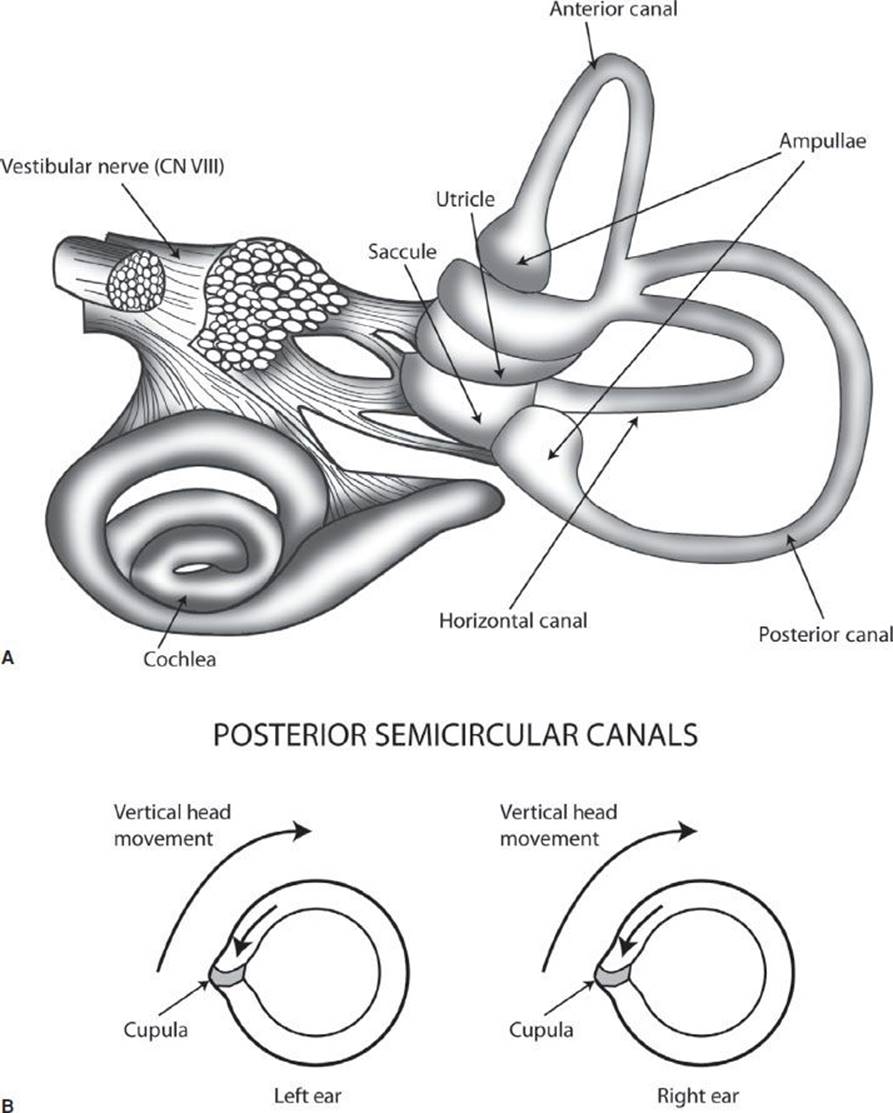
FIGURE 70.1 A: demonstrates the organs of the peripheral vestibular system. B: demonstrates the paired posterior semicircular canals, with vertical head movement causing the more static endolymph inside the canal to cause pressure against the cupula, which stimulates the vestibular nerve.
The central vestibular system receives input through the vestibular nerve to the vestibular nuclei located in the brainstem. From the vestibular nuclei, there are neuronal connections through the medial longitudinal fasciculus to the ocular muscles. This pathway is responsible for the vestibuloocular reflex (VOR), which does not involve the cerebellum, and is tested with the head impulse test (HIT) (see Figure 70.3). The cerebellum is the main organ of the central vestibular system, coordinating vestibular, visual, and somatosensory input to help fine-tune motor activity. The cerebellum is composed of the lateral hemispheres and the midline vermis. The lateral hemispheres are responsible for limb movement, whereas the vermis is responsible for truncal motion. The cerebellum receives its blood supply from the posterior-inferior cerebellar artery (PICA), the anterior-inferior cerebellar artery (AICA), and the superior cerebellar artery (SCA). All originate from the vertebrobasilar system. Importantly, the cerebellum is supplied by the distal branches of each of these arteries, with the proximal parts supplying the lateral brainstem. For this reason, it is common for cerebellar CVA to also present with brainstem findings.
PERIPHERAL VERSUS CENTRAL VERTIGO
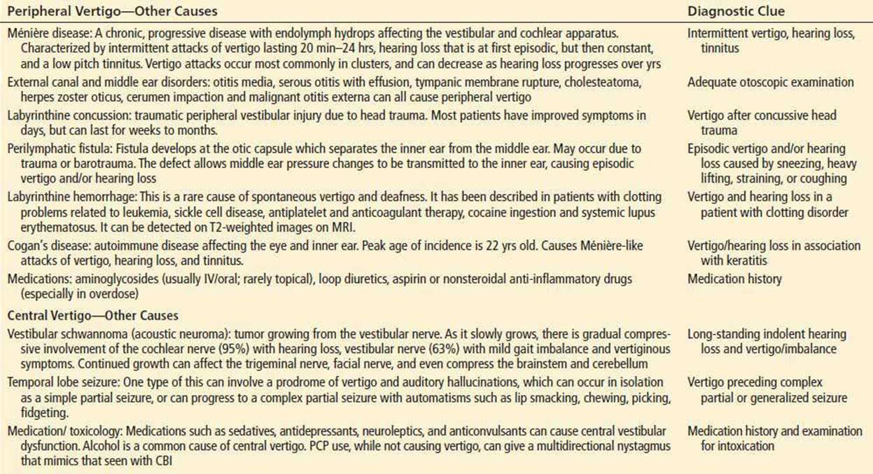
While there are many causes of vertigo, there are four primary diagnoses of significance to the emergency physician evaluating a patient with acute vertigo: (1) benign paroxysmal positional vertigo (BPPV), (2) vestibular neuronitis (VN), (3) migrainous vertigo, and (4) cerebellar/brainstem ischemia (CBI). The discussion will focus on these four diagnoses as they are the most common and important. CBI, which includes cerebellar/brainstem transient ischemic attack/cerebrovascular accident (TIA/CVA) or vertebrobasilar insufficiency, only represents 1% to 3% of vertigo patients, but has a mortality rate of up to 40% (1). It is thus the most important differential diagnosis for any vertigo patient. Other causes of peripheral and central vertigo are listed in Table 70.1.
TABLE 70.1
Less Common Causes of Peripheral and Central Vertigo
BPPV: This is the most common cause of vertigo, representing up to 50% of cases. It has a cumulative incidence of nearly 10% by the age of 80. It is caused by otoconia, calcium carbonate crystals present in the utricle, that become dislodged and migrate into the semicircular canal. The presence of otoconia in the endolymph of one ear will cause increased excitatory input to the vestibular nuclei compared to the unaffected ear on head movement, causing vertigo. BPPV is characterized by intermittent episodes of sudden severe vertigo precipitated by head movement, commonly with nausea and vomiting, which usually last for less than 1 to 2 minutes. When the head stops moving, the vertigo will abate as the movement of the otoconia is the only source of abnormal input; the rest of the vestibular structures are fully functional. As the posterior canal is the most dependent of the semicircular canals and the most likely location for otoconia to settle by gravity (see Fig. 70.1A), 85% of BPPV is caused by posterior canal otoconia. While BPPV is commonly idiopathic, it may also be due to otoconia dislodged due to minor trauma or whiplash injuries, or as a residual effect of Ménière disease.
VN: VN is caused by a selective inflammation of the vestibular nerve, likely due to a post viral cause. Concomitant involvement of the cochlear nerve is termed “labyrinthitis,” and has the same physical examination findings as VN, in addition to hearing loss. Unilateral vestibular nerve involvement will give unequal input to the vestibular nuclei, and cause vertigo. Unlike BPPV, though, keeping the head completely still will not significantly affect the vertigo, as the defect is in the nerve itself, not the canals which give output based on movement. Resolution of symptoms typically takes weeks as recovery is dependent on the vestibular nerve regenerating, and central vestibular compensation mechanisms to develop, as the nerve regeneration is typically incomplete.
Migrainous vertigo: In neurootology clinics, this is the most common cause for referral. Criteria have been developed for the diagnosis of migrainous vertigo and are listed in Table 70.2. Despite these criteria, migrainous vertigo is a diagnosis of exclusion and can be the most difficult diagnosis to make as the vertigo can have either peripheral or central features (2). In addition, the quality and duration of vertigo can range from the mild, indolent quality of CBI which lasts for days, to the severe quality of BPPV or VN, lasting for minutes or over a day, respectively.
TABLE 70.2
Neuhauser Criteria for Migrainous Vertigo

CLINICAL PRESENTATION
A good history is extremely important in ruling out CBI in the vertigo patient. In one study, patients with isolated dizziness and no findings suggestive of CVA had a <1% incidence of CVA (3). Risk factors for CBI are the same as for other types of CVA: previous CVA, diabetes, hypertension, advanced age, atrial fibrillation, hypercholesterolemia. In a case series of patients with vertigo who had a final diagnosis of cerebellar CVA, and who had negative initial diffusion-weighted MRI (DW-MRI), the mean age was 72 years old and all had significant risk factors for CVA (4).
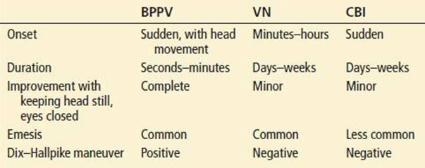
BPPV is usually easy to differentiate from VN and CBI by its sudden onset, short duration, and complete resolution of symptoms in between episodes (Table 70.3). Vertigo that is associated with particular head positioning is almost always due to BPPV (Table 70.3). However, rarely, a patient may have central positional vertigo due to a very localized infarction of CN VIII at the brainstem or of the vestibular nucleus. Either of these may cause a positional vertigo without other neurologic findings. The Dix–Hallpike maneuver (described below) may be particularly helpful in differentiating in this circumstance. Patients with migrainous vertigo usually have a previous history of migraines, with new vestibular symptoms which may or may not occur at the same time as the migraine.
TABLE 70.3
Features of BPPV in Comparison with VN and CBI
DIFFERENTIAL DIAGNOSIS
Traditional teaching emphasizes the differentiation of vertigo from presyncope, disequilibrium, or nonspecific dizziness. However, this approach may be problematic. A large ED-based study found that the incidence of CBI was similar in patients with vertigo versus nonvertiginous dizziness (3). Another ED study found that patients’ descriptions of dizziness were unclear, imprecise, and unreliable (9). Therefore the many causes of vertigo should be at least considered in patients without a clear description of vertigo.
ED EVALUATION
Physical Examination
Standard neurologic examination: All the standard elements of a neurologic examination should be completed, as the presence of any abnormalities can suggest CBI as the cause. Finger–nose testing for dysmetria by itself is not an adequate test of cerebellar function, since it tests only lateral hemispheric function. Large cerebellar vermal CVAs can have a completely normal finger–nose test. Testing of gait is extremely important as patients with peripheral vestibular disorders, while often having significant symptomatology trying to walk, will still be able to walk. Inability to walk (or sit or stand), strongly suggests CBI.
Nystagmus testing should be done by having the patient follow the examiner’s finger in all horizontal and vertical directions. It is also important to hold the finger for 10 to 15 seconds at eccentric vertical and horizontal gaze positions (testing for gaze-evoked nystagmus [GEN]), and then allowing the eyes to return to normal position (testing for rebound nystagmus). When doing this, it is important to not test at the very extremes of gaze as this can evoke a sustained nystagmus even in normal patients. In VN, the nystagmus will be unidirectional, horizontal, with perhaps a slight rotatory component, suppressible by fixating on a target. CBI is more likely if the nystagmus is vertical, purely rotatory, or is not suppressible. Physiologic end-point nystagmus (EPN) occurs upon extreme lateral gaze, when a few beats of low-amplitude nystagmus occur before normal brainstem mechanisms allow the eyes to maintain the eccentric gaze without further nystagmus.
GEN occurs on far eccentric gaze, with sustained, large-amplitude nystagmus. This is caused by an abnormality in the cerebellum or cerebellar pathways. A small lesion of CBI will only manifest at very extremes of gaze. A larger lesion can appear as soon as the eyes deviate from straight-ahead. A vertical GEN, in particular, is diagnostic of cerebellar or brainstem dysfunction. With VN, as the eyes move eccentrically in the direction of the fast phase of nystagmus, the nystagmus will increase until it eventually suppresses with fixation, whereas fixation is unlikely to suppress with CBI.
Rebound nystagmus occurs after holding the eccentric gaze position for 10 to 15 seconds, then returning to the primary gaze position. Nystagmus with the fast phase in the opposite direction of the preceding eccentric gaze indicates CBI. Reversible nystagmus occurs when the direction of the fast phase changes when looking side to side, and is indicative of CBI.
Smooth pursuit is assessed by watching eye movements while performing the nystagmus assessment. It is normal for patients to have slight saccades (fast, jerky eye movements) interrupting smooth pursuit during downward gaze, but should otherwise be smooth without saccades. Significant asymmetry of saccadic motion between each eye is suggestive of a structural brainstem/cerebellar lesion. Saccades due to slow eye movements (eyes have to make saccades to catch up with pursuit) can also be seen with medication/drug usage (alcohol, benzodiazepines).
Skew deviation is tested by asking the patient to fixate on the examiner’s nose, covering one eye, then uncovering that eye while simultaneously covering the other, alternating simultaneous cover and uncover every 1 to 2 seconds. Normally, there is little or no eye movement to regain fixation on the examiner’s nose. With CBI, fixation is not maintained and the eyes make a movement, usually vertical, to regain fixation. For example, as the left eye is uncovered it makes a vertical downward movement to regain fixation, and then, as the right eye is uncovered while the left eye is covered, the right eye makes a vertical upward movement to regain fixation. While not sensitive for CBI, the presence of skew deviation is very specific for CBI.
Dix–Hallpike: The Dix–Hallpike test should be performed if BPPV is a diagnostic consideration. The Dix–Hallpike test positions the head so that otoconia in the posterior semicircular canal move to cause maximal stimulation, with nystagmus and vertigo (Fig. 70.2, panels S and 1). The maneuver has an approximately 50% to 88% sensitivity in detecting BPPV (5).

FIGURE 70.2 Dix–Hallpike test and Epley maneuver. Positioning sequence for left posterior semicircular canal as viewed by operator (behind patient). Small boxes show the labyrinth and migration of particles (large arrow). (S) and (1) represent a Dix–Hallpike test of the left posterior semicircular canal, while the entire sequence demonstrates the Epley maneuver. (S) Start-–patient seated. 1: Place head over end of table, 45 degrees to the left. For Dix–Hallpike test, with a positive test, there will be a short latency period, followed by an upward and torsional nystagmus and vertigo, which fatigue in about 15 to 30 seconds. When the nystagmus fatigues, the patient is returned to the sitting position, at which point the vertigo and nystagmus return briefly, the nystagmus now in the opposite direction. 2: Keeping head tilted downward, rotate to 45 degrees right. 3: Rotate head and body until facing downward 135 degrees from supine. 4: Keeping head turned right, bring patient to sitting position. 5: Turn head forward, chin down to 20 degrees. Pause at each position until induced nystagmus approaches termination, or for approximately 30 seconds if no nystagmus. If able, keep repeating entire series (1 to 5) until no nystagmus in any position.
“Bow and lean” test: Patients who have symptoms consistent with BPPV, but a negative Dix–Hallpike maneuver, should have this maneuver performed to test for horizontal canal otoconia, which can represent up to 10% to 20% of BPPV cases. In this maneuver, the seated patient first bends the head forward (“bow”), aligning the horizontal canal with gravity, and then leans their head backward quickly, flipping the canal 180 degrees. The examiner looks for nystagmus in each position. With right ear horizontal canal otoconia, there will be a right-beating nystagmus on the bow, and a left-beating nystagmus on the lean. Knowing which canal is involved is important in determining the treatment maneuver.
Head impulse testing: The HIT evaluates the VOR, and is vital at differentiating VN from CBI. It is described in Figure 70.3. The sensitivity of HIT for detection of VN is 72% when done by “nonexperts” (6). A study in which the HIT was used in patients at high risk for CVA showed that 31/34 patients with a final diagnosis of CVA had a negative HIT (7). In patients suspected to have VN, the triad of a positive HIT, unilateral nystagmus, and absent skew deviation excluded 91% acute CVAs and identified 78% of peripheral vestibular lesions (8). These studies demonstrate that a positive HIT by itself must be viewed with caution in ruling out CBI, however, in association with the lack of other findings suggesting CBI (see Table 70.4). The ability to rule out CBI is similar to that of MRI, especially within the first 24 hours.

FIGURE 70.3 Head impulse test. This is a test of the VOR. The examiner grasps the sides of the patient’s head and asks the patient to fixate on the examiner’s nose (A). The examiner asks the patient to relax the head, and then performs a low-amplitude, high-velocity, random movement of the head to one side or the other, while having the patient maintain fixation on the nose. With an intact VOR (intact vestibular system), the patient’s eyes will maintain fixation (B). C demonstrates an abnormal VOR: On movement of patients head to the right, the patient briefly loses fixation on the examiner’s nose, and then has a rapid saccade of the eyes to the left to regain fixation (D).
TABLE 70.4
Features Differentiating VN from CBI

Laboratory Testing
Laboratory testing has a very limited role in the evaluation of vertigo. Consider evaluating for hypoglycemia, anemia, thyroid dysfunction, uremia, or electrolyte abnormalities in the patient who has a broader differential diagnosis than vertigo.
Imaging
Imaging has little role in the evaluation of BPPV or VN, except to evaluate for the serious alternative, CBI. CT scan may reveal hemorrhagic CVA. Beyond that, it has no role in the evaluation of CBI since the sensitivity for ischemic CVA is only 40%, and possibly lower for posterior fossa pathology. DW-MRI has become the standard of care in the diagnosis of ischemic CVA. DW-MRI can demonstrate an acute ischemic CVA even in the first few hours. However, the false-negative rate is 19% within 12 hours and 24% within 3 hours (10). The same artifacts that make CT difficult to evaluate the posterior fossa, also make MRI difficult to evaluate in the acute setting. For this reason, a normal DW-MRI is not reliable in ruling out CBI in the first 24 hours of symptom presentation.
KEY TESTING
• Physical examination maneuvers to detect findings consistent with central causes.
• Obtain MRI when central vertigo is suspected, but be cautious of a normal result in the first 24 hours.
ED MANAGEMENT
Vestibular neuritis: In the acute phase, medications that work either as vestibular “sedatives” (decreasing abnormal input from the vestibular system) or antiemetics can be used to decrease symptomatology. These medications include antihistamines, benzodiazepines, antiemetics, and scopolamine. Little evidence supports the use of one treatment over another. One trial examining the IV use of dimenhydrinate 50 mg versus lorazepam 2 mg found less sedation and better symptomatic relief in the dimenhydrinate group (11). Another study showed equivalent effect of IM dimenhydrinate 50 mg and droperidol 2.5 mg (12). All of these classes of drugs can have significant side effects in elderly patients, and the risk–benefit ratio should be carefully weighed in this patient population. For more long-term relief, the use of steroids has been examined. Two studies, one using methylprednisolone in a 22-day taper and another using prednisone 1 mg/kg for 5 days with a 15-day taper, showed no better symptom improvement than placebo at 12-month follow-up (13,14). However, the smaller study using prednisone demonstrated better symptom improvement than placebo at 3- and 6-month follow-up. Therefore, if there is not a contraindication, it is reasonable to give a short course of steroids.
Posterior canal BPPV: Epley and Semont are maneuvers designed to move the head in a way that moves the otoconia out of the semicircular canal and into the utricle, where it can then be washed away. Studies show that Epley and Semont maneuvers perform equally well. As Epley is the most commonly used and most well studied, the discussion will center on this maneuver. The Epley maneuver (Fig. 70.2) has been shown to improve the symptoms in both the short and longer term in randomized controlled trials (RCTs). An ED-based RCT with 11 patients each in Epley and sham maneuver groups, had six Epley-treated patients with complete resolution of symptoms on completion of the maneuvers, compared to only one in the sham arm (15). The average time to complete the maneuvers was 2 to 3 minutes. Another RCT with 67 patients demonstrated 80% improvement in vertigo and nystagmus at 24 hours compared to 10% improvement in patients who did sham maneuver (16). Improvement in Epley-treated patients has also been demonstrated at 1 month post procedure.
Horizontal canal BPPV: Numerous maneuvers have been described, but with little supporting evidence. In the Asprella–Gufoni maneuver, the patient starts in a sitting position, lies down onto the side of the unaffected ear with the head resting on the bed with the eyes looking straight-ahead. Position is held for about 2 minutes, then head is rotated 45 degrees looking toward the bed (toward the unaffected ear). After another 2 minutes, the patient sits up with the head still turned 45 degrees looking toward the unaffected ear. This maneuver is approximately 80% effective in resolving symptoms.
Studies of beneficial medications are scarce. Benzodiazepines have not been shown to be better than placebo. Meclizine is commonly prescribed for this condition but has only demonstrated benefit in one small study (17). Both medication classes have significant side effects, particularly in elderly patients.
Migrainous vertigo: Treatment of migrainous vertigo is similar to that of migraine headache (see Chapter 153), and includes antiemetics (prochlorperazine, metoclopramide, droperidol), and serotonin agonists (triptans, dihydroergotamine).
CRITICAL INTERVENTIONS
• Differentiate BPPV, VN, and CBI with a focused history and physical, with particular concentration on gait and nystagmus testing, Dix–Hallpike test, and HIT
• Obtain MRI in the patient with suspected CBI
• Perform an Epley maneuver and demonstrate home exercises in the patient with BPPV
DISPOSITION
Patients with peripheral vertigo should be instructed to decrease or stop activities such as driving, operating heavy machinery, or work duties until such time as the symptomatology abates. Particular caution needs to be exercised with the elderly, as they can have significant morbidity and mortality from falls due to vertigo/imbalance. Depending on the patient’s functional status, home social situation, and outpatient and ED service capability, consideration should be given for prescribing walking aids, physical therapy referral in the ED to assess gait, home health referral, or admission to hospital or rehabilitation facility. Referral to ENT, a neurootology clinic, or a clinic that specializes in vestibular rehabilitation is indicated as proper rehabilitation can significantly decrease the severity of symptoms and increase functionality. Patients with vestibular neuritis should be counseled that severe symptoms last for 1 to 2 days, with a gradual resolution over weeks to months. Recurrence is rare. BPPV patients should receive instruction on how to do the Epley home maneuvers for the appropriate affected ear. Studies have demonstrated significantly improved resolution rates in patients who were additionally given instruction in how to perform the Epley maneuver at home compared to those who only had the maneuver performed by the physician (18). BPPV does have a significant risk of recurrence: 15% to 18% at 1 year, and 37% to 50% at 5 years. Admission is indicated for workup when CBI is suspected. Neurology consultation is helpful in this setting.
Common Pitfalls
• Using a patient’s description of “dizziness” symptoms to rule out vertigo.
• Using a normal finger–nose test to rule out CBI.
• Failure to use the HIT and nystagmus testing in the differentiation of VN from CBI.
• Failure to perform a Dix–Hallpike test in the patient suspected of BPPV.
• Failure to perform horizontal canal diagnostic maneuvers in patients with BPPV symptoms when the Dix–Hallpike test is negative for posterior semicircular canal involvement.
• Using CT to rule out CBI.
• Failure to recognize that a negative DW-MRI does not rule out CBI in the first 24 hours.
• Treating BPPV with medications of unproven benefit, rather than a potentially curative Epley maneuver.
REFERENCES
1. Savitz SI, Caplan LR, Edlow JA. Pitfalls in the diagnosis of cerebellar infarction. Acad Emerg Med. 2007;14:63–68.
2. von Brevern M, Zeise D, Neuhauser H, et al. Acute migrainous vertigo: Clinical and oculographic findings. Brain. 2005;128:365–374.
3. Kerber KA, Brown DL, Lisabeth LD, et al. Stroke among patients with dizziness, vertigo, and imbalance in the emergency department: A population-based study. Stroke. 2006;37:2484–2487.
4. Morita S, Suzuki M, Iizuka K. False-negative diffusion-weighted MRI in acute cerebellar stroke. Auris Nasus Larynx. 2011;38:577–582.
5. Hoffman RM, Einstadter D, Kroenke K. Evaluating dizziness. Am J Med. 1999;107:
468–478.
6. Jorns-Häderli M, Straumann D, Palla A. Accuracy of the bedside head impulse test in detecting vestibular hypofunction. J Neurol Neurosurg Psychiatry. 2007;78:
1113–1118.
7. Newman-Toker DE, Kattah JC, Alvernia JE, et al. Normal head impulse test differentiates acute cerebellar strokes from vestibular neuritis. Neurology. 2008; 70:2378–2385.
8. Edlow JA, Newman-Toker DE, Savitz SI. Diagnosis and initial management of cerebellar infarction. Lancet Neurol. 2008;7:951–964.
9. Newman-Toker DE, Cannon LM, Stofferahn ME. Imprecision in reports of patient dizziness quality; A cross-sectional study conducted in an acute care setting. Mayo Clin Proc. 2007;82:1329–1340.
10. Chalela JA, Kidwell CS, Nentwich LM, et al. Magnetic resonance imaging and computed tomography in emergency assessment of patients with suspected acute stroke: A prospective comparison. Lancet.2007;369:293–298.
11. Marill KA, Walsh MJ, Nelson BK. Intravenous lorazepam versus dimenhydrinate for treatment of vertigo in the emergency department: A randomized clinical trial. Ann Emerg Med. 2000;36:310–319.
12. Irving C, Richman P, Kaiafas C, et al. Intramuscular droperidol versus intramuscular dimenhydrinate for the treatment of acute peripheral vertigo in the emergency department: A randomized clinical trial. Acad Emerg Med.2002;9:650–653.
13. Strupp M, Zingler VC, Arbusow V, et al. Methylprednisolone, valacyclovir, or the combination for vestibular neuritis. N Engl J Med. 2004;351:354–361.
14. Shupak A, Issa A, Golz A, et al. Prednisone treatment for vestibular neuritis. Otol Neurotol. 2008;29:368–374.
15. Chang AK, Schoeman G, Hill M. A randomized clinical trial to assess the efficacy of the Epley maneuver in the treatment of acute benign positional vertigo. Acad Emerg Med. 2004;11:918–924.
16. von Brevern M, Seelig T, Radtke A, et al. Short-term efficacy of Epley’s manoeuvre: A double-blind randomised trial. J Neurol Neurosurg Psychiatry. 2006;77:980–982.
17. Cohen B, DeJong JM. Meclizine and placebo in treating vertigo of vestibular origin. Relative efficacy in a double-blind study. Arch Neurol. 1972;27:129–135.
18. Tanimoto H, Doi K, Katata K, et al. Self-treatment for benign paroxysmal positional vertigo of the posterior semicircular canal. Neurology. 2005;65:1299–1300.