
Philip Shayne and Edward Stettner
When does an elevated blood pressure become an emergency? The World Health Organization has established that hypertension contributes to one in every eight deaths worldwide. One-quarter of all Americans have hypertension, yet only one-half of those individuals receive treatment, and less than one-third of cases are adequately controlled (6). National treatment guidelines exist based on large studies involving tens of thousands of patients (3); however, there is a dearth of evidence available to guide the acute management of severely elevated blood pressure (1–4,8,14,18,19,21,22). As many as one-third of all patients in the emergency department (ED) with markedly elevated blood pressure have been referred there for blood pressure evaluation from some other clinical setting, making the emergency physician the recognized expert. Yet few of these patients are in crisis or require immediate intervention. In most cases, this requires the emergency physician to determine what to do with the incidental finding of an elevated blood pressure in the patient with no symptoms or with nonspecific complaints. The broad spectrum of disease, from the asymptomatic to the critically ill patient, creates the dilemma for the emergency physician in deciding how and when in the process to intervene.
A hypertensive emergency involves rapid and progressive decompensation or damage of vital-organ function resulting from severely elevated blood pressure. These patients need careful evaluation and monitoring; many, but not all, need immediate parenteral blood pressure control. There is no evidence to suggest that the severe, but asymptomatic, hypertensive patient requires any emergent care. Each patient does require a careful and specific evaluation for signs of critical organ compromise. A rational approach to the elevated blood pressure should be formulated on the basis of the patient’s history and presentation.
CLINICAL PRESENTATION
The presence or risk of target-organ deterioration determines the urgency with which a patient with elevated blood pressure must be treated. A hypertensive emergency is defined as the acute and progressive decompensation or damage of vital organ function caused by an elevated blood pressure. The major organs affected by hypertension are primarily the brain, kidney, heart, and vascular system. Patients determined to have a hypertensive emergency need to be carefully evaluated, be monitored, and usually need to have their blood pressure controlled. The treatment should be dictated by the clinical situation, not the severity of blood pressure elevation. No degree of hypertension by itself defines an emergency.
Many patients present with an elevated blood pressure and with nonspecific symptoms. The emergency physician needs to make a decision as to the etiology of the symptoms to determine how the blood pressure must be managed. If a hypertensive patient’s chest pain is possibly angina, immediate parenteral control might be necessary. However, if that pain is determined not to be cardiovascular in origin, the same patient might need no immediate treatment for the blood pressure. The assessment of the patient determines the need to treat.
The concept of the hypertensive urgency, usually referring to markedly elevated, asymptomatic blood pressures requiring rapid intervention, is no longer widely used. Most patients without acute and progressive target-organ disease can have their severely elevated blood pressure managed on an outpatient basis (19). This approach de-emphasizes the degree to which the blood pressure is elevated. However, the clinician should be aware that certain patients are at higher risk of near-term complications from their uncontrolled hypertension, especially those patients with a history of previous end-organ disease. These patients do require increased vigilance.
Hypertensive Encephalopathy
The triad of severe hypertension, altered mental status, and, often, papilledema characterizes hypertensive encephalopathy. This encephalopathy may be accompanied by the acute or subacute onset of lethargy, confusion, headache, visual disturbances, and seizures. Retinopathy may or may not be present. The diagnosis is confirmed if cerebral function improves with lowering of the blood pressure. The mechanism is cerebral overperfusion; in effect, the pressure overwhelms the brain’s ability to autoregulate cerebral blood flow (CBF). Overperfusion results in vasodilation and increased permeability of cerebral blood vessels, leading in turn to the development of cerebral edema. If not adequately treated, hypertensive encephalopathy can progress to cerebral hemorrhage, coma, and death (1).
Hypertensive encephalopathy is most likely to occur in previously normotensive individuals who experience a rapid rise in blood pressure, such as children with acute glomerulonephritis and young women with preeclampsia or eclampsia. Patients with chronic hypertension usually experience a more gradual rise in blood pressure and therefore develop a right shift in their pressure perfusion autoregulation curve that makes cerebral decompensation less likely. Hypertensive encephalopathy produces characteristic findings on computed tomography (CT), a posterior leukoencephalopathy that predominantly affects the white matter of the parieto-occipital regions bilaterally (17). CT is useful in excluding other causes of altered mental status such as intracranial bleeding.
Accelerated Malignant Hypertension
Accelerated malignant hypertension occurs most commonly in young African American men with underlying renal parenchymal disease or renovascular disease. When endothelial-vasodilator responses are overwhelmed, endothelial decompensation causes further hypertension and endothelial damage, resulting in an inflammatory vasculopathy (22). The diagnosis is based on marked elevation of blood pressure and characteristic eye-ground findings. Flame-shaped hemorrhages occur around the optic disk because of high intravascular pressures, and “soft” exudates are caused by ischemic infarction of the nerve fibers secondary to occlusion of supplying arterioles. Papilledema is considered by many to be the sine qua non of “malignant hypertension.” For this reason, the term “accelerated hypertension” has been used to describe the same condition (hemorrhages and exudates) without papilledema. Because absence of papilledema does not connote different clinical features or a better prognosis, the term “accelerated malignant hypertension” is now recommended (9).
Common symptoms include headache (85%), visual blurring (55%), nocturia (38%), and weakness (30%). Laboratory evidence includes azotemia, proteinuria, hematuria, hypokalemia, and metabolic alkalosis. Accelerated malignant hypertension is most commonly found in patients with long-standing hypertension and usually occurs without encephalopathy. Since the 1950s, the long-term prognosis of accelerated malignant hypertension has improved from a 1-year survival rate of 10% to a 5-year survival rate of >75% (10).
Cerebrovascular Crisis
Hypertension frequently complicates the presentation of cerebrovascular ischemia and hemorrhage (7). An understanding of cerebrovascular physiology is helpful in determining the best treatment strategy (1,9). CBF is a function of the cerebral perfusion pressure (CPP), which is equal to the mean arterial pressure (MAP) minus the intracranial pressure (ICP): CPP = MAP – ICP. CBF is maintained by vasoconstriction and vasodilation of the cerebral vasculature. However, cerebral autoregulation fails at about 25% more or less than the MAP. In addition, changes in ICP or brain injury can result in loss of the brain’s ability to autoregulate blood flow. Increased ICP, commonly seen with hemorrhage or edema, decreases the CPP, making the brain more vulnerable to changes in MAP.
Cardiovascular Crisis
Hypertensive emergencies involving the heart and great vessels include congestive heart failure (CHF), acute coronary syndromes, and dissecting aortic aneurysm. Blood pressure is frequently elevated in patients with acute pulmonary edema, particularly when a high output state is the cause, as in volume-overloaded patients with renal failure, patients with thyrotoxicosis, or those with severe anemia. Acute pulmonary edema with hypertension and CHF may be caused by transient diastolic dysfunction, which may or may not be a direct result of the elevated blood pressure.
Acute coronary syndromes are also frequently accompanied by hypertension. The reduction of myocardial work by lowering the blood pressure and heart rate has been demonstrated to reduce infarct size in patients not receiving thrombolytic therapy. However, Clopidogrel and Metoprolol in Myocardial Infarction Trial/Second Chinese Cardiac Study trial demonstrated an increased incidence of cardiogenic shock with the administration of intravenous metoprolol in patients with suspected myocardial infarction. This effect was greatest in patients >70 years of age, with systolic blood pressure (SBP) <120 mm Hg, a heart rate >110 beats/min, or with evidence of CHF (Killip class greater than one). American Heart Association (AHA) guidelines for the treatment of acute coronary syndrome recommend the administration of oral β-blockers within 24 hours for patients without signs of heart failure, evidence of a low-output state, or other relative contraindications to β-blockers (13). Nitroglycerin remains the agent of choice in patients with ST-elevation myocardial infarction; intravenous β-blockers should be considered only in those patients who are not at risk of developing CHF or hypotension.
Acute aortic dissection is thought to occur via aortic dilatation or high blood pressures superimposed on a structural weakness of the arterial wall. The result is a tear of the intimal layer of the aorta. Pulsatile pressure extends the dissection by separating the layers of the arterial wall. Historical series report a mortality of 1% to 2% per hour. The stresses that extend the dissection are related as much to the pulse pressure (dp/dt or difference between SBP and diastolic blood pressure [DBP] throughout time) as it is to MAP. Factors that contribute to increased pulse pressure include the heart rate, myocardial contractility, and the MAP (15).
Renovascular Crisis
The kidney is unique in being both a target organ as well as the cause of many hypertensive emergencies. Chronic hypertension causes 30% of cases of end-stage renal disease, making it the second most common cause after diabetes. Chronic hypertensive patients may develop nephrosclerosis after 10 to 15 years, which is manifested by damage to the medial layer of capillaries, reduced kidney size, and nonnephrotic levels of proteinuria without hematuria. In contrast, malignant hypertension damages the intimal layer of the renal capillary bed and may result in enlarged kidneys, a cellular urinary sediment, hematuria, and severe proteinuria.
Severe hypertension in a young patient should raise the possibility of intrinsic acute renal disease, such as glomerulonephritis. Immunoglobulin (Ig) A nephropathy has surpassed poststreptococcal glomerulonephritis in frequency, and Henoch–Schönlein purpura is the most likely cause of acute glomerular disease among children.
Stenosis of the renal arteries is present in only 1% of unselected patients with hypertension, but it is present in 4% of blacks and 32% of whites who have severe hypertension (DBP >125 with retinopathy). It is also more common among patients who have a rapidly progressive course. Most patients have atherosclerosis, but a minority of these patients are young women with medial fibroplasia of the renal arteries. An abdominal bruit is present in 46% of patients, and these patients are more likely to have the onset of hypertension after age 50.
Occasionally, a rare but devastating acute renal failure may occur because of intrarenal vasculitis. This disorder is common in the setting of scleroderma and may be responsive to angiotensin-converting enzyme inhibitors (ACEIs).
Catecholamine Excess
The most familiar drugs that cause hypertension in EDs today are sympathomimetic drugs such as phenylephrine, cocaine, and methamphetamine. Tyramine can induce a hypertensive crisis in patients who are taking a monoamine oxidase inhibitor drug, and hypertension can complicate withdrawal syndromes from alcohol, clonidine, and, less frequently, β-blockers.
Pheochromocytomas can cause intermittent hypertensive crises, usually accompanied by headache. Patients with pheochromocytoma can secrete a bewildering variety of catecholamines and even peptide hormones in either a constant or wildly intermittent pattern. Patients can produce many clinical findings other than hypertension, such as headache, sweating, palpitations, pallor, nausea, and, rarely, seizures. Some patients with pheochromocytomas have paroxysms of low blood pressure rather than high blood pressure.
Hypertension in Pregnancy
Hypertension in pregnancy is addressed separately in Chapter 140. Emergencies include eclampsia and preeclampsia. Pregnant women between 20 weeks of gestation and 2 weeks postpartum who have any degree of hypertension (≥140/90 mm Hg) or an increase of more than 30/15 mm Hg above their baseline, accompanied by peripheral edema and proteinuria, should be considered to have preeclampsia. Hypertension is important mainly as a symptom of the underlying disorder rather than a cause. Preeclampsia is important to recognize, because it can progress suddenly to eclampsia, which is defined by the occurrence of convulsions and can rapidly progress to coma or death. Magnesium infusion is more effective than other anticonvulsants in this setting (11). Definitive treatment consists of delivery of the fetus, so the emergency physician usually collaborates with an obstetrician early in the patient’s progress through the department.
Asymptomatic Hypertension
Many ED patients are found to have incidentally elevated blood pressure without active symptoms of target-organ injury. Many of these patients will have a spontaneous decrease in their blood pressure without intervention, though some will remain persistently hypertensive. These patients present a diagnostic and therapeutic challenge to the clinician as there is a dearth of evidence concerning how best to manage them acutely. In general, workup for these patients should be focused on their presenting complaints and symptoms, with therapy directed toward any identified active medical emergencies. There is little evidence to support routine testing in patients with asymptomatic hypertension.
DIFFERENTIAL DIAGNOSIS
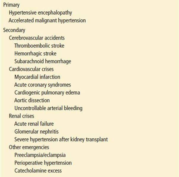
Blood pressure elevation may be primary or secondary to underlying causes (pain, anxiety, drugs). A hypertensive emergency exists if the elevated blood pressure is causing progressive target-organ damage. Two of the entities described above are specific complications of uncontrolled hypertension: hypertensive encephalopathy and malignant accelerated hypertension (9). The other hypertensive emergencies are primary crises of the brain, heart, kidneys, or vasculature that are precipitated or exacerbated by the stress of an elevated blood pressure (Table 90.1). For example, a myocardial infarction may occur in a normotensive as well as hypertensive patient. If the blood pressure is elevated in the setting of an infarct, then it must be managed, but the therapy must be decided primarily in the context of treating the coronary ischemia.
TABLE 90.1
Hypertensive Emergencies
ED EVALUATION
The clinical evaluation should determine the nature, severity, and management of patients in hypertensive crisis. When a patient with markedly elevated blood pressure presents to the ED, attention should first be given to assessing the patient’s airway, breathing, and circulation. This includes accurate measurement of blood pressure. Initially, elevated ED blood pressures frequently decrease spontaneously by the time a second reading is obtained (14). For this reason alone, interventions should be based on the composite of several, separate blood pressure determinations in the ED. To obtain an accurate measurement, the patient should be seated with the arm at the level of the heart and the cuff bladder should cover at least 80% of the arm circumference. Blood pressure should be evaluated in both arms. Blood pressure measurement with an automated cuff is inaccurate in patients with atrial fibrillation and other heart rhythm irregularities, especially tachydysrhythmias. Appropriate pain management and relief of underlying causes (e.g., hypoxia, bladder distention) may resolve hypertension (3). Other medications, over-the-counter preparations, or illicit drugs may exacerbate or mitigate blood pressure (Table 90.2).
TABLE 90.2
Drugs that Can Elevate Blood Pressure or Interfere with the Effectiveness of Antihypertensive Agents

If the patient’s blood pressure is persistently elevated, the history should start with an assessment of symptoms that might be consistent with target-organ compromise. Physicians should ask about specific symptoms such as dyspnea, chest pain, neurologic complaints, and visual changes. Additional important historical details include the duration and severity of pre-existing hypertension, the degree of previous success with blood pressure control, and the presence of target-organ disease (cardiovascular, cerebrovascular, renovascular, and great vessel). The physical examination should be directed toward identifying signs of target-organ damage. A fundoscopic examination demonstrating retinal hemorrhage or papilledema is sufficient to diagnose accelerated malignant hypertension. The cardiovascular examination should focus on identifying signs of heart failure (e.g., increased jugular venous pressure, pulmonary rales, and an S3) and asymmetric pulses. The neurologic examination should assess the level of consciousness, visual fields, and the presence of focal motor and sensory deficits.
Diagnostic studies should be based primarily on the patient’s symptoms. A chest radiograph is indicated for patients with dyspnea and a head CT scan for abnormal neurologic findings. Few studies have assessed the prognostic value of abnormal laboratory findings in patients with severe asymptomatic hypertension (17). If a patient’s blood pressure remains persistently >180/110 mm Hg after treatment of any aggravating symptoms, practitioners should consider a brief assessment of target-organ function. According to the 2013 American College of Emergency Physicians clinical policy, there is no definite role for screening for end-organ damage in adults with markedly elevated blood pressure, though high-risk populations may benefit from a limited evaluation (23). Because renal failure is silent, the measurement of the serum creatinine and/or a urinalysis is used to detect evidence of renal failure or glomerular disease. An ECG is useful in assessing the baseline level of left ventricular hypertrophy, which carries a poor prognosis and should prompt more vigilant follow-up. When renovascular disease or hypercortisolism are suspected causes of hypertension, a serum tube for plasma renin activity and aldosterone should be drawn before medications are administered. A urine screen for cocaine and amphetamines may help confirm common causes of hypertension. The incremental value of obtaining a chest radiograph or complete blood count in the ED patient without relevant symptoms is generally likely to be low.
KEY TESTING
• Base diagnostic evaluation on individual patient presentation and symptoms.
• Consider urinalysis and chemistry panel to assess renal function in patients with markedly elevated blood pressure.
• Patients with suspected cerebrovascular crisis or hypertensive encephalopathy should receive immediate CT of the head.
• Patients with suspected cardiovascular crisis should receive immediate ECG, cardiac enzyme, and chest radiography.
ED MANAGEMENT
The goal of therapy in a hypertensive crisis is a reduction in the mean MAP by 20% to 25% in 1 to 2 hours. Although not well evidence based, this therapeutic guideline is supported by shared practice experience in patients who have done poorly with more aggressive blood pressure reduction. The ideal drug for treating hypertensive emergencies has a rapid onset, rapid maximal effect, and rapid offset for easy titration of blood pressure. These characteristics are found only in parenteral agents. The most common drugs of this group are summarized in Table 90.3 from the Sixth Report of the Joint National Committee on Prevention, Detection, Evaluation, and Treatment of High Blood Pressure (JNC-VII) (8).
TABLE 90.3
Parenteral Drugs for Treatment of Hypertensive Emergencies

Nitroprusside remains the classic treatment for patients with hypertensive emergencies (8). Nitroprusside is a direct arteriovenous vasodilator that decreases both preload and afterload without significant reflex tachycardia. It is quick in onset of action, and its effect lasts for only 2 to 5 minutes after the drug is discontinued. Nitroprusside should be given with close hemodynamic monitoring to prevent inadvertent hypotension. Thiocyanate toxicity may occur if the drug is used for a period of days, particularly in patients with renal failure. Although not strictly contraindicated in pregnancy, nitroprusside does cross the placental barrier and causes elevated fetal cyanide levels. Because there is no oral form, the patient must be switched to another antihypertensive once control is achieved. Despite the many benefits of nitroprusside, there are often agents better suited for individual hypertensive crises.
Nicardipine is a rapid-acting parenteral calcium channel blocker. Nicardipine has a predictable and smooth onset of action but is relatively long acting. Esmolol is a β-blocking agent that is both rapid in onset and of short duration, making it easy to titrate. Labetalol is a combined α- and β-blocker that is more potent than esmolol and is perhaps better at maintaining a consistent CPP. Unlike esmolol, labetalol has a long half-life, making it possible to administer in miniboluses rather than a constant perfusion, but it is more difficult to titrate down. β-Blockers should not be used in patients with asthma, chronic obstructive pulmonary disease, acute CHF, cocaine abuse, or other contraindications to β-blockade. Fenoldopam is a parenteral dopaminergic receptor-blocking agent with an excellent efficacy and safety profile. Fenoldopam holds some promise as being equivalent to nitroprusside in efficacy, without the rare side effects associated with the nitroprusside cyanide moiety and perhaps with less overshoot hypotension. Enalaprilat is a parenteral ACEI, and phentolamine is a pure α-blocking agent.
Cerebrovascular Crisis
Blood pressure control in cerebrovascular hypertensive emergencies should be undertaken with parenteral drugs that have a short half-life and that are easily titrated with minimal effect on cerebral vasculature. Because labetalol does not dilate cerebral capacitance vessels, it is theoretically attractive for use in intracerebral disorders. Direct vasodilators, such as nitroprusside, should be used with caution in the setting of focal brain injury, as they can extend the area of ischemia. Nitroprusside has also been implicated in raising ICP and should be avoided in patients with suspected elevated ICP. Some calcium channel blockers have been linked to this rise as well and therefore are not favored in patients with brain injury, although nicardipine is safe and widely used (20).
The treatment of elevated blood pressure in the setting of ischemic cerebrovascular accidents is controversial. When systemic blood pressure is reduced, cerebral autoregulation may fail, thereby producing an ischemic penumbra surrounding the infarct, leading to stroke extension (7). Alternatively, infarction can lead to edema, which will elevate ICP and reduce CBF further. The current AHA guidelines recommend lowering the blood pressure with stroke only when the DBP is >120 mm Hg or the SBP is >220 mm Hg (12). The guidelines further suggest that for patients deemed to be eligible for thrombolytic therapy, blood pressure should be controlled to a systolic <185 mm Hg and diastolic <110 mm Hg. Recommended agents include intravenous labetalol, intravenous nicardipine, or transdermal nitroglycerin paste. Although no study has examined the use of thrombolytics after acutely lowering arterial hypertension, failure to achieve this level of control is considered an absolute contraindication to the administration of systemic thrombolytic therapy. The AHA guidelines are based on consensus opinion, as there is currently no definitive data that establish the clinical benefit of rapidly lowering blood pressure in patients with ischemic stroke. When the decision is made to lower arterial hypertension, the current guidelines recommend a drop of 15% to 25% in the first 24 hours (12).
The role of antihypertensive therapy in hemorrhagic cerebrovascular accidents and subarachnoid hemorrhage is similarly unclear. Theoretically, treatment for elevated blood pressure in intracranial hemorrhage should be more aggressive than for patients with ischemic stroke. The rationale is to decrease the risk of ongoing bleeding from ruptured small arteries and arterioles; however, the relationship between rebleeding and systemic blood pressure is unproven. As with an ischemic cardiovascular accident, overly aggressive treatment of hypertension may worsen brain injury by decreasing CPP, especially when ICP is increased. There is some evidence to suggest that the tissue immediately surrounding the hemorrhage may be particularly sensitive to changes in the CPP and aggressive lowering of blood pressure may cause tissue ischemia. The most recent AHA guidelines for blood pressure control with hemorrhagic stroke advocate a three-tiered approach. For an SBP >200 mm Hg or MAP >150 mm Hg, it is recommended that clinicians consider aggressive parenteral blood pressure control. The guidelines further suggest that when the SBP is >180 mm Hg or the MAP is >130 mm Hg and there is the possibility of elevated ICP, clinicians should consider using antihypertensive therapy in conjunction with ICP monitoring to maintain a CPP between 60 and 80 mm Hg. Finally, in patients with an SBP >180 mm Hg or a MAP >130 mm Hg and in which elevated ICP is not a concern, the guidelines suggest a modest reduction in blood pressure to a MAP of 110 mm Hg or a blood pressure of 160/90 (12). Nimodipine may be given to decrease the incidence of vasospasm and rebleeding after subarachnoid hemorrhage, but the drug is not recommended for blood pressure control (20).
Cardiovascular Crisis
Nitroglycerin is favored in the treatment of severe hypertension complicating cardiac ischemia. Nitroglycerin is a direct vasodilator that affects the venous vasculature more than the arterial. Nitroglycerin dilates the coronary arteries and, in contrast to nitroprusside, promotes a favorable redistribution of blood flow to ischemic areas. As previously discussed, the role of β-blockers in acute coronary syndromes has changed, and intravenous β-blockers are no longer considered first-line therapy in the ED. Oral β-blockers should be given within 24 hours to patients with acute coronary syndrome who are not at high risk for developing heart failure. The goal of treatment for patients with acute coronary syndromes is reduction of blood pressure to normal or even lower if evidence of ischemia persists. However, this reduction should occur carefully with the patient monitored intensively. Overly vigorous blood pressure lowering may worsen ischemia, because coronary perfusion depends on DBP.
Most critical cases of CHF are treated with a combination of nitroglycerin, furosemide, and an ACEI. For patients with pulmonary edema and hypertension, initiate sublingual nitroglycerin while preparing intravenous nitroglycerin. An ACEI is helpful whenever there is pulmonary congestion and hypertension; captopril may be given orally or sublingually, or enalaprilat can be given intravenously if the patient cannot take oral medications. If systemic fluid overload is present, then administer intravenous furosemide. However, some patients with CHF will not have signs of right-sided heart failure (e.g., jugular venous congestion, peripheral edema, and hepatomegaly) and will in fact be volume depleted because of a pressure diuresis despite the presence of pulmonary edema. In these patients with “dry failure,” diuresis may initially exacerbate the underlying pressure natriuresis and further stimulate the renin–angiotensin axis, leading to hypotension. The decision is based on a clinical judgment of whole-body fluid status. β-Blockers have been found to improve survival in patients with chronic CHF; however, this observation should not be extended to patients with acute pulmonary edema because the negative inotropic effects and bradycardia of β-blockade may precipitate immediate worsening.
In aortic dissection, unlike many presentations of hypertensive emergencies, immediate and aggressive blood pressure control is required. Most guidelines recommend targeting an SBP between 100 and 120 mm Hg, as long as tissue perfusion can be maintained (5). Additionally, it is important to control heart rates in patients with aortic dissection, as tachycardia can increase shearing forces on the damaged aorta, therefore increasing the size of the dissection. β-Blockers should be initiated before starting nitroprusside to avoid the potential adverse effects of reflex tachycardia. As an alternative, labetalol is a unique parenteral agent that achieves its maximal effect within minutes and then remains effective for several hours. This quality allows titration with small boluses, thereby avoiding the constant monitoring required and increased cost incurred with use of nitroprusside (15). Labetalol can also be administered as a parenteral infusion rather than as individual boluses, but careful monitoring is required with this strategy.
Renovascular Crisis
Although malignant hypertension may precipitate acute renal failure by injuring the kidney’s microvasculature, this causal chain is often reversed with hypertension being a manifestation, rather than a cause, of the renal failure. Because this distinction cannot be made in the ED, these patients should have their blood pressure lowered. Both nitroprusside and labetalol are excellent choices in this setting. ACEI drugs definitely improve prognosis among chronic hypertensive patients with mild proteinuria; however, they should be used cautiously in hyperkalemic patients with renal failure. Nitroprusside can cause thiocyanate poisoning in several days in patients with renal failure and should be used only briefly or if the patient is to have dialysis soon.
Treatment with an ACEI may reverse high blood pressure dramatically in patients with unilateral stenosis, but it may provoke acute renal failure and severe hyperkalemia in patients with bilateral stenosis, particularly if these patients are taking supplemental potassium or a potassium-sparing diuretic. This complication can be completely reversed by discontinuing the ACEI (16).
Catecholamine Excess
Patients with severe hypertension resulting from pheochromocytoma are commonly treated with the pure α-blocker phentolamine, given intravenously. This drug may be accompanied by a β-blocker if needed for tachycardia. Administration of β-blockers alone in the setting of any sympathomimetic (e.g., cocaine) may leave the α-receptors “open,” with subsequent worsening of hypertension. Therefore, an attractive alternative to β-blockers is labetalol, a β-blocker with some α-antagonist properties. However, the α- and β-blockade with labetalol may not be equally effective. Although several studies report reductions in blood pressure and heart rate when labetalol is given to patients with cocaine toxicity, case reports indicate that some individuals may get worse. The clinical importance of the “unopposed α” effect of labetalol increasing blood pressure requires further investigation.
Benzodiazepine therapy is considered by some to be the first-line treatment for hypertension caused by catecholamine excess, especially in the setting of sympathomimetic (e.g., cocaine or methamphetamine) toxicity. It is believed that the central nervous sedation may blunt the sympathomimetic effects caused by central stimulation, therefore lowering blood pressure. Regardless of which benzodiazepine is selected as the therapeutic agent, it is important to titrate the dose to clinical efficacy while carefully observing for respiratory depression.
Hypertension in Pregnancy
The mainstay of antihypertensive treatment for hypertensive crisis in pregnancy in many institutions is hydralazine administered intravenously in boluses of 5 to 10 mg every 20 to 30 minutes. This dose is in addition to magnesium therapy for seizure control. If treatment is refractory to hydralazine, second-line agents are diazoxide and β-blockers. Calcium channel blockers have been studied in chronic hypertension among pregnant patients, but they may not be effective with proteinuric hypertension.
Asymptomatic Hypertension
In patients with asymptomatic elevations in blood pressure and no evidence of target-organ damage, there is no role for immediate lowering of blood pressure. The 2013 American College of Emergency Physicians clinical policy states explicitly that routine intervention is not required, though clinicians can consider beginning therapy in selected high-risk populations, and all patients should be referred for follow-up (level C recommendation).
CRITICAL INTERVENTIONS
• Base clinical decisions on correctly measured and repeated blood pressure.
• Search for and correct underlying causes of an elevated blood pressure (e.g., pain, hypoxia, bladder distention, ingestion, or withdrawal).
• Investigate complaints carefully for evidence of target-organ disease.
• Treat the blood pressure according to specific indications (e.g., ischemia).
• Avoid relative hypotension or dropping blood pressure in the absence of an indication.
DISPOSITION
All hypertensive emergencies require admission to a monitored setting. These patients generally require emergent involvement of an appropriate specialist for management of a neurologic, cardiovascular, or renovascular crisis. Close blood pressure monitoring, preferably with an arterial line, is indicated. Patients with preeclampsia/eclampsia require emergent obstetric consultation.
Most patients presenting with elevated blood pressures are not in crisis. In patients with asymptomatic elevated blood pressure with no evidence of target-organ disease, the most important intervention is to ensure proper follow-up (3,18). The goal should be life-long control of the blood pressure. When the elevated blood pressure may be the artifact of a systemic process such as pain or infection, the best strategy is to refer the patient for reevaluation of the blood pressure once the primary problem has resolved. If the patient has discontinued their blood pressure medications, the regimen should be restarted, barriers to compliance evaluated, and a primary care physician contacted to ensure reevaluation in a week. The JNC-VII guidelines recommend a thiazide-type diuretic as an initial agent, usually in combination with drugs from other classes (3). The second agent may be from a number of categories and is best chosen in relation to any compelling indications in the patient’s history (Table 90.4).
TABLE 90.4
Compelling Indications for Individual Drug Classes for Hypertension Therapy

In principle, if there has never been a previous measurement of elevated blood pressure, the blood pressure needs to be rechecked during another visit before the diagnosis of hypertension can be made. However, in individuals with readings persistently >180/110 mm Hg in the ED, the latest national guidelines recommend starting a combination therapy immediately (3). Ideally, the physician who will provide ongoing supervision should be the one to pick the initial agents with which they are comfortable. The best role of the emergency physician is to identify a primary physician for the patient, encourage that physician to select an initial antihypertensive agent, and provide a follow-up appointment within the week.
There is an intermediate group of patients with a severely elevated blood pressure and with known target-organ disease but without active decompensation. Examples include a severely hypertensive patient with a previous history of myocardial infarction or stroke. The immediacy arises because the patient with known target-organ disease may be considered at higher risk for a hypertension-related adverse event in the short term. There is no good evidence basis for how these patients should best be managed. These patients should have a treatment strategy initiated from the ED although the blood pressure does not necessarily need to be lowered during the visit. These patients do require an increased level of vigilance. It may be reasonable to treat these patients as outpatients, although some may need to be held for short-term observation if there is question as to medication compliance or blood pressure monitoring. The decision depends on the clinical judgment of the physician.
Common Pitfalls
• Diagnosing a hypertensive emergency when one does not exist. Patients with hypertensive emergencies have evidence of acute end-organ dysfunction.
• Reducing the blood pressure too quickly or to too low a level. In patients with chronic hypertension whose autoregulation curve has been reset, this reduction can lead to cerebral or cardiac ischemia.
• Lowering a patient’s blood pressure acutely without an urgent indication.
• Failing to diagnose hypertension or preeclampsia in pregnant patients with blood pressures >140/90 mm Hg or with an increase in blood pressure of more than 30/15 mm Hg.
• Neglecting to match the antihypertensive agent to the clinical scenario.
REFERENCES
1. Blumenfeld JD, Laragh JH. Management of hypertensive crises: The scientific basis for treatment decisions. Am J Hypertens. 2001;14:1154–1167.
2. Calhoun DA, Oparil S. Treatment of hypertensive crisis. N Engl J Med. 1990; 323:1177–1183.
3. Chobanian AV, Bakris GL, Black HR, et al. The seventh report of the Joint National Committee on Prevention, Detection, Evaluation, and Treatment of High Blood Pressure: The JNC 7 Report. JAMA.2003;289(19):2560–2572.
4. Gifford RW. Management of hypertensive crises. JAMA. 1991;266:829–835.
5. Gilmore RM, Miller SJ, Stead LG. Severe hypertension in the emergency department patient. Emerg Med Clin North Am. 2005;23:1141–1158.
6. Hyman DJ, Pavlik VN. Characteristics of patients with uncontrolled hypertension in the United States. N Engl J Med. 2001;345:479–486.
7. Jauch EC, Saver JL, Adams HP Jr, et al. Guidelines for the early management of adults with acute ischemic stroke: A guideline for healthcare professionals from the American Heart Association/American Stroke Association, American Heart Association. Stroke. 2013;44:870–947.
8. Joint National Committee on Prevention, Detection, and Treatment of High Blood Pressure. The sixth report of the Joint National Committee on Prevention, Detection, Evaluation, and Treatment of High Blood Pressure. Arch Intern Med. 1997;157:2413–2446.
9. Kaplan NM. Management of hypertensive emergencies. Lancet. 1994;344:1335–1338.
10. Lip GY, Beevers M, Beevers DG. Complications and survival of 315 patients with malignant-phase hypertension. J Hypertens. 1995;13:915–924.
11. Lucas MJ, Leveno KJ, Cunningham FG. A comparison of magnesium sulfate with phenytoin for the prevention of eclampsia. N Engl J Med. 1995;333:201–205.
12. Morgenstern LB, Hemphill JC III, Anderson C, et al. Guidelines for the management of spontaneous intracerebral hemorrhage: In adults: A guideline for healthcare professionals from the American Heart Association/American Stroke Association. Stroke. 2010;41:2108–2129.
13. O’Gara PT, Kushner FG, Ascheim DD, et al. 2013 ACCF/AHA guideline for the management of ST-elevation myocardial infarction. Circulation. 2013;127:e362–e425.
14. Pitts SR, Adams RP. Emergency department hypertension and regression to the mean. Ann Emerg Med. 1998;31:214–218.
15. Pretre R, Von Segesser LK. Aortic dissection. Lancet. 1997;349:1461–1464.
16. Safian RD, Textor SC. Renal-artery stenosis. N Engl J Med. 2001;344:431–442.
17. Schwartz RB, Jones KM, Kalina P, et al. Hypertensive encephalopathy: Findings on CT, MR imaging, and SPECT imaging in 14 cases. AJR Am J Roentgenol. 1992; 159:379–383.
18. Shayne P, Pitts S. Severe hypertension in the emergency department: A state of the art review. Ann Emerg Med. 2003;41:513–529.
19. Thach AM, Schultz PJ. Nonemergent hypertension: New perspectives for the emergency medicine physician. Emerg Med Clin North Am. 1995;13:1009–1035.
20. Tietjen CS, Hurn PD, Ulatowski JA, et al. Treatment modalities for hypertensive patients with intracranial pathology: Options and risks. Crit Care Med. 1996; 24:311–322.
21. Varon J, Marik PE. The diagnosis and management of hypertensive crises. Chest. 2000;118:214–227.
22. Vaughan CJ, Delanty N. Hypertensive emergencies. Lancet. 2000;356:411–417.
23. Wolf SJ, Lo B, Shih RD, et al. American College of Emergency Physicians Clinical Policy: Critical issues in the evaluation and management of adult patients in the emergency department with asymptomatic elevated blood pressure. 2013, http://www.acep.org/clinicalpolicies/