
Richard Byyny and Howard A. Bessen
Pericarditis is present in approximately 5% of chest pain patients presenting to the ED without myocardial infarction (MI) (1) and can complicate nearly any class of disease, including infectious, neoplastic, rheumatologic, and metabolic diseases, as well as trauma. In the majority of cases the cause remains unclear despite testing (i.e., idiopathic pericarditis) (2,3). Many of these cases are thought to be viral in origin.
However, pericarditis may also occur as a complication of a systemic or intrathoracic infection. It is the most prominent cardiac complication of neoplastic disease, and nontraumatic pericardial tamponade is usually of metastatic origin (4,5). In addition, patients receiving radiation treatment as therapy for their cancer may develop postirradiation pericarditis. Pericarditis may occur as a complication of chronic renal failure or of connective tissue diseases such as systemic lupus erythematosus, scleroderma, or rheumatoid arthritis. Medications may also be implicated.
Pericarditis frequently occurs during the first few days after acute MI (6,7) and may also present as late-onset postinfarction pericarditis (Dressler syndrome). Aortic dissection, cardiac trauma, invasive procedures that involve the great vessels and the heart (central venous catheter insertion and cardiac catheterization), and thoracic surgery may also cause pericarditis and pericardial tamponade within hours to weeks. The common causes of acute pericarditis are listed in Table 89.1.
TABLE 89.1
Causes of Acute Pericarditis

Acute pericarditis is a diagnostic challenge because its presentation is similar to that of several other disorders and because it may have life-threatening complications. Management ranges from symptomatic treatment to lifesaving invasive procedures.
CLINICAL PRESENTATION
There are no predefined diagnostic criteria for pericarditis. However, presence of two of the following four criteria is highly suggestive: (1) characteristic chest pain, (2) pericardial friction rub, (3) suggestive ECG changes, and (4) new or worsening pericardial effusion (1,8–10). The prevalence of symptoms is presented in Table 89.2.
TABLE 89.2
Symptom Prevalence with Acute Pericarditis

Chest pain is the most common presenting complaint with acute pericarditis. The pain is usually sharp, substernal, and often pleuritic, but it may also be dull, constrictive, or aching. Pain may radiate to any part of the chest and back as well as to the ridge of the trapezius (11). Typically, the chest discomfort is described as being positional with the pain being eased by sitting up and leaning forward and increased with recumbency. The pain may be associated with shortness of breath, which may be caused by coexisting pleuritis or by cardiac tamponade. Patients may also present with hypotension and other signs and symptoms of tamponade.
The classic diagnostic physical finding is the pericardial friction rub, which may consist of one, two, or three components (1). An audio file can be found in the work by Carlson et al. (12). It is important to recognize that the rub is often transient and may not be heard at all times. However, it is present at some point in up to 85% of patients. The rub is often described as being high-pitched like leather squeaking or scratching. It is typically heard best along the left sternal border and is increased by having the patient sit or lean forward. The rub may be confused with a heart murmur or a pleural friction rub. However, having the patient hold their breath helps distinguish a pleural friction rub from a pericardial rub.
The major complications of acute pericarditis are pericardial effusion (potentially leading to cardiac tamponade), recurrent pericarditis, and chronic constrictive pericarditis. Dysrhythmias are unusual in pericarditis but may occur in patients with associated myocarditis or other underlying heart disease.
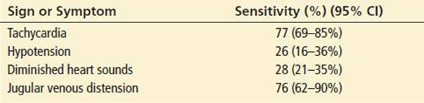
Cardiac tamponade is an acute life-threatening complication resulting from the accumulation of fluid within the pericardial space. The pericardium is poorly distensible; therefore, the fluid can cause an impairment of cardiac filling and thus a decrease in cardiac output. Beck’s triad (jugular venous distension [JVD], hypotension, and distant heart sounds) has been taught as the classic presentation, but the sensitivity of these findings is poor (Table 89.3). The presentation may be subtle, potentially leading to a delayed or missed diagnosis and a poor outcome. Close attention to physical findings is essential to make the diagnosis. Clinical decompensation can be rapid. Tamponade is often mistaken for congestive heart failure (CHF) because patients present with dyspnea, orthopnea, JVD, and hepatic enlargement.
TABLE 89.3
Sensitivity of Presenting Signs and Symptoms of Cardiac Tamponade
DIFFERENTIAL DIAGNOSIS
The differential diagnosis includes other conditions that cause chest pain and dyspnea, such as acute MI, CHF, pulmonary embolism, pneumonia, pneumothorax, pneumomediastinum, pleuritis, and aortic dissection. Cardiac tamponade may be confused with CHF, cardiogenic shock, or massive pulmonary embolism.
Acute pericarditis has many overlapping features with acute MI. Both cause chest pain, cause electrocardiographic abnormalities, and can cause elevations of cardiac markers (13). In contrast to patients with MI, patients with pericarditis may describe a viral prodrome, may present with fever, and typically have sharper pain that increases with body motion and varies with position. ECG findings may be useful in distinguishing MI from pericarditis (see section “ED Evaluation”). Patients with pericarditis (even with tamponade) have clear lung fields, unlike patients with CHF with pulmonary edema or those with pneumonia.
ED EVALUATION
Initial assessment and therapy consists of measures appropriate for any patient who presents with shortness of breath and chest pain: supplemental oxygen administration, intravenous access placement, cardiac monitoring, and a 12-lead ECG.
The ECG can be diagnostic. The most common ECG finding is concave-upward ST-segment elevation seen in most of the leads; PR-segment depression may also be seen (Fig. 89.1). Although the ECG is usually abnormal, diagnostic changes may be absent in patients with clinically evident pericarditis, particularly in patients with post-MI pericarditis.
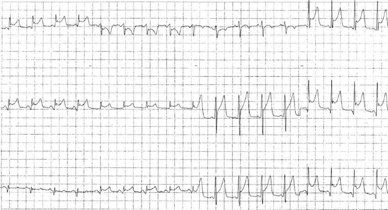
FIGURE 89.1 Electrocardiogram of a 35-year-old man with acute (stage 1) pericarditis. Note the diffuse, concave upward ST-segment elevation.
Early repolarization, a normal electrocardiographic variant characterized by ST-segment elevation, can be difficult to distinguish from pericarditis (1,14–16). If the clinical presentation suggests pericarditis, serial ECGs may be required to differentiate pericarditis from the benign early repolarization variant. On a single ECG, an ST/T ratio (the amount of ST-segment elevation divided by the height of the T wave) of 0.25 or more in lead V6 is suggestive of pericarditis.
MI and pericarditis may be difficult to differentiate. In pericarditis, ST-segment elevation is more diffuse, usually is upward sloping and is without reciprocal ST depression (16), and the Q waves that evolve in acute infarction do not develop. Pericarditis is also more likely to have associated PR depression. Having PR depression in both limb and precordial leads has a positive predictive value of 96.7% and a negative predictive value of 90% (17). Additionally ST-segment elevation in patients with acute MI is typically focal (in the distribution of a coronary artery), usually is convex or downward sloping and may be associated with ST-segment depression in reciprocal leads.
The chest radiograph is often normal. The cardiac silhouette may be enlarged secondary to a pericardial effusion, but the heart size may be normal even if an effusion is present (18).
The white blood cell count can range from normal to markedly elevated and is usually not helpful in making or excluding the diagnosis. Similarly, the erythrocyte sedimentation rate is usually elevated, but this is a nonspecific finding. Cardiac marker levels may be significantly elevated because of associated myocarditis (13,19,20).
Echocardiography can provide useful information in suspected pericarditis. In the absence of significant associated myocarditis, most patients with pericarditis have normal cardiac wall motion. In contrast, patients with chest pain caused by cardiac ischemia often have focal wall motion abnormalities. Echocardiography is also the best method for detecting and monitoring pericardial effusions (discussion follows).
Cardiac Tamponade
The classic findings of cardiac tamponade are muffled heart sounds, diminished arterial pressure, and JVD. However, these findings are seen only with fully developed tamponade and are much more likely to occur when pericardial fluid accumulates quickly, precluding pericardial distension (5,21). The most common presenting signs and symptoms associated with tamponade are summarized in Table 89.3 (22).
An important clue to the diagnosis is the presence of pulsus paradoxus, an inspiratory decrease in systolic blood pressure of >10 mm Hg (23). Other clinical entities associated with pulsus paradoxus include emphysema, asthma, obesity, pulmonary embolism, cardiogenic shock, right ventricular infarction, and restrictive cardiomyopathy. Patients with acute traumatic tamponade may not have a pulsus paradoxus.
Patients with tamponade may have relatively normal ECGs. With large effusions, reduction of the QRS amplitude or electrical alternans (alternating beat-to-beat variation in QRS amplitude) may be seen. Central venous pressure measurements reveal an elevation of right-sided pressures and can help confirm the diagnosis, although this may also be present in patients with pulmonary embolism or CHF.
A definitive diagnosis can be made with cardiac ultrasound. Echocardiography can detect small amounts of pericardial fluid; in cardiac tamponade, it may demonstrate diffuse hypokinesis, right atrial collapse, and right ventricular diastolic collapse (21,24). If possible, echocardiography should be performed emergently in any patient with suspected tamponade, because delays in evaluating the presence and size of a pericardial effusion can be disastrous (4).
The diagnosis of tamponade can also be definitively established by measuring pressures in the cardiac chambers. Right atrial, right ventricular diastolic, pulmonary artery diastolic, and pulmonary capillary wedge pressures are equalized in this condition.
Recurrent Pericarditis
After recovering from acute pericarditis, patients may have one or several episodes of recurrent pericarditis, weeks to months after the initial episode (25–28). Evaluation and management of recurrences are, for the most part, the same as for a first presentation of acute pericarditis. Choice of initial therapy may affect the frequency of recurrence (see the ED management section of this chapter).
Constrictive Pericarditis
Constrictive pericarditis, an uncommon complication of acute pericarditis, results when healing of acute pericarditis leads to encasement of the heart in fibrous tissue, impeding ventricular filling (1,9). Patients often present with dyspnea on exertion, ascites, peripheral edema, and hepatic enlargement. The lung fields are clear in these patients, in contrast to patients with CHF. The presence of distended neck veins in constrictive pericarditis helps to separate these patients from those with severe liver disease, who may appear clinically similar. Pericardial calcification on the chest radiograph and pericardial thickening on echocardiography may also provide clues to this diagnosis. The definitive treatment is resection of the pericardium.
KEY TESTING

ED MANAGEMENT
The treatment of uncomplicated pericarditis is largely symptomatic, but specific therapy is available for a few causes of pericarditis. Most studies have evaluated either aspirin at moderate to high doses or other nonsteroidal anti-inflammatory drugs in particular ibuprofen (29). There is no standard duration of therapy but many studies suggest a course of several weeks with a gradually tapering dose during that time period. Because of the high doses recommended, physicians should consider giving a proton pump inhibitor as prophylaxis against gastrointestinal discomfort or hemorrhage. In patients with suspected coronary artery disease indomethacin should be avoided because it can decrease coronary blood flow (1,30). In patients with post-MI pericarditis nonsteroidal anti-inflammatory drugs in general should be avoided because they impair myocardial remodeling and it is recommended that patients should be on aspirin therapy at baseline (31).
Colchicine can also be used to treat patients with idiopathic, viral, or autoimmune pericarditis. In a randomized trial of aspirin alone versus aspirin plus colchicine, patients who received both drugs had both a faster resolution of symptoms (36.7% of patients in the aspirin group were still symptomatic at 72 hours versus only 11.7% in the combination group) and a lower rate of recurrence (32.3% in the aspirin only group versus 10.7% in the combination group) (9). The most common reason for discontinuation of colchicine is diarrhea. Colchicine is generally safe but should be avoided in severe liver or kidney disease.
Bacterial pericarditis must be treated with drainage and appropriate antimicrobial therapy. Uremic pericarditis is usually an indication for dialysis. Corticosteroids should be avoided in most patients, because steroids have been associated with an increased rate of recurrence of pericarditis (28). The exception to this contraindication are patients in whom pericarditis is secondary to rheumatologic causes, where treating the underlying rheumatologic etiology with steroids may be indicated.
The definitive treatment for pericardial tamponade is pericardiocentesis. However, to “buy time” until pericardiocentesis can be performed, volume infusion is the treatment of choice. Ideally, pericardiocentesis should be done with echocardiographic or fluoroscopic guidance (often in the cardiac catheterization laboratory), but emergent “blind” pericardiocentesis may be necessary for hypotensive patients who do not respond to volume infusion.
The pericardium may be accessed by a left parasternal or subxiphoid puncture. In the parasternal approach, the needle is inserted perpendicular to the skin in the fifth intercostal space, just lateral to the sternum. In the subxiphoid approach, the puncture is made about 2 cm below and 1 cm to the left of the xiphoid process; the needle enters the skin at a 30° to 45° angle and is aimed toward the sternal notch or the left shoulder. The needle is advanced with constant syringe aspiration. As much fluid is aspirated as is needed to improve the clinical condition or to obtain specimens for diagnostic use.
After completing the procedure, a chest radiograph should be obtained to look for complications such as pneumothorax or pleural effusion secondary to laceration of the lung or myocardium. Serial physical examinations and central venous pressure monitoring are critical to detect reaccumulation of fluid. The admitting consultant may choose to observe the patient for fluid reaccumulation, may place a soft catheter in the pericardial space for continued drainage, or may perform surgical drainage in the operating room.
CRITICAL INTERVENTIONS
• Perform pericardiocentesis (with echocardiographic or fluoroscopic guidance if possible) to treat pericardial tamponade.
• Obtain an echocardiogram to exclude pericardial effusion before discharging patients with pericarditis.
DISPOSITION
Many patients with pericarditis can be successfully treated on an outpatient basis. Patients should be considered for admission if they exhibit any high-risk features (Table 89.4) (32,33). Before making a decision for outpatient therapy, it is prudent to obtain an echocardiogram to rule out a large effusion, tamponade, or myocarditis. Patients without high-risk clinical features on history, physical examination, or echocardiogram can be treated as outpatients if close follow-up can be assured; otherwise, they should be admitted with a cardiology consultation.
TABLE 89.4
High-Risk Features of Pericarditis

Common Pitfalls
• Failing to consider pericarditis in the differential diagnosis of chest pain.
• Mistakenly diagnosing pericarditis in patients with other causes of chest pain, such as MI, pulmonary embolism, or aortic dissection.
• Misdiagnosing acute pericarditis as acute MI, potentially leading to the inappropriate administration of thrombolytics or activation of the catheterization laboratory.
• Failing to consider the etiology of pericarditis. Many of the causes require specific therapy.
• Failing to consider cardiac tamponade in the differential diagnosis of hypotension, especially in patients with cancer.
REFERENCES
1. Spodick DH. Acute pericarditis: Current concepts and practice. JAMA. 2003; 289(9):1150–1153.
2. Permanyer-Miralda G, Sagrista-Sauleda J, Soler-Soler J. Primary acute pericardial disease: A prospective series of 231 consecutive patients. Am J Cardiol. 1985; 56(10):623–630.
3. Zayas R, Anguita M, Torres F, et al. Incidence of specific etiology and role of methods for specific etiologic diagnosis of primary acute pericarditis. Am J Cardiol. 1995;75(5):378–382.
4. Markiewicz W, Borovik R, Ecker S. Cardiac tamponade in medical patients: Treatment and prognosis in the echocardiographic era. Am Heart J. 1986;111(6):1138–1142.
5. Guberman BA, Fowler NO, Engel PJ, et al. Cardiac tamponade in medical patients. Circulation. 1981;64(3):633–640.
6. Correale E, Maggioni AP, Romano S, et al. Pericardial involvement in acute myocardial infarction in the post-thrombolytic era: Clinical meaning and value. Clin Cardiol. 1997;20(4):327–331.
7. Sugiura T, Iwasaka T, Takehana K, et al. Clinical significance of pericardial effusion associated with pericarditis in acute Q-wave anterior myocardial infarction. Chest. 1993;104(2):415–418.
8. Khandaker MH, Espinosa RE, Nishimura RA, et al. Pericardial disease: Diagnosis and management, Mayo Clinic proceedings. Mayo Clinic. 2010;85(6):572–593.
9. Imazio M, Bobbio M, Cecchi E, et al. Colchicine in addition to conventional therapy for acute pericarditis: Results of the COlchicine for acute PEricarditis (COPE) trial. Circulation. 2005;112(13):2012–2016.
10. Troughton RW, Asher CR, Klein AL, et al. 2004;363(9410):717–727.
11. Hoit BD. Pericardial heart disease. Curr Probl Cardiol. 1997;22(7):353–400.
12. Carlson DW, Atwood JE. Images in clinical medicine. An audible case of acute pericarditis. N Engl J Med. 2012;367(15):e20.
13. Roongsritong C, Warraich I, Bradley C. Common causes of troponin elevations in the absence of acute myocardial infarction: Incidence and clinical significance. Chest. 2004;125(5):1877–1884.
14. Mehta M, Jain AC, Mehta A. Early repolarization. Clin Cardiol. 1999;22(2):59–65.
15. Bhardwaj R, Berzingi C, Miller C, et al. Differential diagnosis of acute pericarditis from normal variant early repolarization and left ventricular hypertrophy with early repolarization: An electrocardiographic study. Am J Med Sci.2013; 345(1):28–32.
16. Chan TC, Brady WJ, Pollack M. Electrocardiographic manifestations: Acute myopericarditis. J Emerg Med. 1999;17(5):865–872.
17. Porela P, Kyto V, Nikus K, et al. PR depression is useful in the differential diagnosis of myopericarditis and ST elevation myocardial infarction. Ann Noninvasive Electrocardiol. 2012;17(2):141–145.
18. Eisenberg MJ, Dunn MM, Kanth N, et al. Diagnostic value of chest radiography for pericardial effusion. J Am Coll Cardiol. 1993;22(2):588–593.
19. Hooper AJ, Celenza A. A descriptive analysis of patients with an emergency department diagnosis of acute pericarditis. Emerg Med J. 2013;30(12):1003–1008.
20. Imazio M, Demichelis B, Cecchi E, et al. Cardiac troponin I in acute pericarditis. J Am Coll Cardiol. 2003;42(12):2144–2148.
21. Fowler NO. Cardiac tamponade. A clinical or an echocardiographic diagnosis?. Circulation. 1993;87(5):1738–1741.
22. Roy CL, Minor MA, Brookhart MA, et al. Does this patient with a pericardial effusion have cardiac tamponade? JAMA. 2007;297(16):1810–1818.
23. McGregor M. Current concepts: Pulsus paradoxus. N Engl J Med. 1979;301(9):480–482.
24. Hoit BD. Pericardial disease and pericardial tamponade. Crit Care Med. 2007; 35(8 Suppl):S355–S364.
25. Adler Y, Finkelstein Y, Guindo J, et al. Colchicine treatment for recurrent pericarditis. A decade of experience. Circulation. 1998;97(21):2183–2185.
26. Imazio M, Bobbio M, Cecchi E, et al. Colchicine as first-choice therapy for recurrent pericarditis: Results of the CORE (COlchicine for REcurrent pericarditis) trial. Arch Intern Med. 2005;165(17):1987–1991.
27. Imazio M, Demichelis B, Parrini I, et al. Management, risk factors, and outcomes in recurrent pericarditis. Am J Cardiol. 2005;96(5):736–739.
28. Imazio M, Trinchero R, Shabetai R. Pathogenesis, management, and prevention of recurrent pericarditis. J Cardiovasc Med (Hagerstown). 2007;8(6):404–410.
29. Lange RA, Hillis LD. Clinical practice. Acute pericarditis. N Engl J Med. 2004; 351(21):2195–2202.
30. Schifferdecker B, Spodick DH. Nonsteroidal anti-inflammatory drugs in the treatment of pericarditis. Cardiol Rev. 2003;11(4):211–217.
31. Hammerman H, Kloner RA, Schoen FJ, et al. Indomethacin-induced scar thinning after experimental myocardial infarction. Circulation. 1983;67(6):1290–1295.
32. Imazio M, Cecchi E, Demichelis B, et al. Indicators of poor prognosis of acute pericarditis. Circulation. 2007;115(21):2739–2744.
33. Imazio M, Demichelis B, Parrini I, et al. Day-hospital treatment of acute pericarditis: A management program for outpatient therapy. J Am Coll Cardiol. 2004;43(6):1042–1046.