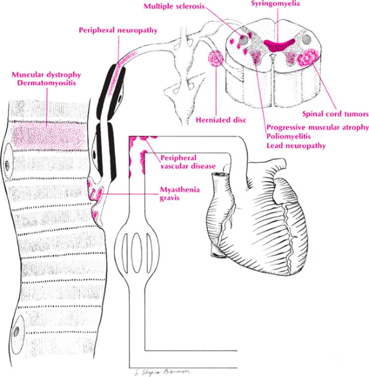
This symptom, as opposed to generalized weakness and fatigue (see page 454), is almost invariably due to a neurologic disorder. Consequently, a comprehensive list of causes is developed using neuroanatomy. Muscle weakness or paralysis may be due to disease of the muscle, myoneural junction, peripheral nerve, nerve roots and anterior horn cells, and pyramidal tract involvement in the spinal cord, brainstem, or cerebrum. Table 61 has been constructed with these anatomic components cross-indexed with the various etiologies suggested by the mnemonic VINDICATE. The most important of these will be covered in the following discussion.
|
TABLE 61. Weakness or Paralysis of One or More Extremities |
|||||||||||||||||||||||||||||||||||||||||||||||||||||||||||||||||||||||||||||||
|
|||||||||||||||||||||||||||||||||||||||||||||||||||||||||||||||||||||||||||||||
1. Muscle. This should suggest muscular dystrophy and dermatomyositis.
2. Myoneural junction. Primary and symptomatic myasthenia gravis are promptly brought to mind here. The toxic effects of succinylcholine chloride (Anectine), cholinergic drugs, and antispasmodics should also be mentioned.
3. Nerve. The many causes of peripheral neuropathy should be recalled here. The most important are diabetic neuropathy, alcoholic and nutritional neuropathies, Guillain–Barré syndrome, Buerger disease, periarteritis nodosa, porphyria, peroneal muscular atrophy, and lacerations or contusions from blunt trauma or surgery.
4. Nerve root or anterior horn. Poliomyelitis, lead neuropathy, and progressive muscular atrophy are a few diseases that specifically attack the anterior horn and roots; the roots may also be compressed by herniated disks, fractures, tuberculosis, or metastatic carcinomas of the spine. The spinal cord is often involved in the compression, too. Cervical spondylosis and spondylolisthesis may also compress the nerve root.
5. Spinal cord. The pyramidal tracts are involved in malformations such as syringomyelia, arteriovenous anomalies, and Friedreich ataxia; in inflammatory diseases like syphilis, tuberculosis of the spine, and transverse myelitis; in neoplasms (both primary and metastatic); and in traumaticlesions such as fractures, herniated discs, and hematomas. Thus, the mnemonic MINT is helpful in recalling these lesions. Cervical spondylosis, amyotrophic lateral sclerosis, syringomyelia, pernicious anemia, and multiple sclerosis may be forgotten, however, if only this mnemonic is used.
6. Brainstem. Brainstem gliomas and multiple sclerosis are important causes of pyramidal tract disease, but vascular occlusions of the basilar artery and its branches far exceed these in number.
7. Cerebrum. Any space-occupying lesions such as neoplasms, cerebral abscesses, subdural hematomas, and large aneurysms may cause focal monoplegia, hemiplegia, or paraplegia (parasagittal meningioma). Occlusions and hemorrhages of the cerebral arteries, however, are much more common causes of focal paralysis. Diffuse paralysis may result from the toxic and inflammatory encephalitides, presenile dementia, lipoidosis, and diffuse sclerosis. Multiple sclerosis and lupus erythematosus may also attack the cerebral peduncles.
Approach to the Diagnosis
The site of weakness is determined by associated symptoms and signs. Fasciculations suggest nerve root or anterior horn cell involvement, whereas sensory changes suggest peripheral nerve or spinal cord involvement. A combination of spasticity in the lower extremities and flaccid and atrophic weakness in the upper extremities suggests cervical cord involvement. Cranial nerve lesions in association with paraplegia or quadriplegia usually indicate a brainstem lesion.
The workup will depend on the site in which the pathology is suspected to be located. If muscle is the site, then an EMG or biopsy is indicated. If the myoneural junction is involved, a Tensilon test is done. Peripheral nerve lesions require a more extensive workup, including a glucose tolerance test, blood lead level, urine for porphobilinogens, EMG, nerve conduction velocity (NCV) test, and possibly a muscle biopsy. Spinal cord lesions may require x-ray of the spine, CT scan or MRI, myelography, discography, and spinal fluid analysis. Brainstem and cerebral lesions are best screened with a skull x-ray, MRI, or CT scan before a spinal tap or arteriogram is considered.
Other Useful Tests
1. CBC (pernicious anemia with neurologic involvement)
|
|
|
Weakness or paralysis of one or more extremities |
2. Chemistry panel (muscle disease, liver or kidney disease with neurologic involvement)
3. ANA analysis (collagen disease with neuropathy or myopathy)
4. Acetylcholine receptor antibody titer (myasthenia gravis)
5. Urine creatine and creatinine levels (muscular dystrophy)