DOORWAY INFORMATION
Opening Scenario
Carol Holland, a 67-year-old female, comes to the office complaining of neck pain.
Vital Signs
BP: 115/75 mm Hg Temp: 98.0°F (36.7°C)
RR: 16/minute HR: 74/minute, regular
Examinee Tasks
1. Take a focused history.
2. Perform a focused physical exam (do not perform rectal, genitourinary, or female breast exam).
3. Explain your clinical impression and workup plan to the patient.
4. Write the patient note after leaving the room.
Checklist/SP Sheet
Patient Description
Patient is a 67 yo F who lives with her husband.
Notes for the SP
■ Sit still with your back slightly hunched and head straight ahead; avoid turning your neck, and instead just move your eyes to make eye contact with the examinee.
■ Show pain when moving your neck and when the examinee palpates your neck.
■ Pretend to have numbness in the back of your left forearm.
Challenging Questions to Ask
“I’m supposed to visit my sister in Florida in 3 days. Will I still be able to go?”
Sample Examinee Response
“Before I am comfortable with you traveling, I want to make sure you don’t have a serious injury, like a broken bone or a nerve compression in your spine. I would like to see the results of some tests first to make sure you’ll be safe.”
Examinee Checklist
Building the Doctor-Patient Relationship Entrance
□ Examinee knocked on the door before entering.
□ Examinee introduced self by name.
□ Examinee identified his/her role or position.
□ Examinee correctly used patient’s name.
□ Examinee made eye contact with the SP.
Reflective Listening
□ Examinee asked an open-ended question and actively listened to the response.
□ Examinee asked the SP to list his/her concerns and listened to the response without interrupting.
□ Examinee summarized the SP’s concerns, often using the SP’s own words.
Information Gathering
□ Examinee elicited data efficiently and accurately.


Connecting with the Patient
□ Examinee recognized the SP’s emotions and responded with PEARLS.
Physical Examination
□ Examinee washed his/her hands.
□ Examinee asked permission to start the exam.
□ Examinee used respectful draping.
□ Examinee did not repeat painful maneuvers.
Closure
□ Examinee discussed initial diagnostic impressions.
□ Examinee discussed initial management plans:
□ Follow-up tests.
□ Examinee asked if the SP had any other questions or concerns.
Sample Closure
Mrs. Holland, given your symptoms, I am concerned that you may have a pinched nerve in your neck. Since you have a history of low bone density, I want to make sure your symptoms weren’t caused by a fracture. And although it’s unlikely, certain cancers may spread to the neck and spine and cause similar symptoms. I want to run some tests to rule out this possibility. I would like to start by getting an x-ray of your neck. Do you have any other questions for me?

History
HPI: 67 yo F with 2 days of neck pain and left upper extremity numbness.
■ Started after quick rotation to the left.
■ Sharp pain 2/10 at rest, 8/10 with motion.
■ Associated left arm numbness. Denies weakness.
■ 10-lb weight loss in past 6 months attributed to poor appetite.
■ No recent trauma or heavy lifting.
■ No dyspnea, fevers, night sweats.
■ Screenings up to date.
ROS: Negative except as above.
Allergies: NKDA.
Medications: Calcium and vitamin D supplements.
PMH: Osteopenia on last DEXA.
PSH: None.
SH: Social alcohol use, no tobacco or drugs. Retired magazine editor.
FH: Mother with osteoporosis, father with MI at 68.
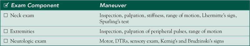
Physical Examination
Patient sitting rigid and still, avoiding moving neck.
VS: WNL.
Neck: No scars or deformities, limited ROM 2/2 pain. Tenderness to palpation on cervical spinous processes. Q Lhermitte and Spurling tests.
Extremities: No scars or deformities, brachial and radial pulses full. Full range of motion.
Neuro: Motor: Strength 5/5 throughout upper extremities. DTRs: 2+ symmetric, Q Babinski bilaterally. Sensation: Loss of pinprick sensation noted on dorsum of left hand and posterior left arm and forearm; all other sensation normal.
Differential Diagnosis


CASE DISCUSSION
Patient Note Differential Diagnoses
■ Disk herniation: As with other areas of the spine, pain at the site of compression with the addition of signs of nerve compression suggests radiculopathy caused by disk herniation.
■ Cervical fracture: Cervical fractures are dangerous, acute findings that can compromise innervation to the diaphragm if they interrupt the phrenic nerve. The exam would presumably show tenderness to palpation, but it is critical to include this in the differential given the patient’s history of osteopenia.
■ Neck muscle strain: Many people experience neck strains caused by quick turning of the head. The patient’s radiculopathy suggests that this is more than a simple strain.
Additional Differential Diagnoses
■ Osteoarthritis: Degenerative disease of the spine could cause the findings seen by the same routes as herniation and fracture—compression of the nerves.
■ Cervical spondylosis: A spondylosis would be caused by the same channels as degenerative disk disease.
■ Metastatic cancer: Breast and lung cancers, among others, can metastasize to the bone and cause cord compression. A possible spinal lesion in conjunction with weight loss in an older woman should raise concern for metastatic disease.
■ Multiple myeloma: Although a rarer malignancy, multiple myeloma is a cause of spinal lesions in both men and women. Associated findings may include symptoms of anemia, renal failure, and hypercalcemia in addition to the constitutional symptoms typically found in malignancy.
Diagnostic Workup
■ XR—C-spine: The first test to order for pain that raises concern for fracture or radiculopathy. Check for space narrowing or fractures.
■ MRI—C-spine: MRI is indicated for patients who have neck pain with neurologic signs or symptoms regardless of plain film findings. MRI is the most sensitive method with which to diagnose disk, spine, and spinal cord pathology. Because of its high sensitivity, MRI may detect clinically insignificant abnormalities.
■ Nerve conduction studies: Nerve stimulation will determine if the patient’s loss of sensation is due to a conduction issue in the peripheral nerve. Although they are specific, nerve conduction studies are not necessarily sensitive for cervical pathology.
■ CBC, calcium, BUN/Cr: To detect anemia, hypercalcemia, and renal failure, all of which may be clues to underlying multiple myeloma.
■ Serum and urine protein electrophoresis: To detect a monoclonal paraprotein in myeloma.