EQUIPMENT
 Tangential light source
Stethoscope with bell and diaphragm
Sphygmomanometer
Centimeter ruler
EXAMINATION
|
TECHNIQUE |
FINDINGS |
|
PERIPHERAL ARTERIES |
|
|
Palpate arterial pulses in neck and extremities |
|
|
Palpate carotid, brachial, radial, femoral, popliteal, dorsalis pedis, and posterior tibial arteries, using distal pads of second and third fingers, as shown in figures below and on p. 125. |
|
|
Palpation of arterial pulses. A, Carotid. B, Brachial.C, Radial. D, Femoral. E, Popliteal. F, Dorsalis pedis. G, Posterior tibial. |
|
|
Compare characteristics bilaterally, as well as between upper and lower extremities. |
EXPECTED:Femoral pulse as strong as or stronger than radial pulse. |
|
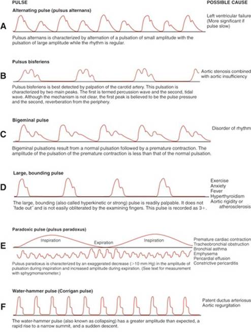
UNEXPECTED:Femoral pulse weaker than radial pulse or absent. Alternating pulse (pulsus alternans), pulsus bisferiens, bigeminal pulse (pulsus bigeminus), bounding pulse, labile pulse, paradoxical pulse (pulsus paradoxus), pulsus differens, tachycardia, trigeminal pulse (pulsus trigeminus), or water-hammer pulse (Corrigan pulse). |
|
|
A-F, Pulse abnormalities. Modified from Barkauskas, 1998. |
|
|
|
EXPECTED:60 to 90 beats per minute. |
|
UNEXPECTED:Rate different from that observed during cardiac examination. |
|
|
|
EXPECTED:Regular. |
|
UNEXPECTED:Irregular, either in a pattern or patternless. |
|
|
|
EXPECTED:Smooth, rounded, or dome shaped. |
|
|
UNEXPECTED:Bounding, full, diminished, or absent. Describe on scale of 0 to 4: 0 = Absent, not palpable 1 = Diminished 2 = Expected 3 = Full, increased 4 = Bounding |
|
Auscultate temporal, carotid, and subclavian arteries; abdominal aorta; and renal, iliac, and femoral arteries for bruits |
|
|
When auscultating the carotid vessels, you may at times need to ask patient to hold breath for a few heartbeats. Auscultate with bell of stethoscope. |
UNEXPECTED:Transmitted murmurs, bruits. |
|
Assess for arterial occlusion and insufficiency |
|
|
Assess for pain distal to possible occlusion. |
UNEXPECTED:Dull ache accompanied by fatigue and often crampiness; possible constant or excruciating pain. Weak, thready, or absent pulses; systolic bruits over arteries; loss of body warmth; localized pallor or cyanosis; delay in venous filling; or thin, atrophied skin, muscle atrophy, and loss of hair. |
|
Ask patient to lie supine. Elevate extremity, note degree of blanching, then ask patient to sit on edge of table or bed to lower extremity. Note time for maximal return of color when extremity is lowered. |
EXPECTED:Slight pallor on elevation and return to full color as soon as leg becomes dependent. |
|
UNEXPECTED:Delay of more than 2 seconds. |
|
|
Measure blood pressure |
|
|
Measure in both arms at least once. Patient’s arm should be slightly flexed and comfortably supported on table, pillow, or your hand. |
EXPECTED:100 to 140 mm Hg systolic and 60 to 90 mm Hg diastolic, with pulse pressure of 30 to 40 mm Hg (sometimes to 50 mm Hg). Reading between arms may vary by as much as 10 mm Hg; usually higher in right arm. Prehypertension is now defined as a blood pressure between 120 and 139 mm Hg systolic or 80 and 89 mm Hg diastolic. |
|
UNEXPECTED:Hypertension (see table below). |
|
Classification of Blood Pressure for Adults Ages 18 Years and Older*

|
PERIPHERAL VEINS |
|
|
Assess jugular venous pressure |
|
|
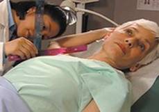
Ask patient to recline at 45-degree angle. With tangential light, observe the jugular vein. As shown in figure below, use a centimeter ruler to measure vertical distance between midaxillary line and highest level of jugular vein distention. |
EXPECTED:Pressure 9 cm H2O or less, bilaterally symmetric. |
|
UNEXPECTED:Abnormal elevation, distention or distention on one side. |
|
|
|
|
|
Assess for venous obstruction and insufficiency |
|
|
Inspect extremities, with patient both standing and supine. |
|
|
|
UNEXPECTED:Constant pain with swelling and tenderness over muscles, engorgement of superficial veins, cyanosis. |
|
Flex patient’s knee slightly with one hand, and with other, dorsiflex foot to test for Homans sign. |
UNEXPECTED:Redness, thickening, tenderness along superficial vein. Calf pain with test for Homans sign. |
|
Press index finger over bony prominence of tibia or medial malleolus for several seconds. |
UNEXPECTED:Orthostatic (pitting) edema; thickening and ulceration of skin possible. |
|
Grade edema from 1+ to 4+ as follows: 1+ = Slight pitting, no visible distortion, disappears rapidly 2+ = Deeper than 1+ and disappears in 10 to 15 seconds 3+ = Noticeably deep and may last more than 1 minute, with dependent extremity full and swollen 4+ = Very deep and lasts 2 to 5 minutes, with grossly distorted dependent extremity |
|
|
If suspected, have patient stand on toes 10 times in succession. |
EXPECTED:Pressure from toe standing disappears in seconds. |
|
UNEXPECTED:Veins dilated and swollen; often tortuous when extremities are dependent and pressure does not quickly disappear. |
|
|
Ask patient to lie supine, lift leg above heart level until veins empty, then quickly lower leg. |
UNEXPECTED:Rapid filling of veins. |
|
|
UNEXPECTED:Superficial veins fail to empty. |
|
|
UNEXPECTED:Stripped vessel fills before pressure is released by distal finger, or blood refills entire vein when pressure is released by proximal finger. |
AIDS TO DIFFERENTIAL DIAGNOSIS
|
ABNORMALITY |
DESCRIPTION |
|
Arterial aneurysm |
Subjective Data:Generally asymptomatic until they dissect or compress an adjacent structure. With dissection the patient may describe a severe ripping pain. |
|
Objective Data:Pulsatile swelling along the course of an artery. Occurs most commonly in the aorta, although renal, femoral, and popliteal arteries are also common sites. A thrill or bruit may be evident over the aneurysm. |
|
|
Venous thrombosis |
Subjective Data:Tenderness along the iliac vessels or the femoral canal, in the popliteal space, or over the deep calf veins. Deep vein thrombosis in the femoral and pelvic circulations may be asymptomatic. Pulmonary embolism may occur without warning. |
|
Objective Data:Swelling may be distinguished only by measuring and comparing the circumference of the upper and lower legs bilaterally. There may be minimal ankle edema; low-grade fever; and tachycardia. Homans sign can be helpful but is not absolutely reliable in suggesting deep vein thrombosis. |
|
|
Raynaud phenomenon |
Subjective Data:Involved areas will feel cold and achy, which improves on rewarming. In secondary Raynaud there can be intense pain and digital ischemia with necrosis at the tips. |
|
Objective Data:With primary Raynaud phenomenon there is triphasic demarcated skin pallor (white), cyanosis (blue), and reperfusion (red) within the extremities. The vasospasm may last from minutes to less than an hour. In secondary Raynaud ulcers may appear on the tips of the digits, and eventually the skin over the digits can appear smooth, shiny, and tight from loss of subcutaneous tissue. |
Pediatric Variations
EXAMINATION
|
TECHNIQUE |
FINDINGS |
|
|
Palpate arterial pulses in distal extremities |
||
|
|
EXPECTED: |
|
|
Age |
Beats per Minute |
|
|
Newborn |
120-170 |
|
|
1 year |
80-160 |
|
|
3 years |
80-120 |
|
|
6 years |
75-115 |
|
|
10 years |
70-110 |
|
|
Auscultate arteries for bruits |
||
|
EXPECTED:In children it is not unusual to hear a venous hum over internal jugular veins. There is usually no pathologic significance. |
||
|
Measure blood pressure |
||
|
When measuring an infant’s blood pressure, use flush technique if needed. |
EXPECTED:Calculation of systolic blood pressure for children older than 1 year can be estimated with following formula: |
|
|
Example: Calculation of expected systolic blood pressure of 5-year-old child: |
||
|
Although this calculation gives a figure below the expected mean, it is still considered within normal limits for a 5-year-old child. |
||
|
UNEXPECTED:Hypertension (see tables on pp. 135-138). |
||
AIDS TO DIFFERENTIAL DIAGNOSIS
|
ABNORMALITY |
DESCRIPTION |
|
Coarctation of the aorta |
Subjective Data:Most patients are asymptomatic unless severe hypertension or vascular insufficiency develops. In those settings patients may develop symptoms of heart failure or vascular insufficiency of an involved upper extremity with activity. |
|
Objective Data:Differences in systolic blood pressure readings when the radial and femoral pulses are palpated simultaneously. |
SAMPLE DOCUMENTATION
Vessels.Neck veins not distended. Both A and V waves are visualized. Jugular venous pressure (JVP) is 4 cm water at 45 degrees. Arterial pulses equal and symmetric, testing on a scale of 1/4.

Vessels soft. No bruits are audible.
Extremities.No edema, skin, or nail changes. Superficial varicosities noted in both lower extremities. No areas of tenderness to palpation.
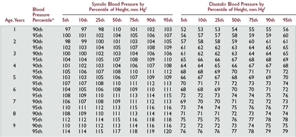
Blood Pressure Levels for the 90th and 95th Percentiles of Blood Pressure for Boys 1 to 17 Years of Age by Percentile of Height


Blood Pressure Levels for the 90th and 95th Percentiles of Blood Pressure for Girls 1 to 17 Years of Age by Percentile of Height


 Characteristics