EQUIPMENT
 Tangential light source
Skin-marking pencil
Stethoscope with bell and diaphragm
Centimeter ruler
EXAMINATION
|
TECHNIQUE |
FINDINGS |
|
HEART |
|
|
Inspect precordium |
|
|
Have patient supine, and keep light source tangential. |
EXPECTED:Visible about midclavicular line in fifth left intercostal space. Sometimes visible only with patient sitting. |
|
UNEXPECTED:Visible in more than one intercostal space; exaggerated lifts or heaves. |
|
|
Palpate precordium |
|
|
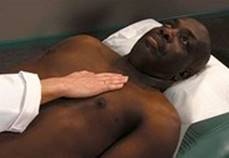
Have patient supine. With warm hands, gently feel precordium, using proximal halves of fingers held together or whole hand. As shown in figure on p. 114, methodically move from apex to left sternal border, base, right sternal border, epigastrium, axillae. Locate sensation in terms of its intercostal space and relationship to midsternal, midclavicular, axillary lines. |
EXPECTED:Gentle, brief impulse, palpable within radius of 1 cm or less, although often not felt. UNEXPECTED:Heave or lift, loss of thrust, displacement to right or left; thrill. |
|
|
|
|
Percuss precordium (optional) |
|
|
Begin by tapping at anterior axillary line, moving medially along intercostal spaces toward sternal borders until tone changes from resonance to dullness. Mark skin with marking pen. |
EXPECTED:No change in tone before right sternal border; on left, loss of resonance generally close to point of maximal impulse at fifth intercostal space. Loss of resonance may outline left border of heart at second to fifth intercostal spaces. |
|
Auscultate heart |
|
|
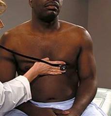
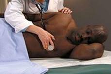
Make certain patient is warm and relaxed. Isolate each sound and each pause in cycle, and then inch along with stethoscope. Approach each of the five precordial areas shown in figure on p. 115 systematically, base to apex or apex to base, using each position shown in figures at right and below. Use diaphragm of stethoscope first, with firm pressure, then bell, with light pressure. |
|
|
|
|
|
|
|
|
|
|
|
|
|
|
Assess overall rate and rhythm. |
EXPECTED:Rate 60 to 90 beats per minute, regular rhythm. UNEXPECTED:Bradycardia, tachycardia, dysrhythmia. |
|
Ask patient to breathe comfortably, then hold breath in expiration. Listen for S1 (best heard toward apex) while palpating carotid pulse. Note intensity, variations, effect of respiration, splitting. Concentrate on systole, then diastole. |
EXPECTED:S1 usually heard as one sound and coincides with rise of carotid pulse. See table on p. 116. UNEXPECTED:Extra sounds or murmurs. |
|
Ask patient to breathe comfortably as you listen for S2 (best heard in aortic and pulmonic areas) to become two components during inspiration. Ask patient to inhale and hold breath. |
EXPECTED:S2 to become two components during inspiration. S2 to become an apparent single sound as breath is exhaled. See table on p. 116. |
|
|
EXPECTED:S2 splitting—greatest at peak of inspiration—varying from easily heard to nondetectable. |
|
If needed, ask patient to raise a leg to increase venous return or to grip your hand vigorously and repeatedly to increase venous return. |
EXPECTED:Both S3 and S4 quiet and difficult to hear. S3 has rhythm of Ken-tuc-ky; S4, Tenn-es-see. UNEXPECTED:Increased intensity (and ease of hearing) of either. |
|
|
UNEXPECTED:Extra heart sounds—snaps, clicks, friction rubs, murmurs. See table on p. 117. |
|
Assess characteristics of murmurs |
|
|
Timing and duration, pitch, intensity, pattern, quality, location, radiation, respiratory phase variations |
|
Heart Sounds According to Auscultatory Area

Extra Heart Sounds
|
Sound |
Detection |
Description |
|
Increased S3 |
Bell at apex; patient left lateral recumbent |
Early diastole, low pitch |
|
Increased S4 |
Bell at apex; patient supine or semilateral |
Late diastole or early systole, low pitch |
|
Gallops |
Bell at apex; patient supine or left lateral recumbent |
Presystole, intense, easily heard |
|
Mitral valve opening snap |
Diaphragm medial to apex, may radiate to base; any position, second left intercostal |
Early diastole briefly, before S3; high pitch, sharp snap or click; not affected by respiration; easily confused with S2 |
|
Ejection clicks |
Diaphragm; patient sitting or supine |
|
|
Aortic valve |
Diaphragm, right second intercostal space |
Early systole, intense, high pitch; radiates, not affected by respirations |
|
Pulmonary valve |
Diaphragm; left second intercostal right space |
Early systole, less intense than aortic click; intensifies on expiration, decreases on inspiration |
|
Pericardial friction rub |
Diaphragm, widely heard, sound clearest toward apex |
May occupy all of systole and diastole; intense, grating, machine-like; may have three components and obliterate heart sounds; if only one or two components, may sound like murmur |
AIDS TO DIFFERENTIAL DIAGNOSIS
|
ABNORMALITY |
DESCRIPTION |
|
Left ventricular hypertrophy |
Subjective Data:Initially asymptomatic, may cause shortness of breath or chest pain. |
|
Objective Data:Vigorous sustained lift palpable during ventricular systole, sometimes over broader area than usual (by 2 cm or more). Displacement of apical impulse can be well lateral of midclavicular line and downward. |
|
|
Right ventricular hypertrophy |
Subjective Data:Fatigue, shortness of breath, syncope may indicte more severe disease. |
|
Objective Data:Lift along left sternal border in third and fourth left intercostal spaces accompanied by occasional systolic retraction at apex. Left ventricle displaced and turned posteriorly by enlarged right ventricle. |
|
|
Congestive heart failureMay be left or right sided: Left sided is either systolic or diastolic |
Subjective Data:Fatigue, orthopnea, shortness of breath, edema. |
|
Objective Data:Congestion in pulmonary or systemic circulation. Can develop gradually or suddenly with acute pulmonary or systemic edema. |
|
|
Cor pulmonale |
Subjective Data:Tachypnea, fatigue, exertional dyspnea, cough hemoptysis. |
|
Objective Data:Left parasternal systolic lift and loud S2 in pulmonic region, evidence of pulmonary disease. |
|
|
Myocardial infarction |
Subjective Data:Deep substernal or visceral pain, often radiating to jaw, neck, left arm (although discomfort is sometimes mild); women may experience milder and different symptoms. |
|
Objective Data:Dysrhythmias; S4 often present. Heart sounds distant, with soft, systolic, blowing murmur; pulse possibly thready; varied blood pressure (although hypertension usual in early phases). |
|
|
Myocarditis |
Subjective Data:Initially symptoms vague; fatigue, dyspnea, fever, palpitations. Symptoms may progress. |
|
Objective Data:Cardiac enlargement, murmur, gallop rhythms, tachycardia, dysrhythmias, pulsus alternans. |
|
|
Conduction disturbances |
Subjective Data:Transient weakness, syncope, strokelike episodes, palpitations. |
|
Objective Data:Labile heart rates. |
|
|
Atherosclerotic heart disease |
Subjective Data:Maybe be asymptomatic or cause angina pectoris, shortness of breath, and palpitations. |
|
Objective Data:May cause myocardial insufficiency, dysrhythmias, congestive heart failure. |
|
|
Angina |
Subjective Data:Substernal pain or intense pressure radiating at times to neck, jaws, arms, particularly left arm, often accompanied by shortness of breath, fatigue, diaphoresis, faintness, syncope. Cessation of activity may relieve pain. |
|
Objective Data:No pathognomonic exam findings, tachycardia, hypertension, diaphoresis, decresed S1 intensity, S4. |
Chest Pain
|
Type of Chest Pain |
Characteristics |
|
Anginal |
Substernal; provoked by effort, emotion, eating; relieved by rest and/or nitroglycerin |
|
Pleural |
Precipitated by breathing or coughing; usually described as sharp |
|
Esophageal |
Burning, substernal, occasional radiation to shoulder; nocturnal occurrence, usually when lying flat; relief with food, antacids, sometimes nitroglycerin |
|
From a peptic ulcer |
Almost always infradiaphragmatic and epigastric; nocturnal occurrence and daytime attacks; should not be relieved by food; unrelated to activity |
|
Biliary |
Usually under right scapula, prolonged in duration; will trigger angina more often than mimic it |
|
From arthritis/bursitis |
Usually of hours-long duration; local tenderness and/or pain with movement |
|
Cervical |
Associated with injury; provoked by activity, persists after activity; painful on palpation and/or movement |
|
Musculoskeletal (chest) |
Intensified or provoked by movement, particularly twisting or costochondral bending; long lasting; often associated with local tenderness |
|
Psychoneurotic |
Associated with or occurring after anxiety; poorly described, located in intramammary region |
Data from Samiy et al, 1987; Harvey et al, 1988.
Pediatric Variations
EXAMINATION
|
TECHNIQUE |
FINDINGS |
|
Assess characteristics of murmurs |
|
|
Timing and duration, intensity, pattern, quality, location, radiation, respiratory phase variations. |
In children it is necessary to distinguish innocent murmurs from organic murmurs caused by congenital defect or rheumatic fever. |
AIDS TO DIFFERENTIAL DIAGNOSIS
|
ABNORMALITY |
DESCRIPTION |
|
Chest pain |
Subjective Data:Unlike in adults, chest pain in children and adolescents is seldom caused by a cardiac problem. It is very often difficult to find a cause, but trauma, exercise-induced asthma and use of cocaine, even in a somewhat younger child, as in the adolescent and adult, should be among the considerations. |
|
Objective Data:Exam usually normal. |
|
|
Congenital defects |
|
|
Tetralogy of Fallot |
Subjective Data:Dyspnea with feeding, poor growth, exercise intolerance, tetralogy spells. |
|
Objective Data:Parasternal heave and precordial prominence, cyanosis, systolic ejection murmur heard over third intercostal space, sometimes radiating to left side of neck. Single S2. |
|
|
Ventricular septal defect |
Subjective Data:Tachypnea, symptoms of right-sided congestive heart failure, poor growth, recurrent respiratory infections. |
|
Objective Data:Arterial pulse small, jugular venous pulse unaffected, regurgitation occurs through septal defect, resulting in holosystolic murmur that is frequently loud, coarse, high-pitched, best heard along left sternal border in third to fifth intercostal spaces. Distinct lift often discernible along left sternal border and apical area. Does not radiate to neck. |
|
|
Patent ductus arteriosus (PDA) |
Subjective Data:Asymptomic if small, larger PDAs cause dyspnea on exertion. |
|
Objective Data:Neck vessels dilated and pulsate, and pulse pressure wide. Harsh, loud, continuous murmur with machine-like quality, heard at first to third intercostal spaces and lower sternal border. Murmur usually unaltered by postural change. |
|
|
Atrial septal defect |
Subjective Data:Often asymptomatic, congestive heart failure in adults. |
|
Objective Data:Systolic ejection murmur, best heard over pulmonic area that is diamond-shaped, often loud, high in pitch, and harsh. May be accompanied by brief, rumbling, early diastolic murmur. Does not usually radiate beyond precordium. Systolic thrill may be felt over area of murmur along with palpable parasternal |
|
|
thrust. S2 may be split fairly widely. Particularly significant with palpable thrust and occasional radiation through to back. |
|
SAMPLE DOCUMENTATION
Heart. No visible pulsations over precordium. Point of maximal impulse (PMI) palpable at the fifth intercostals (ICS) in the midclavicular line (MCL), 1 cm in diameter. No lifts, heaves, or thrills felt on palpation. S1 is crisp. Split S2 increases with inspiration. No audible S3, S4, murmur, click, or rub.
 Apical impulse