EQUIPMENT
 Drapes
Speculum
Gloves
Water-soluble lubricant
Lamp or light source
Specimen collection equipment such as:
• Sterile cotton swabs
• Glass slides
• Wooden or plastic spatula
• Cervical brush devices
• Cytologic fixative
• Culture plates or media, if needed
• DNA probe kits for chlamydia and gonorrhea, if needed
EXAMINATION
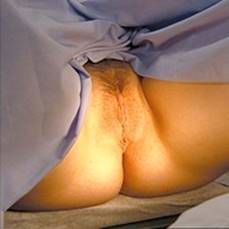
Have patient in lithotomy position, draped for minimal exposure.
|
TECHNIQUE |
FINDINGS |
|
EXTERNAL GENITALIA |
|
|
Wear gloves on both hands |
|
|
Ask patient to separate or drop open her knees. Tell patient you are beginning the examination, then touch either lower thigh and—without breaking contact—move hand along thigh to external genitalia. |
|
|
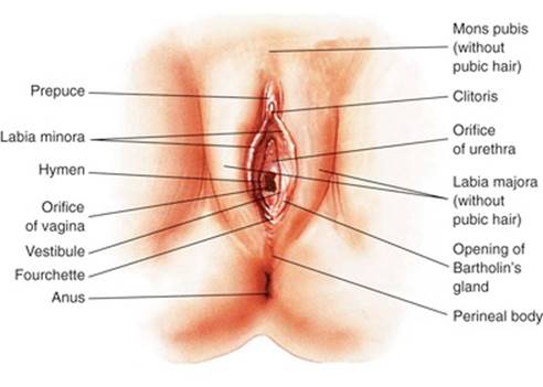
Inspect and palpate mons pubis |
|
|
|
EXPECTED:Skin smooth and clean. |
|
UNEXPECTED:Improper hygiene. |
|
|
|
EXPECTED:Regularly distributed female pubic hair. |
|
UNEXPECTED:Nits or lice. |
|
|
From Lowdermilk and Perry, 2004.
|
|
|
Inspect and palpate labia |
|
|
|
EXPECTED:Gaping or closed, dry or moist, shriveled or full, tissue soft and homogeneous, usually symmetric. |
|
UNEXPECTED:Swelling, redness, tenderness, discoloration, varicosities, obvious stretching, or signs of trauma or scarring. If excoriation, rashes, or lesions are present, ask patient whether she has been scratching. |
|
|
Separate labia majora with fingers of one hand. With other hand, palpate labia minora between thumb and second finger. |
EXPECTED:Moist, dark pink inner surface. Tissue soft and homogeneous. |
|
UNEXPECTED:Tenderness, inflammation, irritation, excoriation, caking of discharge in tissue folds, discoloration, ulcers, vesicles, irregularities, or nodules. Hyperemia of fourchette not related to recent sexual activity. |
|
|
Inspect clitoris |
|
|
|
EXPECTED:Length 2 cm or less; diameter 0.5 cm. |
|
UNEXPECTED:Enlargement, atrophy, inflammation, or adhesions. |
|
|
Inspect urethral meatus and vaginal opening |
|
|
|
EXPECTED:Slit or irregular opening, close to or in vaginal introitus, usually midline. |
|
UNEXPECTED:Discharge, polyps, caruncles, fistulas, lesions, irritation, inflammation, or dilation. |
|
|
|
EXPECTED:Thin vertical slit or large orifice with irregular edges. Tissue moist. |
|
UNEXPECTED:Swelling, discoloration, discharge, lesions, fistulas, or fissures. |
|
|
Milk Skene glands |
|
|
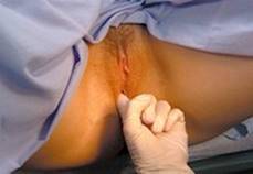
Tell patient you will be inserting one finger into her vagina and pressing forward with it. With palm up, insert index finger to second joint, press upward, and milk Skene glands by moving finger outward. Perform on both sides of urethra and directly on urethra. |
UNEXPECTED:Discharge or tenderness. Note color, consistency, odor of any discharge; obtain culture. |
|
|
|
|
Palpate Bartholin glands |
|
|
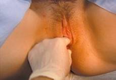
Tell patient she will feel you pressing around the entrance to her vagina. Palpate lateral tissue between index finger and thumb, then palpate entire area bilaterally, particularly posterolateral portion of labia majora. |
EXPECTED:No swelling. |
|
UNEXPECTED:Swelling, tenderness, masses, heat, fluctuation, or discharge. Note color, consistency, odor of any discharge; obtain culture. |
|
|
Test vaginal muscle tone if indicated |
|
|
Ask patient to squeeze vaginal opening around your finger. |
EXPECTED:Fairly tight squeezing by some nulliparous women, less so by some multiparous women. |
|
UNEXPECTED:Protrusion of cervix or uterus. |
|
|
Locate the cervix |
|
|
With your finger still in place you can reach in farther to locate the cervix and note the direction in which it points. This may help you locate the cervix when you insert the speculum. |
EXPECTED:Midline, may point horizontally, anteriorly, or posteriorly. |
|
UNEXPECTED:Deviates to right or left. |
|
|
Inspect for bulging and urinary incontinence if indicated |
|
|
Ask patient to bear down. |
EXPECTED:No bulging. |
|
UNEXPECTED:Bulging of anterior or posterior wall, or urinary incontinence. |
|
|
Inspect and palpate perineum |
|
|
Compress perineal tissue between finger and thumb. |
EXPECTED:Perineum surface smooth—generally thick and smooth in a nulliparous woman, thinner and rigid in a multiparous woman. Possible episiotomy scarring in women who have borne children. |
|
UNEXPECTED:Tenderness, inflammation, fistulas, lesions, or growths. |
|
|
|
|
|
Inspect anus |
|
|
|
EXPECTED:Skin darkly pigmented and possibly coarse. |
|
UNEXPECTED:Scarring, lesions, inflammation, fissures, lumps, skin tags, or excoriation. |
|
|
INTERNAL GENITALIA—SPECULUM EXAMINATION |
|
|
If you touched the perineum or anal skin while examining the external genitalia, change gloves before beginning internal examination. |
|
|
Lubricate speculum and gloved fingers with water or water-soluble gel lubricant. Water is preferred if obtaining a Pap smear. |
|
|
Insert speculum |
|
|
Tell patient she will feel you touching her again, then insert two fingers of hand not holding the speculum just inside vaginal introitus and gently press downward. Ask patient to breathe slowly and try to consciously relax her muscles. |
|
|
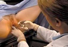
Use fingers of that hand to separate labia minora widely so that the vaginal opening becomes clearly visible. Then slowly insert speculum along path of least resistance, often slightly downward, avoiding trauma to urethra and vaginal walls. Some clinicians insert speculum blades at an oblique angle; others prefer to keep blades horizontal. In either case avoid touching clitoris, catching pubic hair, or pinching labial skin. Insert speculum the length of the vaginal canal. Maintaining downward pressure, open speculum by pressing on thumb piece. Sweep speculum slowly upward until cervix comes into view. Adjust light, then manipulate speculum farther into vagina so that cervix is well exposed between anterior and posterior blades. Stabilize distal spread of blades, and adjust proximal spread as needed. |
|
|
Inspect cervix |
|
|
|
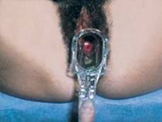
EXPECTED:Evenly distributed pink. Symmetric, circumscribed erythema around os can be expected. From Edge and Miller, 1994.
|
|
UNEXPECTED:Bluish, pale, or reddened cervix (especially if patchy or with irregular borders). |
|
|
|
EXPECTED:In midline, horizontal or pointing anteriorly or posteriorly. Protruding into vagina 1 to 3 cm. |
|
UNEXPECTED:Deviation to right or left. Protrusion into vagina greater than 1 to 3 cm. |
|
|
|
EXPECTED:3 cm in diameter. |
|
UNEXPECTED:Larger than 3 cm. |
|
|
|
EXPECTED:Uniform. |
|
UNEXPECTED:Distorted. |
|
|
|
EXPECTED:Surface smooth. Possible symmetric, reddened circle around os (squamocolumnar epithelium). Possible small, white, or yellow, raised round areas on cervix (nabothian cysts). |
|
UNEXPECTED:Friable tissue, red patchy areas, granular areas, or white patches. |
|
|
Note any discharge. Determine origin—cervix or vagina. |
EXPECTED:Odorless, creamy or clear, thick, thin, or stringy (often heavier at midcycle or immediately before menstruation). |
|
UNEXPECTED:Odorous and white to yellow, green, or gray. |
|
|
Follow standard precautions for safe collection of human secretions. |
EXPECTED:Nulliparous woman: Small, round, oval. Multiparous woman: Usually a horizontal slit or irregular and stellate. |
|
UNEXPECTED:Slit resulting from trauma from induced abortion, difficult removal of intrauterine device (IUD), or sexual abuse. |
|
|
Withdraw speculum and inspect vaginal walls |
|
|
Unlock speculum, and remove it slowly, rotating it so vaginal walls can be inspected. Maintain downward pressure, and hook index finger over anterior blade as it is removed. Note odor of any discharge pooled in posterior blade, and obtain specimen if not already obtained. |
EXPECTED:Vaginal wall color same pink as cervix or lighter; moist, smooth or rugated; and homogeneous. Thin, clear or cloudy, odorless secretions. |
|
UNEXPECTED:Reddened patches, lesions, pallor, cracks, bleeding, nodules, swelling. Secretions that are profuse; thick, curdy, or frothy; gray, green, or yellow; or malodorous. |
|
|
INTERNAL GENITALIA—BIMANUAL EXAMINATION |
|
|
Change gloves, and then lubricate index and middle fingers of examining hand. |
|
|
Tell patient you are going to examine her internally with your fingers. Prevent thumb from touching clitoris during examination. |
|
Obtaining Vaginal Smears and Cultures
Vaginal specimens are obtained while the speculum is in place in the vagina but after the cervix and its surrounding tissue have been inspected. Collect specimens as indicated for a Papanicolaou (Pap) smear, sexually transmitted disease screening, and wet mount. Label the specimen with the patient’s name and a description of the specimen (e.g., cervical smear, vaginal smear, and culture). Be sure to follow standard precautions for the safe collection of human secretions.
Pap Smear
Brushes and brooms are now being used in conjunction with or instead of the conventional spatula to improve the quality of cells obtained. The cylindric-type brush (e.g., a Cytobrush) collects endocervical cells only. First, collect a sample from the ectocervix with a spatula. Insert the longer projection of the spatula into the cervical os. Rotate it 360 degrees, keeping it flush against the cervical tissue. Withdraw the spatula, and spread the specimen on a glass slide. A single light stroke with each side of the spatula is sufficient to thin out the specimen over the slide. Fix the specimen and label as ectocervical. Then introduce the brush device into the vagina, and insert it into the cervical os until only the bristles closest to the handle are exposed. Slowly rotate one half to one full turn. Remove and prepare the slide. A single light stroke with each side of the spatula is sufficient to thin out the specimen over the slide. Fix the specimen and label as ectocervical. Then introduce the brush device into the vagina, and insert it into the cervical os until only the bristles closest to the handle are exposed. Slowly rotate one half to one full turn. Remove and prepare the endocervical smear by rolling the brush with moderate pressure across a glass slide. Fix the specimen and label as endocervical. Alternatively, both specimens can be placed on a single slide.
The broom-type brush is used for collecting ectocervical and endocervical cells at the same time. The broom has flexible plastic bristles, which are reported to cause less blood spotting after the examination. Introduce the brush into the vagina, and insert the central long bristles into the cervical os until the lateral bristles bend fully against the ectocervix. Maintain gentle pressure, and rotate the brush by rolling the handle between the thumb and forefinger three to five times to the left and right. Withdraw the brush, and transfer the sample to a glass slide with two single “paint” strokes. Apply first one side of the bristle, then turn the brush over, and paint the slide again in exactly the same area. Apply fixative and label as the ectocervical and endocervical specimen.
For the liquid preparation technology, using the broom-type device, insert the central bristles of the broom into the endocervical canal deep enough to allow the shorter bristles to fully contact the ectocervix. Push gently, and rotate the broom clockwise five times. Rinse the broom into the solution vial by pushing the broom into the bottom of the vial 10 times, forcing the bristles apart. As a final step, swirl the broom vigorously to further release material. Discard the collection device. Alternatively, deposit the broom end of the device directly into the collection vial. With any collection device, be sure to follow the manufacturer’s and laboratory instructions to collect and preserve the specimen appropriately. Close the vial tightly to prevent leakage and loss of the sample during transport. The liquid sample is also used to test for the HPV virus.
Gonococcal Culture Specimen
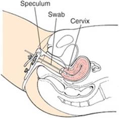
Immediately after the Pap smear is obtained, introduce a sterile cotton swab into the vagina, and insert it into the cervical os. Hold it in place for 10 to 30 seconds. Withdraw the swab, and spread the specimen in a large z pattern over the culture medium, rotating the swab at the same time. Label the tube or plate, and follow agency routine for transporting and warming the specimen. If indicated, an anal culture can be obtained after the vaginal speculum has been removed. Insert a fresh, sterile cotton swab about 2.5 cm into the rectum, and rotate it in a full circle. Hold it in place for 10 to 30 seconds. Withdraw the swab, and prepare the specimen as described for the vaginal culture. Gonococcal cultures are now used less frequently than the combined DNA probe for chlamydia and gonorrhea.
DNA Probe for Chlamydia and Gonorrhea
This test involves the construction of a nucleic acid sequence (called a probe) that will match a sequence in the DNA or RNA of the target tissue. Results are rapid and sensitive. Use a Dacron swab (with a plastic or wire shaft) when collecting the specimen; wooden, cotton-tipped applicators may interfere with test results. Also be sure to check the expiration date so as not to use out-of-date materials. Insert the swab into the cervical os, and rotate the swab in the endocervical canal for 30 seconds to ensure adequate sampling and absorption by the swab. Avoid contact with the vaginal mucous membranes, which would contaminate the specimen. Remove the swab, and place it in the tube containing the specimen reagent.
Wet Mount and Potassium Hydroxide (KOH) Procedures
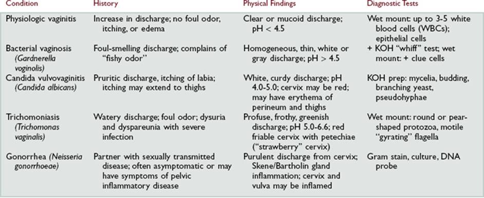
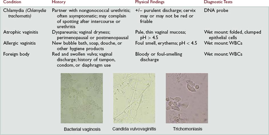
In a woman with vaginal discharge, these microscope examinations can demonstrate the presence of Trichomonas vaginalis, bacterial vaginosis, or candidiasis. For the wet mount, obtain a specimen of vaginal discharge using a swab. Smear the sample on a glass slide, and add a drop of normal saline solution. Place a coverslip on the slide, and view under the microscope. The presence of trichomonads indicates T. vaginalis. The presence of bacteria-filled epithelial cells (clue cells) indicates bacterial vaginosis. On a separate glass slide, place a specimen of vaginal discharge, apply a drop of aqueous 10% KOH, and put a coverslip in place. The presence of a fishy odor (the “whiff test”) suggests bacterial vaginosis. The KOH dissolves epithelial cells and debris and facilitates visualization of the mycelia of a fungus. View under the microscope for the presence of mycelial fragments, hyphae, and budding yeast cells, which indicate candidiasis.
|
TECHNIQUE |
FINDINGS |
|
Palpate vaginal wall while inserting fingers into vagina |
|
|
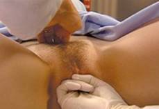
Insert tips of index and middle fingers into vaginal opening and press downward, waiting for muscles to relax. Gradually insert fingers full length while palpating vaginal wall. |
EXPECTED:Smooth and homogeneous. |
|
UNEXPECTED:Tenderness, lesions, cysts, nodules, masses, or growths. |
|
|
Palpate cervix |
|
|
Locate cervix with palmar surface of fingers, feel end, and run fingers around circumference to feel fornices. |
|
|
|
EXPECTED:Consistent with speculum examination. |
|
|
EXPECTED:Firm in nonpregnant woman; softer in pregnant woman. |
|
UNEXPECTED:Nodules, hardness, or roughness. |
|
|
|
EXPECTED:In midline horizontal or pointing anteriorly or posteriorly. Protruding into vagina 1 to 3 cm. |
|
UNEXPECTED:Deviation to right or left. Protrusion into vagina greater than 1 to 3 cm. |
|
|
Grasp cervix gently between fingers and move from side to side. Observe patient’s facial expression. |
EXPECTED:1 to 2 cm movement in each direction. Minimal discomfort. |
|
UNEXPECTED:Pain on movement (“cervical motion tenderness”). |
|
|
Palpate uterus |
|
|
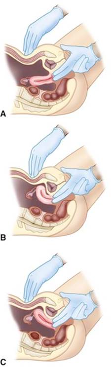
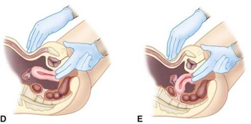
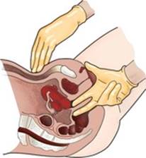
Place palmar surface of outside hand on abdominal midline, halfway between umbilicus and symphysis pubis, and place intravaginal fingers in anterior fornix. As shown in figure on p. 186, |
EXPECTED:In midline, horizontal, or pointing anteriorly or posteriorly. Protruding into vagina 1 to 3 cm. |
|
UNEXPECTED:Deviation to right or left. Protrusion into vagina greater than 1 to 3 cm. |
|
|
slowly slide outside hand toward pubis while pressing down and forward with flat surface of fingers; at the same time, push inward and up with fingertips of intravaginal hand while pushing down on cervix with backs of fingers. If uterus is anteverted or anteflexed, you should feel fundus between fingers of two hands at level of pubis. If uterus cannot be felt with this maneuver, place intravaginal fingers together in posterior fornix and outside hand immediately above symphysis pubis. Press firmly down with outside hand while pressing inward against cervix with intravaginal hand. If uterus is retroverted or retroflexed, you should feel fundus. If uterus cannot be felt with either of these maneuvers, move intravaginal fingers to each side of cervix, and while keeping contact with cervix, press inward and feel as far as possible. Slide fingers so they are on top and bottom of cervix and continue pressing in while moving fingers to feel as much of uterus as possible (when uterus is in midposition, you will not be able to feel it with outside hand). |
|
|
A, Anteverted. B, Anteflexed. C, Retroverted. D, Retroflexed. E, Midposition of uterus. |
|
|
|
EXPECTED:Pear-shaped and 5.5 to 8 cm long (larger in all dimensions in multiparous women). Contour rounded and, in nonpregnant women, walls firm and smooth. |
|
UNEXPECTED:Larger than expected or interrupted contour or smoothness. |
|
|
Gently move uterus between intravaginal fingers and outside hand. |
EXPECTED:Mobile in anteroposterior plane. |
|
UNEXPECTED:Fixed uterus or tenderness on movement. |
|
|
Palpate ovaries |
|
|
Place fingers of outside hand on lower right quadrant. With intravaginal hand facing up, place both fingers in right lateral fornix. Press intravaginal fingers deeply in and up toward abdominal hand, while sweeping flat surface of fingers of outside hand deeply in and obliquely down toward symphysis pubis. Palpate entire area by firmly pressing outside hand and intravaginal fingers together. Repeat on left side. |
|
|
|
EXPECTED:If palpable, ovaries should feel firm, smooth, slightly to moderately tender. |
|
UNEXPECTED:Marked tenderness or nodularity. Palpable fallopian tubes. |
|
|
|
EXPECTED:About 3 × 2 × 1 cm. |
|
UNEXPECTED:Enlargement. |
|
|
|
EXPECTED:Ovoid. |
|
Palpate adnexal areas |
|
|
Use hand positions for palpating ovaries. |
EXPECTED:Adnexa difficult to palpate. |
|
UNEXPECTED:Masses and tenderness. If adnexal masses are found, characterize by size, shape, location, consistency, tenderness. |
|
|
INTERNAL GENITALIA—RECTOVAGINAL EXAMINATION |
|
|
Change gloves. This examination may be uncomfortable for the patient. Assure her that although she may feel the urgency of a bowel movement, she will not have one. Ask her to breathe slowly and try to relax her sphincter, rectum, and buttocks. |
|
|
Insert index finger into vagina and middle finger into anus |
|
|
To insert middle finger into anus, press against anus and ask patient to bear down. As she does, slip tip of finger into rectum just past sphincter. |
|
|
Assess sphincter tone |
|
|
Palpate area of anorectal junction and just above it. Ask patient to tighten and relax anal sphincter. |
EXPECTED:Even sphincter tightening. |
|
UNEXPECTED:Extremely tight, lax, or absent sphincter. |
|
|
Palpate anterior rectal wall and rectovaginal septum |
|
|
Slide both fingers in as far as possible, then ask patient to bear down. Rotate rectal finger to explore anterior rectal wall and palpate rectovaginal septum. |
EXPECTED:Smooth and uninterrupted. Uterine body and uterine fundus sometimes felt with retroflexed uterus. |
|
UNEXPECTED:Masses, polyps, nodules, strictures, irregularities, tenderness. |
|
|
Palpate posterior aspect of uterus |
|
|
Place outside hand just above symphysis pubis and press firmly and deeply down, while positioning intravaginal finger in posterior vaginal fornix and pressing strongly upward against posterior side of cervix, as shown in figure below. Palpate as much of posterior side of uterus as possible. |
EXPECTED:Consistent with bimanual examination regarding location, position, size, shape, contour. |
|
UNEXPECTED:Tenderness. |
|
|
From Lowdermilk and Perry, 2004.
|
|
|
Palpate posterior rectal wall |
|
|
As you withdraw fingers, rotate intrarectal finger to evaluate posterior rectal wall. |
EXPECTED:Smooth and uninterrupted. |
|
UNEXPECTED:Masses, polyps, nodules, strictures, irregularities, tenderness. |
|
|
Note characteristics of feces when gloved finger removed |
|
|
EXPECTED:Light to dark brown. |
|
|
UNEXPECTED:Blood. Note color and prepare specimen for fecal occult blood test (FOBT) if indicated. |
|
|
Wipe patient’s perineum, using front-to-back stroke and clean tissue for each stroke |
|
AIDS TO DIFFERENTIAL DIAGNOSIS
|
ABNORMALITY |
DESCRIPTION |
|
Premenstrual syndrome (PMS) |
Subjective Data:Breast swelling and tenderness, acne, bloating and weight gain, headache or joint pain, food cravings, irritability, difficulty concentrating, mood swings, crying spells, depression. Symptoms occur 5 to 7 days before menses (luteal phase) and subside with onset of menses. |
|
Objective Data:None; diagnosis based on symptoms. |
|
|
Endometriosis |
Subjective Data:Pelvic pain, dysmenorrhea, heavy or prolonged menstrual flow. |
|
Objective Data:May have no physical findings; on bimanual examination, tender nodules may be palpable along the uterosacral ligaments. Diagnosis confirmed by laparoscopy. |
|
|
Condylomata acuminata (genital warts) |
Subjective Data:Warty lesions on labia, within vestibule, or in perianal region. |
|
Objective Data:Flesh-colored, whitish pink to reddish brown, discrete, soft growths on labia, vestibule or perianal area; may occur singly or in clusters and may enlarge to cauliflower masses. |
|
|
Genital herpes |
Subjective Data:Painful lesions in genital area; history of sexual contact; may report burning or pain with urination. |
|
Objective Data:Small, red vesicles in genital area. |
|
|
Vaginal infections |
Subjective Data:Vaginal discharge, possibly accompanied by urinary symptoms. Sometimes asymptomatic. |
|
Objective Data:See table on pp. 193-194. |
|
|
Cervical carcinoma |
Subjective Data:Often asymptomatic; sometimes vaginal bleeding. |
|
Objective Data:Hard granular surface at or near cervical os. Lesion can evolve to form extensive, irregular, easily bleeding cauliflower growth. Precancerous and early cancer changes detected by Pap smear, not by physical examination. |
|
|
Uterine bleeding |
See table on p. 195. |
|
Pelvic inflammatory disease (PID) |
Subjective Data:Painful intercourse, painful urination, irregular menstrual bleeding, pain in the right upper abdomen. |
|
Objective Data:Acute PID: Very tender bilateral adnexal areas. Chronic PID: Bilateral tender, irregular, fairly fixed adnexal areas. |
|
|
Ovarian cancer |
Subjective Data:Persistent and unexplained vague gastrointestinal symptoms such as generalized abdominal discomfort and/or pain, gas, indigestion, pressure, swelling, bloating, cramps or feeling of fullness even after a light meal |
|
Objective Data:May have no physical findings; on bimanual examination an enlarged ovary in premenopausal woman or a palpable ovary in post menopausal women. |
Differential Diagnosis of Vaginal Discharges and Infections
Types of Uterine Bleeding and Associated Causes
|
Type |
Common Causes |
|
Midcycle spotting |
Midcycle estradiol fluctuation associated with ovulation |
|
Delayed menstruation |
Anovulation or threatened abortion with excessive bleeding |
|
Frequent bleeding |
Chronic pelvic inflammatory disease, endometriosis, dysfunctional uterine bleeding (DUB), anovulation |
|
Profuse menstrual bleeding |
Endometrial polyps, DUB, adenomyosis, submucous leiomyomas, intrauterine device |
|
Intermenstrual or irregular bleeding |
Endometrial polyps, DUB, uterine or cervical cancer, oral contraceptives |
|
Postmenopausal bleeding |
Endometrial hyperplasia, estrogen therapy, endometrial cancer |
Modified from Thompson et al, 1997.
Pediatric Variations
EXAMINATION
|
TECHNIQUE |
FINDINGS |
|
Inspect external genitalia |
|
|
Examine infant using the frog-leg position. |
EXPECTED:Genitalia of newborn reflect influence of maternal hormones. Labia majora and minora may be swollen, with labia minora often more prominent. |
|
Inspect clitoris |
|
|
|
EXPECTED:The clitoris of a term infant is usually covered by labia minora and may appear relatively large. |
|
Inspect urethral meatus and vaginal opening |
|
|
|
EXPECTED:Mucoid whitish discharge is frequently seen during newborn period and sometimes as late as 4 weeks after birth. Discharge may be mixed with blood. |
|
UNEXPECTED:Mucoid discharge from irritation by diapers or powder; any discharge in children. |
|
|
Assess pubertal development |
|
|
Assess Tanner stages of female pubic hair development. |
|
|
Six stages of pubic hair development in females. From Van Wieringen et al, 1971.
|
|
Modified from Koop, 1988; McClain et al, 2000; Hornor, 2004.
“Red Flags” for Sexual Abuse
The following signs and symptoms in children or adolescents should raise your suspicion for sexual abuse. Remember, however, that any sign or symptom by itself is of limited significance; it may be related to sexual abuse, or it may be from another cause altogether. This is an area in which good clinical judgment is imperative. Each sign or symptom must be considered in context with the particular child’s health status, stage of growth and development, and entire history.
Medical Complaints and Findings
• Evidence of general physical abuse or neglect
• Evidence of trauma and/or scarring in genital, anal, and perianal areas
• Unusual changes in skin color or pigmentation in genital or anal area
• Presence of sexually transmitted disease (oral, anal, genital)
• Anorectal problems such as itching, bleeding, pain, fecal incontinence, poor anal sphincter tone, bowel habit dysfunction
• Genitourinary problems such as rash or sores in genital area, vaginal odor, pain (including abdominal pain), itching, bleeding, discharge, dysuria, hematuria, urinary tract infections, enuresis
Examples of Nonspecific Behavioral Manifestations
• Problems with school
• Dramatic weight changes or eating disturbances
• Depression
• Sleep problems or nightmares
• Sudden change in personality or behavior
• Aggression or destructiveness
• Sudden avoidance of certain people or places
Examples of Sexual Behaviors That are Concerning
• Use of sexually provocative mannerisms
• Excessive masturbation or sexual behavior that cannot be redirected
• Age-inappropriate sexual knowledge or experience
• Repeated object insertion into vagina and/or anus
• Child asking to be touched/kissed in genital area
• Sex play between children with 4 years or more age difference
• Sex play that involves the use of force, threats, or bribes
Early Signs of Pregnancy
|
Following are physical signs that occur early in pregnancy. These signs, along with internal ballottement, palpation of fetal parts, and positive test results for urine or serum human chorionic gonadotropin, are probable indicators of pregnancy. They are considered “probable” because clinical conditions other than pregnancy may cause any one of them. Their occurrence together, however, creates a strong case for the presence of a pregnancy. |
||
|
Sign |
Finding |
Approximate Weeks of Gestation |
|
Goodell |
Softening of cervix |
4-6 |
|
Hegar |
Softening of uterine isthmus |
6-8 |
|
McDonald |
Easy flexing of fundus on cervix |
7-8 |
|
Braun von Fernwald |
Fullness and softening of fundus near site of implantation |
7-8 |
|
Piskacek |
Palpable lateral bulge or soft prominence of one uterine cornu |
7-8 |
|
Chadwick |
Bluish color of cervix, vagina, vulva |
8-12 |
SAMPLE DOCUMENTATION
Subjective.A 45-year-old female with vaginal discharge and itching for past week. Has had yeast infections before. Completed course of antibiotics for sinusitis 2 days ago. Last menstrual period 2 weeks ago. Sexually active, one partner, mutually monogamous. No unusual vaginal bleeding. Does not douche.
Objective.External: Female hair distribution; no masses, lesions, or swelling. Urethral meatus intact without erythema or discharge. Perineum intact with healed episiotomy scar present. No lesions.
Internal: Vaginal mucosa pink and moist with rugae present. No unusual odors. Profuse thick, white, curdy discharge. Cervix pink with horizontal slit midline; no lesions or discharge.
Bimanual: Cervix smooth, firm, mobile. No cervical motion tenderness. Uterus midline, anteverted, firm, smooth, nontender; not enlarged. Ovaries not palpable. No adnexal tenderness.
Rectovaginal: Septum intact. Sphincter tone intact; anal ring smooth and intact. No masses or tenderness.
 Characteristics