EQUIPMENT
 Snellen chart or Lea cards, Landolt C or HOTV chart
Eye cover, gauze, or opaque card
Rosenbaum or Jaeger near-vision card
Penlight
Cotton wisp
Ophthalmoscope
EXAMINATION
Ask patient to sit or stand.
|
TECHNIQUE |
FINDINGS |
|
VISUAL TESTING |
|
|
Measure visual acuity in each eye separately |
|
|
Use Snellen chart, Landolt C or HOTV chart. If testing with and without corrective lenses, test without lenses first and record readings separately. |
EXPECTED:Vision 20/20 with or without lenses with near and far vision in each eye. |
|
UNEXPECTED:Myopia, amblyopia, or presbyopia. |
|
|
Use near-vision card. |
EXPECTED:Vision 20/20. |
|
UNEXPECTED:Fields of vision more limited than temporally, 50 degrees superiorly, 70 degrees inferiorly. |
|
|
|
|
|
|
|
|
EXTERNAL EXAMINATION |
|
|
Inspect eyebrows |
|
|
|
EXPECTED:Unusually thin if plucked. |
|
UNEXPECTED:End short of temporal canthus. |
|
|
|
UNEXPECTED:Coarse. |
|
Inspect orbital area |
|
|
UNEXPECTED:Edema, puffiness not related to aging, or sagging tissue below orbit. Xanthelasma. |
|
|
Inspect eyelids |
|
|
|
UNEXPECTED:Ectropion or entropion. |
|
Examine with eyes lightly closed, closed tightly, open wide. |
EXPECTED:Superior eyelid covering a portion of iris when open. |
|
UNEXPECTED: Fasciculations when lightly closed. Ptosis. Lagophthalmos. |
|
|
|
UNEXPECTED:Flakiness, redness, or swelling. Hordeola. |
|
|
EXPECTED:Present on both lids. Turned outward. |
|
Palpate eyelids |
|
|
UNEXPECTED:Nodules. |
|
|
Palpate eye |
|
|
EXPECTED:Can be gently pushed into orbit without discomfort. |
|
|
UNEXPECTED:Firm and resists palpation. |
|
|
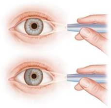
Pull down lower lids and inspect conjunctivae and sclerae |
|
|
Inspect upper tarsal conjunctivae only if presence of foreign body is suspected. |
EXPECTED:Conjunctivae clear and inapparent. Sclerae white and visible above irides only when eyelids are wide open. |
|
UNEXPECTED:Conjunctivae with erythema. Sclerae yellow or green. Sclerae with dark, rust-colored pigment anterior to insertion of medial rectus muscle. |
|
|
|
UNEXPECTED:Exudate. Pterygium. Corneal arcus senilis or opacities. |
|
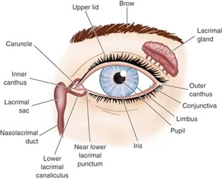
Inspect lacrimal gland region |
|
|
Palpate lower orbital rim near inner canthus. If temporal aspect of upper lid feels full, evert lid and inspect gland. |
EXPECTED:Slight elevations with central depression on both upper and lower lid margins. |
|
UNEXPECTED:Enlarged glands. Dry eyes. |
|
|
Test corneal sensitivity |
|
|
Touch wisp of cotton to cornea. |
EXPECTED:Bilateral blink reflex. |
|
Inspect external eyes |
|
|
Shine light tangentially on cornea. |
UNEXPECTED:Blood vessels present. |
|
|
EXPECTED:Clearly visible pattern. Similar color. |
|
|
EXPECTED:Round, regular, equal in size. |
|
UNEXPECTED:Miosis, mydriasis, anisocoria, or coloboma. |
|
|
|
EXPECTED:Constricting with consensual response of opposite pupil. |
|
|
EXPECTED:Constricting when pupils focus on near object or dilating when focus changes from near to distant object. |
|
|
EXPECTED:The pupil toward which the light is moving dilates and then constricts as the light shines onto it. |
|
UNEXPECTED:The pupil continues to dilate when the light shines into it. |
|
|
EXTRAOCULAR EYE MUSCLES |
|
|
Evaluate muscle balance and movement of eyes |
|
|
Hold patient’s chin, and ask patient to watch finger or penlight. |
EXPECTED:A few horizontal nystagmic beats. Smooth, full, coordinated movement of eyes. |
|
UNEXPECTED:Sustained or jerking nystagmus. Exposure of sclera from lid lag. Inability of eye to move in all directions. |
|
|
|
|
|
Direct light at nasal bridge from 30 cm (12 inches). Have patient look at nearby object. |
EXPECTED:Light reflected symmetrically from both eyes. |
|
Perform if imbalance found with corneal light reflex test. Have patient stare ahead at near, fixed object. Cover one eye and observe other; remove cover and observe uncovered eye. Repeat with other eye. |
UNEXPECTED:Movement of covered or uncovered eye. |
|
OPHTHALMOSCOPIC EXAMINATION |
|
|
Inspect internal eye |
|
|
|
UNEXPECTED:Shallow chamber. If observed, avoid mydriatics. |
|
Shine focused light tangentially at limbus. Note illumination of iris nasally. |
|
|
|
|
|
With patient looking at distant object, direct light at pupil from about 30 cm (12 inches). Move toward patient, observing: |
|
|
|
UNEXPECTED:Opacities. |
|
|
EXPECTED:Yellow or pink background, depending on race. Possible crescents or dots of pigment at disc margin, usually temporally. |
|
UNEXPECTED:Discrete areas of pigmentation away from disc. Lesions. Drusen bodies. Hemorrhages. |
|
|
|
|
|
Follow blood vessels distally in each quadrant, noting crossings of arterioles and venules. |
EXPECTED:Possible venous pulsations (should be documented). Arteriole/venule (A/V) ratio 3 : 5 or 2 : 3. |
|
UNEXPECTED:Nicking, tortuosity. |
|
|
|
|
|
|
EXPECTED:Yellow to creamy pink, varying by race. Sharp, well-defined margin, especially in temporal region; 1.5-mm diameter. |
|
UNEXPECTED:Myelinated nerve fibers. Papilledema. Glaucomatous cupping. |
|
|
Ask patient to look directly at light. |
EXPECTED:Yellow dot surrounded by deep pink. |
AIDS TO DIFFERENTIAL DIAGNOSIS
|
ABNORMALITY |
DESCRIPTION |
|
Strabismus (paralytic and nonparalytic) |
Subjective Data:Eyes cannot focus simultaneously. Can focus separately in nonparalytic type. |
|
Objective Data:Eye movement on the cover-uncover test, strabismic eye will fixate on the object after the “straight” eye is covered. |
|
|
Episcleritis |
Subjective Data:Acute onset of mild to moderate discomfort or photophobia. Painless injection and/or watery discharge. |
|
Objective Data:Injection of the bulbar conjunctiva purplish elevation of a few millimeters. |
|
|
Cataracts |
Subjective Data:Cloudy or blurry vision, faded colors; halo may appear around lights. |
|
Objective Data:Opacity of lens, generally central, occasionally peripheral. |
|
|
Diabetic retinopathy (Background) |
Subjective Data:Initially asymptomatic. Blurred vision, distortion, or visual acuity loss in more advanced stages. |
|
Objective Data:Dot hemorrhages, microaneurysms, hard exudates. |
|
|
(Proliferative) |
Subjective Data:Generally asymptomatic, floaters, blurred vision, or progressive visual acuity loss in advanced stages. |
|
Objective Data:New vessel formation; extension out of the retina, hemorrhage. |
Pediatric Variations
EXAMINATION
|
TECHNIQUE |
FINDINGS |
|
VISUAL TESTING |
|
|
Measure visual acuity |
|
|
|
EXPECTED:Infants should be able to focus on and track a face or light through 60 degrees. Ages 3-5 years: 20/40 or better Age 6: 20/30 or better |
|
Visual acuity is tested, when child is cooperative, with Lea cards, Landolt C or HOTV chart, usually at about 3 years of age. |
|
|
EXTRAOCULAR EYE MUSCLES |
|
|
Evaluate muscle balance and movement of eyes |
|
|
Evaluation of six cardinal fields of gaze is performed as with adults. You may, however, need to hold child’s head still. |
|
SAMPLE DOCUMENTATION
Eyes.Near vision 20/40 in each eye uncorrected, corrected to 20/20 with glasses. Distant vision 20/20 by Snellen. Visual fields full by confrontation. Extraocular movements intact and full, no nystagmus. Corneal light reflex equal.
Lids and globes symmetric. No ptosis. Eyebrows full, no edema or lesions evident.
Conjunctivae pink, sclerae white. No discharge evident. Cornea clear, corneal reflex intact. Irides brown; pupils equal, round, reactive to light and accommodation.
Ophthalmoscopic examination reveals red reflex. Discs cream colored, borders well defined with temporal pigmentation in both eyes. No venous pulsations evident at disc. Arteriole/venule ratio 3 : 5; no nicking or crossing changes, hemorrhages, or exudates noted. Maculae are yellow in each eye.
 Distance vision