
CYTOMEGALOVIRUS (CMV)
Microbiology
CMV is a herpesvirus, has double-stranded DNA, and renders infected cells 2–4 times the size of surrounding cells. These cytomegalic cells contain an eccentrically placed intranuclear inclusion surrounded by a clear halo, with an “owl’s-eye” appearance.
Epidemiology
CMV disease is found worldwide. Perinatal and early childhood infections are common; ~1% of U.S. newborns are infected.
• The virus can be spread in breast milk, saliva, feces, and urine.
• Transmission requires repeated or prolonged contact as opposed to casual contact. Sexual transmission is common among adolescents and adults, and CMV has been identified in semen and cervical secretions.
• Latent CMV infection persists throughout life unless reactivation is triggered by depressed cell-mediated immunity (e.g., in transplant recipients or HIV-infected pts).
Pathogenesis
Primary CMV infection is associated with a vigorous T lymphocyte response; activated CD8+ T cells predominate among atypical lymphocytes.
• Latent infection occurs in multiple cell types and various organs. Chronic antigen stimulation in the presence of immunosuppression (e.g., in the transplantation setting) and certain immunosuppressive agents (e.g., antithymocyte globulin) promote CMV reactivation.
• CMV disease increases the risk of infection with opportunistic pathogens by depressing T lymphocyte responsiveness.
Clinical Manifestations
The most common presentation is CMV mononucleosis in immunocompetent pts, but disease can be more severe in immunocompromised pts (including newborns).
Congenital CMV Infection
Cytomegalic inclusion disease occurs in ~5% of infected fetuses in the setting of primary maternal CMV infection in pregnancy.
• Petechiae, hepatosplenomegaly, and jaundice are present in 60–80% of cases; microcephaly with or without cerebral calcifications, intrauterine growth retardation, prematurity, and chorioretinitis are less common.
• Laboratory findings include elevated values in LFTs, thrombocytopenia, conjugated hyperbilirubinemia, hemolysis, and increased CSF protein levels.
• The mortality rate is 20–30% among infants with severe disease; survivors have intellectual or hearing difficulties.
Perinatal CMV Infection
Perinatal infection with CMV is acquired by breast-feeding or contact with infected maternal secretions (e.g., in the birth canal). Although most pts are asymptomatic, disease similar to—but less severe than—congenital CMV can occur.
CMV Mononucleosis
Symptoms last 2–6 weeks and include high fevers, profound fatigue and malaise, myalgias, headache, and splenomegaly; in contrast to EBV infection, exudative pharyngitis and cervical lymphadenopathy are rare in CMV infection.
• Laboratory findings include relative lymphocytosis with >10% atypical lymphocytes, transaminitis, and immunologic abnormalities (e.g., the presence of cryoglobulins, rheumatoid factor, or cold agglutinins).
• The incubation period ranges from 20 to 60 days.
• Recovery is generally complete, but postviral asthenia can persist for months.
CMV Infection in the Immunocompromised Host
CMV is the most common and important viral pathogen complicating organ transplantation, with the greatest risk of infection at 1–4 months after transplantation. HIV-infected pts with CD4+ T cell counts of <50–100/μL also are at risk for severe CMV disease.
• Primary CMV infection (including reinfection with a new, donor-derived strain) is more likely than reactivation to cause severe disease with high viral loads.
– Reactivation infection is common but less important clinically.
– The transplanted organ is at particular risk; e.g., CMV pneumonitis tends to follow lung transplantation.
– The risk of severe disease is reduced by antiviral prophylaxis or preemptive therapy.
• Pts present initially with prolonged fever, malaise, anorexia, fatigue, night sweats, and arthralgias or myalgias but can ultimately have multi-organ involvement.
– Respiratory involvement is evidenced by tachypnea, hypoxia, unproductive cough, and chest radiographs demonstrating bilateral interstitial or reticulonodular infiltrates.
– GI involvement often includes hepatitis and ulcer formation.
– CMV encephalitis, particularly in HIV-infected pts, can occur as either progressive dementia or ventriculoencephalitis characterized by cranial nerve deficits, disorientation, and lethargy.
– CMV retinitis is an important cause of blindness in pts with advanced AIDS.
Diagnosis
Diagnosis requires isolation of CMV or detection of its antigens or DNA in appropriate clinical specimens in conjunction with a compatible clinical syndrome. Immunofluorescence assays for CMV antigens (pp65), PCR, viral culture, and serology are all useful means of detection.
TREATMENT Cytomegalovirus Infections
• When possible, seronegative donors should be used for seronegative transplant recipients.
• Ganciclovir (5 mg/kg IV bid for 14–21 days followed by 5 mg/kg IV qd) or valganciclovir (the oral prodrug of ganciclovir; 900 mg PO bid for 14–21 days followed by 900 mg PO qd) produces response rates of 70–90% among HIV-infected pts with CMV retinitis or colitis.
– In severe infections, ganciclovir is often combined with CMV immune globulin.
– Neutropenia is an adverse reaction to ganciclovir treatment that may require administration of colony-stimulating factors.
– Prophylactic or suppressive treatment can be given to high-risk transplant recipients (those who are seropositive before transplantation or culture positive without symptoms afterward).
– Resistance to ganciclovir is common among pts treated for >3 months and is usually related to mutations in the CMV UL97 gene.
– For CMV retinitis, ganciclovir can be administered via a slow-release pellet sutured into the eye, but this intervention does not provide treatment for the contralateral eye or for systemic disease.
• Foscarnet (180 mg/kg qd divided into 2 or 3 doses for 2 weeks, followed by 90–120 mg/kg IV qd) inhibits CMV DNA polymerase and is active against most ganciclovir-resistant CMV isolates. The primary adverse events include electrolyte disturbances and renal dysfunction.
• Cidofovir (5 mg/kg IV per week for 2 weeks followed by 3–5 mg/kg IV every 2 weeks) is a nucleotide analogue that is also effective against CMV; however, it can cause severe nephrotoxicity by proximal tubular cell injury. The use of saline hydration and probenecid reduces this adverse effect.
• CMV immune or hyperimmune globulin may reduce the risk of CMV disease in seronegative renal transplant recipients and prevent congenital CMV infection in infants born to women with primary CMV infection during pregnancy.
EPSTEIN-BARR VIRUS (EBV)
Epidemiology
EBV is a DNA virus in the family Herpesviridae that infects >90% of persons by adulthood.
• Infectious mononucleosis (IM) is a disease of young adults and is more common in areas with higher standards of hygiene; infection occurs at a younger age in areas with deficient standards of hygiene.
• EBV is spread by contact with oral secretions (e.g., by transfer of saliva during kissing) and is shed in oropharyngeal secretions by >90% of asymptomatic seropositive individuals.
Pathogenesis
EBV infects the epithelium of the oropharynx and salivary glands as well as B cells in tonsillar crypts prior to a period of viremia.
• There is polyclonal activation of B cells, and memory B cells form the reservoir for EBV. Reactive T cells proliferate, with up to 40% of CD8+ T cells directed against EBV antigens during acute infection.
• Cellular immunity is more important than humoral immunity in controlling infection. If T cell immunity is compromised, EBV-infected B cells may proliferate—a step toward neoplastic transformation.
Clinical Manifestations
The nature of disease depends on the pt’s age and immune status: young children typically develop asymptomatic infections or mild pharyngitis, adolescents and adults develop an IM syndrome, and immunocompromised pts can develop lymphoproliferative disease.
• In IM, a prodrome of fatigue, malaise, and myalgia may last for 1–2 weeks before the onset of fever, exudative pharyngitis, and lymphadenopathy with tender, symmetric, and movable nodes; splenomegaly is more prominent in the second or third week.
– The incubation period is ~4–6 weeks.
– Most pts treated with ampicillin develop a rash that does not represent a true penicillin allergy.
– Illness lasts for 2–4 months, but malaise and difficulty with concentration can persist for months longer. EBV is not, however, a cause of chronic fatigue syndrome.
– Lymphocytosis occurs in the second or third week, with >10% atypical lymphocytes (enlarged cells with abundant cytoplasm and vacuoles); abnormal liver function is common.
– Complications include CNS disease (e.g., meningitis, encephalitis), Coombs-positive autoimmune hemolytic anemia, splenic rupture, and upper airway obstruction due to hypertrophy of lymphoid tissue.
• Lymphoproliferative disease—i.e., infiltration of lymph nodes and multiple organs by proliferating EBV-infected B cells—occurs in pts with deficient cellular immunity (e.g., pts with AIDS, those with severe combined immunodeficiency, and those receiving immunosuppressive medications). Pts develop fever and lymphadenopathy or GI symptoms.
• Oral hairy leukoplakia—raised, white, corrugated, EBV DNA–containing lesions on the tongue—is an early manifestation of infection with HIV in adults.
• EBV-associated malignancies include Burkitt’s lymphoma (~90% of cases in Africa and ~15% of cases in the U.S.), anaplastic nasopharyngeal carcinoma in southern China, Hodgkin’s disease (especially the mixed-cellularity type), and CNS lymphoma (especially HIV-related).
Diagnosis
Serologic testing is the mainstay of diagnostic assessment. PCR analysis can be useful in monitoring EBV DNA levels in blood from pts with lymphoproliferative disease.
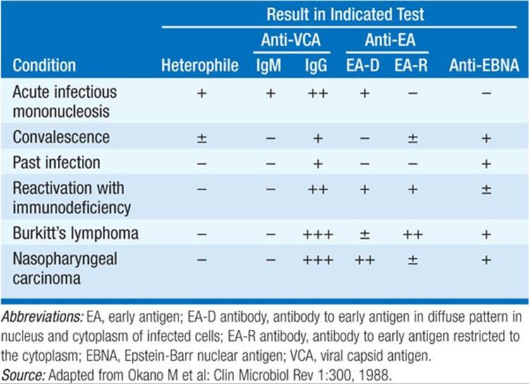
• Heterophile antibodies (Table 109-1) form the basis of most rapid testing, which assesses the ability of serum to agglutinate sheep, horse, or cow erythrocytes after adsorption with guinea pig kidney.
TABLE 109-1 SEROLOGIC FEATURES OF EBV-ASSOCIATED DISEASES
– The antibodies can persist for up to 1 year after infection.
– The monospot test for heterophile antibodies is ~75% sensitive and ~90% specific in comparison with EBV-specific serologies.
– Pts <5 years old and elderly pts usually do not develop heterophile antibodies.
• EBV-specific antibody testing (Table 109-1) can be used in heterophile-negative pts and pts with atypical disease. Antibodies to viral capsid antigen occur in >90% of cases, with elevated IgM titers present only during the first 2–3 months of disease.
• Antibodies to Epstein-Barr nuclear antigen are not detected until 3–6 weeks after symptom onset and then persist for life.
TREATMENT Epstein-Barr Virus Infections
• IM is treated with supportive measures, including rest and analgesia.
– Excessive physical activity should be avoided in the first month of illness to reduce the possibility of splenic rupture, which necessitates splenectomy.
– Administration of glucocorticoids may be indicated for some complications of IM; e.g., these agents may be given to prevent airway obstruction or to treat autoimmune hemolytic anemia.
– Antiviral therapy (e.g., with acyclovir) is generally not effective for IM but is effective for oral hairy leukoplakia.
• Treatment of posttransplantation EBV lymphoproliferative syndrome is generally directed toward reduction of immunosuppression, although other treatments—e.g., with interferon α, antibody to CD20 (rituximab), and donor lymphocyte infusions—have been used with varying success.

For a more detailed discussion, see Baden LR, Dolin R: Antiviral Chemotherapy, Excluding Antiretroviral Drugs, Chap. 178, p. 1442; Cohen JI: Epstein-Barr Virus Infections, Including Infectious Mononucleosis, Chap. 181, p. 1467; and Hirsch MS: Cytomegalovirus and Human Herpesvirus Types 6, 7, and 8, Chap. 182, p. 1471, in HPIM-18.