
INFLUENZA
Microbiology and Pathogenesis
Influenza A, B, and C viruses are RNA viruses and members of the family Orthomyxoviridae with different nucleoprotein (NP) and matrix (M) protein antigens. Influenza A and B viruses are major human pathogens and are morphologically similar; influenza B infection is associated with less severe disease than influenza A infection, and influenza C virus causes subclinical disease.
• Influenza A viruses are subtyped by surface hemagglutinin (H) and neuraminidase (N) antigens.
– Virus attaches to sialic acid cell receptors via the hemagglutinin. Neuraminidase degrades the receptor and plays a role in the release of virus from infected cells after replication has occurred.
– Antibodies to the H antigen are the major determinants of immunity, while antibodies to the N antigen limit viral spread and contribute to reduction of the infection.
• Influenza is acquired from aerosolized respiratory secretions of acutely ill individuals and possibly by hand-to-hand contact or other personal or fomite contact. Viral shedding usually stops 2–5 days after disease onset.
Epidemiology
Influenza outbreaks occur each year but vary in extent and severity. Influenza A epidemics occur almost exclusively during the winter months in temperate climates but occur year-round in the tropics. These epidemics begin abruptly, peak over 2–3 weeks, last 2–3 months, and then subside rapidly.
• Global pandemics (the most recent of which took place in 2009 and was due to an A/H1N1 virus) occur, by definition, at multiple locations; they carry high attack rates (10–20% of the general population), extend beyond normal seasonality patterns, and are due in part to the propensity of the H and N antigens to undergo periodic antigenic variation.
– Major changes (which are restricted to influenza A viruses) are called antigenic shifts and are associated with pandemics. Minor variations are called antigenic drifts.
– The avian influenza strain A/H5N1, first detected in 1997, has not resulted in a pandemic because efficient person-to-person transmission has not occurred; infection is linked to direct contact with infected poultry.
• The segmented genome of influenza A and B viruses allows reassortment of strains between different animal species. The pandemic A/H1N1 virus of 2009–2010 represented a quadruple reassortment among swine, avian, and human influenza viruses.
• Interpandemic outbreaks of influenza are associated with economic costs exceeding $87 billion in the U.S. Chronic cardiopulmonary disease and old age are the most prominent risk factors for severe illness.
Clinical Manifestations
Influenza has a wide spectrum of clinical presentations, ranging from a mild illness resembling the common cold to severe prostration with relatively few respiratory symptoms. The classic description involves the abrupt onset of headache, fever, chills, myalgia, and malaise in the setting of respiratory symptoms (e.g., cough, sore throat).
• Pts typically defervesce within 2–3 days, but respiratory symptoms accompanied by substernal pain can persist for ≥1 week. Postinfluenzal asthenia may persist for weeks, particularly in the elderly.
• Complications of influenza (pneumonia and extrapulmonary manifestations) are more common among pts >64 years old, pregnant women, and pts with chronic disorders (e.g., cardiopulmonary disease, diabetes, renal diseases, hemoglobinopathies, or immunosuppression).
– Pneumonia: Primary influenza pneumonia is the least common but most severe of the pneumonic complications, most often affecting pts with mitral stenosis and pregnant women. Pts have progressive pulmonary disease and high titers of virus in respiratory secretions.
• Secondary bacterial pneumonia is usually due to Streptococcus pneumoniae, Staphylococcus aureus, or Haemophilus influenzae and presents as the reappearance of fever and respiratory symptoms after 2–3 days of clinical improvement.
• The most common pneumonic complication involves aspects of viral and bacterial pneumonia.
– Extrapulmonary complications: Reye’s syndrome, myositis, rhabdomyolysis, myoglobinuria, and CNS disease (e.g., encephalitis, transverse myelitis, Guillain-Barré syndrome) can occur as complications of influenza infection.
• Reye’s syndrome is a serious complication in children that is associated with influenza B virus (and less commonly with influenza A virus), varicella-zoster virus, and aspirin therapy for the antecedent viral infection.
Laboratory Findings
Most commonly, a laboratory diagnosis is made with a rapid test that detects viral antigens from throat swabs, nasopharyngeal washes, or sputum. These tests are relatively specific but variably sensitive.
• Reverse-transcription PCR of respiratory samples is most sensitive and specific for detecting influenza. Viral culture is also possible and gives a positive result within 48–72 h.
• Serologic testing requires the availability of acute- and convalescent-phase sera and is useful only retrospectively.
TREATMENT Influenza
• See Table 110-1 for specific treatment of influenza.
TABLE 110-1 ANTIVIRAL MEDICATIONS FOR TREATMENT AND PROPHYLAXIS OF INFLUENZA

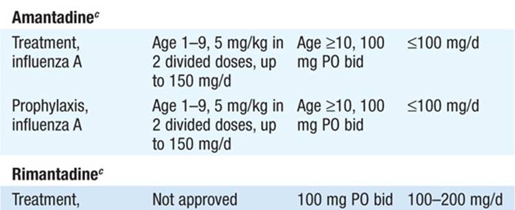
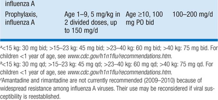
– Antiviral agents have been tested in healthy adults with uncomplicated influenza but not in the treatment or prevention of complications associated with influenza.
– If started within 2 days of illness due to a susceptible virus, the neuraminidase inhibitors (oseltamivir and zanamivir) and the adamantane agents (amantadine and rimantadine) reduce the duration of signs and symptoms by 1–1.5 days and ~50%, respectively.
– IV formulations of neuraminidase inhibitors (peramivir and zanamivir) are currently in clinical trials but may be accessed through the FDA’s Emergency Investigational New Drug (E-IND) application procedures.
– Zanamivir may exacerbate bronchospasm in asthmatic pts, while oseltamivir has been associated with nausea and vomiting (reactions whose incidence is reduced if the drug is given with food) and with neuropsychiatric side effects in children.
– Amantadine causes mild CNS side effects (e.g., jitteriness, anxiety, insomnia, difficulty concentrating) in ~5–10% of pts; rimantadine has fewer CNS side effects.
• For uncomplicated influenza in individuals at low risk for complications, symptom-based rather than antiviral therapy may be considered.
Prophylaxis
Annual vaccination with either an inactivated or a live attenuated vaccine is the main public health measure for prevention of influenza.
• Vaccine strains are generated from influenza A and B viruses that have circulated during the previous influenza season and whose circulation during the upcoming season is predicted.
– For inactivated vaccines, 50–80% protection against influenza is expected if the vaccine virus and the currently circulating viruses are closely related.
– Influenza vaccination is currently recommended for all individuals >6 months of age.
• Chemoprophylaxis against influenza (see Table 110-1 for regimens) should be reserved for individuals at high risk of complications who have had close contact with a pt sick with influenza. Chemoprophylaxis can be administered simultaneously with inactivated—but not with live—vaccine.
OTHER COMMON VIRAL RESPIRATORY INFECTIONS
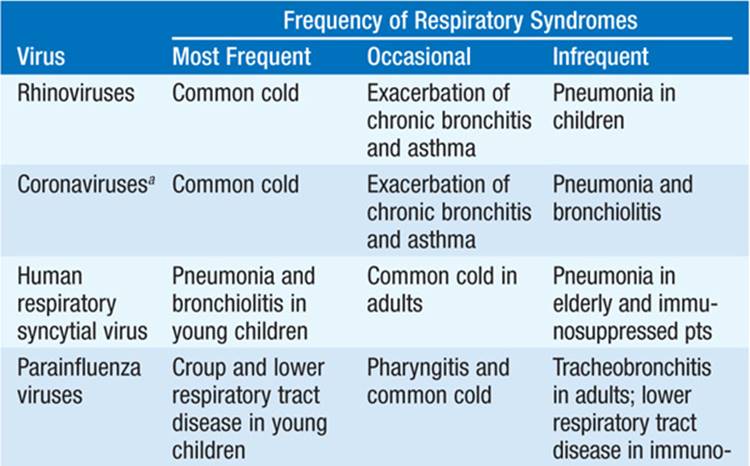
Acute viral respiratory illnesses account for ≥50% of all acute illnesses; adults have 3–4 cases per person per year. Clinical presentations for viral infections are generally not specific enough to allow an etiologic diagnosis, and viral illnesses are typically grouped into clinical syndromes (e.g., the “common cold,” pharyngitis, tracheitis, pneumonia). This section will cover the six major groups of respiratory viruses; see Table 110-2 for an overview and Chap. 64for additional details on viral respiratory infections.
TABLE 110-2 ILLNESSES ASSOCIATED WITH RESPIRATORY VIRUSES


RHINOVIRUSES
Microbiology
Rhinoviruses are nonenveloped, single-stranded RNA viruses in the family Picornaviridae that together are the major cause of the “common cold” (up to 50% of cases).
Epidemiology
Rhinoviruses are spread by direct contact with infected secretions, usually respiratory droplets.
Clinical Manifestations
After an incubation period of 1–2 days, pts develop rhinorrhea, sneezing, nasal congestion, and sore throat that lasts 4–9 days. Fever and other systemic symptoms are unusual.
• Severe disease, including fatal pneumonia, is rare but has been described in immunocompromised pts, particularly bone marrow transplant recipients.
Diagnosis
An etiologic diagnosis usually is not attempted, given that the disease is generally mild and self-limited. PCR and tissue culture methods are available.
Treatment
Treatment is limited to symptom relief (e.g., with antihistamines, decongestants).
CORONAVIRUSES
Microbiology
Coronaviruses are pleomorphic, single-stranded RNA viruses.
Epidemiology and Clinical Manifestations
Coronaviruses often result in the common cold (accounting for 10–35% of cases) with symptoms similar to those caused by rhinoviruses.
• Compared with rhinoviruses, the incubation period for coronaviruses is slightly longer (3 days) and the duration of illness is slightly shorter (6–7 days).
• In 2002–2003, a coronavirus-induced severe acute respiratory syndrome (SARS) developed in >8000 pts in 28 countries (with 90% of cases in China and Hong Kong) and was associated with a ~9.5% case-fatality rate; no cases were reported in 2005–2009.
– SARS has an incubation period of 2–7 days, after which pts develop fever, malaise, headache, myalgias, and then (1–2 days later) a nonproductive cough and dyspnea.
– Respiratory function may worsen in the second week of illness and can progress to ARDS and multiorgan dysfunction.
Diagnosis
Laboratory diagnosis of coronavirus-induced colds is rarely required, but ELISA, immunofluorescence, and RT-PCR assays can detect the virus in clinical specimens.
• The coronavirus associated with SARS (SARS-CoV) can be detected by RT-PCR or viral culture from respiratory samples and serum early in illness and from urine and stool later on.
• SARS is also associated with lymphopenia (mostly CD4+ cells) in 50% of cases.
TREATMENT Coronaviruses
• For the common cold, no treatment beyond symptom relief is generally needed.
• For SARS, aggressive supportive care is most important. No specific therapy (e.g., ribavirin, glucocorticoids) has been established as efficacious.
HUMAN RESPIRATORY SYNCYTIAL VIRUS
Microbiology
Human respiratory syncytial virus (HRSV) is an enveloped, single-stranded RNA virus and a member of the family Paramyxoviridae.
Epidemiology
With an attack rate approaching 100% among susceptible individuals, HRSV is a major respiratory pathogen among young children (particularly those 2–3 months of age) and the foremost cause of lower respiratory disease among infants.
• HRSV accounts for 20–25% of hospital admissions of young children for pneumonia and for up to 75% of cases of bronchiolitis in this age group.
• The virus is transmitted efficiently via contact with contaminated fingers or fomites and by spread of coarse aerosols. The incubation period is ~4–6 days.
Clinical Manifestations
Infants typically develop rhinorrhea, low-grade fever, cough, and wheezing; 20–40% of infections result in lower tract disease, including pneumonia, bronchiolitis, and tracheobronchitis.
• In adults, HRSV typically presents as the common cold, but it can cause lower respiratory tract disease with fever, including severe pneumonia in elderly or immunosuppressed pts. HRSV pneumonia has a case-fatality rate of 20–80% among transplant pts.
Diagnosis
Rapid viral diagnosis is available by immunofluorescence, ELISA, or RT-PCR of nasopharyngeal washes, aspirates, or (less satisfactorily) swabs.
TREATMENT Human Respiratory Syncytial Virus
• Treatment is symptom-based for upper tract disease and mild lower tract disease.
• For severe lower tract disease, intubation and ventilatory assistance should be given as needed.
– Aerosolized ribavirin has a demonstrated modest beneficial effect for infants with severe HRSV pneumonia, but its efficacy in older children and adults (including immunocompromised pts) has not been established.
– No benefit has been found in any pt population for IV immunoglobulin (IVIg), immunoglobulin with high titers of antibody to HRSV (RSVIg), or a monoclonal IgG antibody to HRSV (palivizumab).
Prevention
Monthly administration of palivizumab is approved for prophylaxis in children <2 years of age who have bronchopulmonary dysplasia or cyanotic heart disease or who were born prematurely. In settings with high transmission rates (e.g., pediatric wards), contact precautions are useful to limit spread of the virus.
HUMAN METAPNEUMOVIRUS
Microbiology
Human metapneumovirus (HMPV) is a pleomorphic, single-stranded RNA virus of the family Paramyxoviridae.
Epidemiology
HMPV accounts for 1–5% of childhood upper respiratory tract infections and for 2–4% of acute respiratory illnesses in ambulatory adults.
Clinical Manifestations
Disease manifestations are similar to those caused by HRSV.
Diagnosis
Diagnosis is made by immunofluorescence, PCR, or tissue culture of nasal aspirates or respiratory secretions.
Treatment
Treatment is primarily supportive and symptom-based.
PARAINFLUENZA VIRUS
Microbiology and Epidemiology
This enveloped, single-stranded RNA virus of the family Paramyxoviridae ranks second only to HRSV as a cause of lower respiratory tract disease among young children and is the most common cause of croup (laryngotracheobronchitis).
Clinical Manifestations
Infections are milder among older children and adults, but severe, prolonged, and fatal infection has been reported among pts with severe immunosuppression, including transplant recipients.
Diagnosis
Tissue culture, rapid testing with immunofluorescence or ELISA (both of which are less sensitive), or PCR of respiratory tract secretions, throat swabs, or nasopharyngeal washings can detect the virus.
Treatment
Treatment of upper respiratory tract disease is symptom-based. Humidified air may be helpful for mild cases of croup.
• For cases of croup with respiratory distress, intermittent racemic epinephrine and glucocorticoids are beneficial.
• Anecdotal reports describe the use of ribavirin (particularly in immuno-suppressed pts), but its clinical utility is still unclear.
ADENOVIRUSES
Microbiology and Epidemiology
Adenoviruses are double-stranded DNA viruses that cause ~10% of acute respiratory infections among children and <2% of respiratory illnesses among civilian adults. Some serotypes are associated with outbreaks among military recruits. Transmission takes place primarily from fall to spring via inhalation of aerosolized virus, through inoculation of the conjunctival sacs, and probably via the fecal-oral route.
Clinical Manifestations
In children, adenovirus causes acute upper and lower respiratory tract infections and outbreaks of pharyngoconjunctival fever (a syndrome of fever, bilateral conjunctivitis, sore throat, and cervical adenopathy typically due to types 3 and 7).
• In adults, adenovirus types 4 and 7 cause an acute respiratory disease consisting of a prominent sore throat, fever on the second or third day of illness, cough, coryza, and regional adenopathy. Pharyngeal edema and tonsillar hypertrophy with little or no exudate may be seen.
• In addition to respiratory disease, adenovirus can cause diarrheal illness, hemorrhagic cystitis, and epidemic keratoconjunctivitis. In pts who have received a solid-organ transplant, adenovirus can affect the transplanted organ and disseminate to other organs.
Diagnosis
Definitive diagnosis can be made by isolation of the virus in tissue culture; by rapid testing (with immunofluorescence or ELISA) of nasopharyngeal aspirates, conjunctival or respiratory secretions, urine, or stool; or by PCR testing.
Treatment
Treatment is supportive. Ribavirin and cidofovir exhibit in vitro activity against adenovirus and therefore are used occasionally in disseminated adenovirus infections, but definitive efficacy data are not available.

For a more detailed discussion, see Baden LR, Dolin R: Antiviral Chemotherapy, Excluding Antiretroviral Drugs, Chap. 178, p. 1442; Dolin R: Common Viral Respiratory Infections, Chap. 186, p. 1485; and Dolin R: Influenza, Chap. 187, p. 1493, in HPIM-18.