
Pneumocystis, an opportunistic fungal pulmonary pathogen, is an important cause of pneumonia in immunocompromised hosts.
MICROBIOLOGY
• P. jirovecii infects humans, whereas P. carinii—the original species described—infects rats.
• In contrast to most fungi, Pneumocystis lacks ergosterol and thus is not susceptible to antifungal drugs that inhibit ergosterol synthesis.
• Developmental stages include the small trophic form, the cyst, and the intermediate precyst stage.
EPIDEMIOLOGY
• Pneumocystis is found worldwide, and most healthy children have been exposed to the organism by 3–4 years of age.
• Both airborne transmission and person-to-person transmission have been demonstrated; the cyst is the transmissible form.
• Defects in cellular and humoral immunity (including immunosuppressive medications) predispose to Pneumocystis pneumonia. The risk in HIV-infected pts rises dramatically when CD4+ T cell counts fall below 200/μL.
PATHOGENESIS
• The organisms are inhaled and attach tightly to type I cells in alveoli, although they remain extracellular.
• On histology, alveoli are seen to be filled with foamy, vacuolated exudates.
• Severe disease may cause interstitial edema, fibrosis, and hyaline membrane formation.
CLINICAL MANIFESTATIONS
• Pts develop dyspnea, fever, and nonproductive cough.
– HIV-infected pts are usually ill for several weeks or longer with subtle manifestations.
– Pts without HIV infection often become symptomatic after their glucocorticoid dose has been tapered, and their symptoms are of shorter duration.
• Physical exam reveals tachypnea, tachycardia, and cyanosis out of proportion to the few abnormalities present on chest auscultation.
• Nonspecific laboratory findings include elevated serum concentrations of lactate dehydrogenase and β-D-glucan, a component of the fungal cell wall.
• CXR classically reveals bilateral diffuse infiltrates beginning in the perihilar regions. Other findings (e.g., nodular densities, cavitary lesions) have been described.
• Rare cases of disseminated infection have been described, generally involving lymph nodes, spleen, liver, and bone marrow.
DIAGNOSIS
• Histopathologic staining makes the definitive diagnosis.
– Cell-wall stains (e.g., methenamine silver) are used for Pneumocystis cysts and Wright-Giemsa stains for the nuclei of all developmental stages.
– Immunofluorescence with monoclonal antibodies increases diagnostic sensitivity.
• DNA amplification by PCR is most sensitive but may not distinguish colonization from infection.
• Proper specimens are key.
– Fiberoptic bronchoscopy with bronchoalveolar lavage remains the mainstay of diagnosis.
– Sputum induction and oral washes are noninvasive options that are gaining popularity.
– Transbronchial biopsy and open lung biopsy are used only when a diagnosis cannot be made by bronchoalveolar lavage.
TREATMENT Pneumocystis Infections
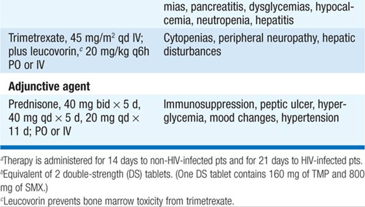
• Trimethoprim-sulfamethoxazole (TMP-SMX) is the drug of choice for all pts. For doses and adverse effects of TMP-SMX and alternative regimens, see Table 116-1.
TABLE 116-1 TREATMENT OF PNEUMOCYSTOSIS
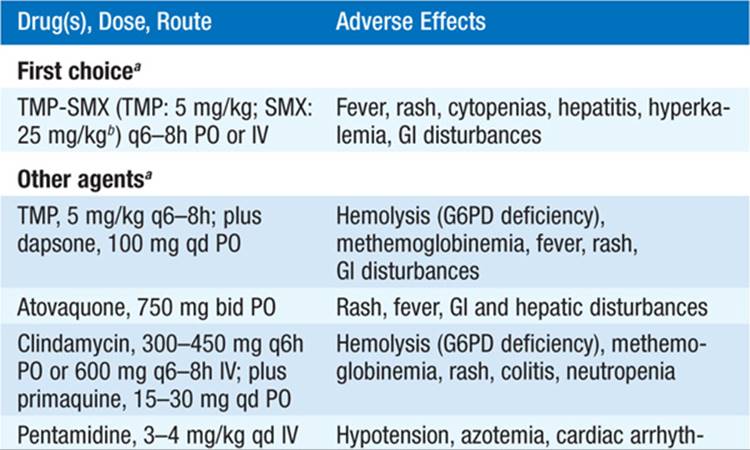
• For mild to moderate cases (a Pao2 >70 mmHg or a PAo2 – Pao2 gradient <35 mmHg on room air), alternatives include TMP plus dapsone and clindamycin plus primaquine. Atovaquone is less effective than TMP-SMX but is better tolerated.
• For moderate to severe cases (a Pao2 ≤70 mmHg or a Pao2 – Pao2 gradient ≥ 35 mmHg), alternatives include IV pentamidine, IV clindamycin plus primaquine (potentially more effective than pentamidine), and trimetrexate plus leucovorin.
• Adjunctive administration of tapering doses of glucocorticoids to HIV-infected pts with moderate to severe disease reduces the risk of respiratory function deterioration shortly after initiation of treatment. Use of glucocorticoids in other pts remains to be evaluated.
PROGNOSIS
• Therapy is most effective if started early, before there is extensive alveolar damage.
• The mortality rate among HIV-infected pts is 0–15%.
• The risk of early death remains high among people who need mechanical ventilation (60%) and among non-HIV-infected pts (40%).
PREVENTION
• Prophylaxis is indicated for HIV-infected pts with CD4+ T cell counts <200/μL or a history of oropharyngeal candidiasis and for any pt with a history of Pneumocystis pneumonia. Guidelines for other compromised hosts are less clear.
• In HIV infection, once CD4+ counts have risen to >200/μL and have remained above that cutoff for ≥3 months, prophylaxis may be stopped.
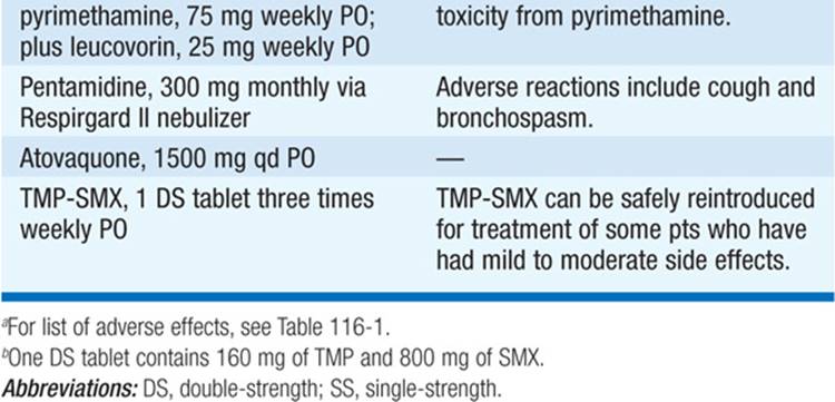
• For prophylactic regimens, see Table 116-2. TMP-SMX is the drug of choice for both primary and secondary prophylaxis and also protects against toxoplasmosis and some bacterial infections.
TABLE 116-2 PROPHYLAXIS OF PNEUMOCYSTOSISa


For a more detailed discussion, see Smulian AG, Walzer PD: Pneumocystis Infections, Chap. 207, p. 1671, in HPIM-18.