
MALARIA
Microbiology
Five major species of Plasmodium cause nearly all cases of human disease: P. falciparum, P. vivax, P. ovale, P. malariae, and P. knowlesi.
• P. falciparum, the cause of most cases of severe disease and most deaths, predominates in Africa, New Guinea, and Hispaniola.
• P. vivax is more common in Central America.
• P. falciparum and P. vivax are equally prevalent in South America, the Indian subcontinent, eastern Asia, and Oceania.
• P. ovale is unusual outside of Africa, where it makes up ~1% of isolates.
• P. malariae is found in most areas (especially throughout sub-Saharan Africa) but is less common.
• P. knowlesi (the monkey malaria parasite) can reliably be identified only by molecular techniques and is present in Borneo and Southeast Asia.
Epidemiology
Malaria is the most important parasitic disease in humans, causing ~1 million deaths each year.
Pathogenesis
After introduction of sporozoites into the bloodstream by female anopheline mosquitoes, the parasite travels to the liver and reproduces asexually to form merozoites that infect RBCs. The merozoites transform into trophozoites, feed on intracellular proteins (principally hemoglobin), multiply 6- to 20-fold every 48–72 h, and cause the RBCs to rupture, releasing daughter merozoites. The process then repeats.
• Some parasites develop into long-lived sexual forms called gametocytes, which can be taken up by another female anopheline mosquito, allowing transmission.
• In P. vivax or P. ovale infection, dormant forms called hypnozoites remain in liver cells and may cause disease 3 weeks to >1 year later.
• RBCs infected with P. falciparum may exhibit cytoadherence (attachment to venular and capillary endothelium), rosetting (adherence to uninfected RBCs), and agglutination (adherence to other infected RBCs). The result is sequestration of P. falciparum in vital organs, with consequent underestimation (through parasitemia determinations) of parasite numbers in the body. Sequestration is central to the pathogenesis of falciparum malaria but is not evident in the other three “benign” forms.
• In nonimmune individuals, infection triggers nonspecific host defense mechanisms such as increased splenic filtration.
– With repeated exposure to malaria, pts develop resistance to high-level parasitemia and disease but not to infection.
– Hemoglobinopathies (e.g., sickle cell disease, ovalocytosis, thalassemia) and G6PD deficiency are more common in endemic areas and protect against death from malaria.
Clinical Manifestations
Pts initially develop nonspecific symptoms (e.g., headache, fatigue, myalgias) that are followed by fever.
• Febrile paroxysms at regular intervals are unusual and suggest infection with P. vivax or P. ovale.
• Splenomegaly, hepatomegaly, mild anemia, and jaundice may develop.
• The diagnosis of severe falciparum malaria requires ≥1 of the following: impaired consciousness/coma, severe normocytic anemia, renal failure, pulmonary edema, ARDS, circulatory shock, DIC, spontaneous bleeding, acidosis, hemoglobinuria, jaundice, repeated generalized convulsions, and a parasitemia level of >5%.
– Cerebral malaria manifests as diffuse symmetric encephalopathy, typically without focal neurologic signs.
– Coma is an ominous sign associated with mortality rates of ~20%.
• Pregnant women have unusually severe illness. Premature labor, fetal distress, stillbirth, and delivery of low-birth-weight infants are common.
• Tropical splenomegaly (hyperreactive malarial splenomegaly) may result as a chronic complication of malaria and is characterized by massive splenomegaly, hepatomegaly, and an abnormal immunologic response to infection.
Diagnosis
Although antibody-based diagnostic tests are being used with increasing frequency, demonstration of asexual forms of the parasite on peripheral-blood smears is required for diagnosis.
• Thick and thin smears should be examined; thick smears and the less sensitive thin smears detect parasitemia levels as low as 0.001% and ~0.05%, respectively.
• If the level of clinical suspicion is high and smears are initially negative, they should be repeated q12–24h for 2 days.
• Other laboratory findings generally include normochromic, normocytic anemia; elevated inflammatory markers; and thrombocytopenia (~105/μL).
TREATMENT Malaria
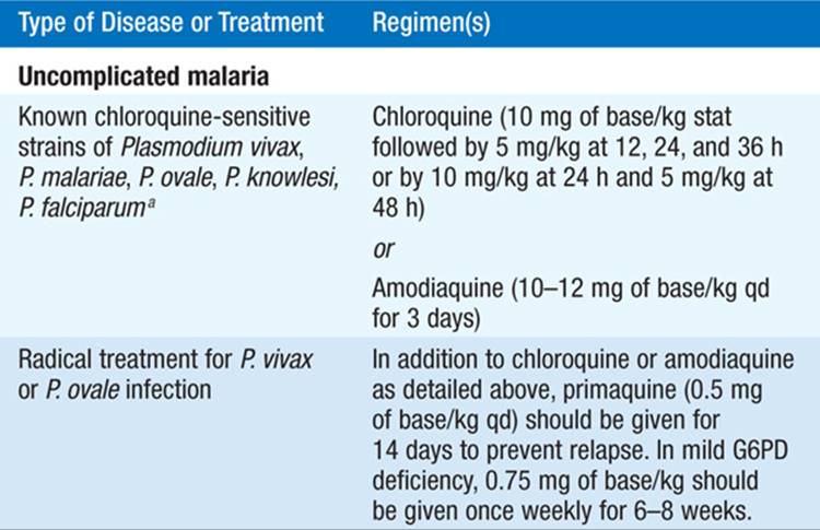
• See Table 117-1 for treatment regimens. IV artesunate is approved by the U.S. Food and Drug Administration for emergency use against severe malaria through the CDC [Malaria Hotline: 770-488-7788; Emergency Operations Center (after hours): 770-488-7100].
TABLE 117-1 REGIMENS FOR THE TREATMENT OF MALARIA

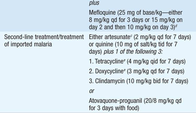


• Pts receiving quinidine should undergo cardiac monitoring; a total plasma level of >8 μg/mL, increased QT intervals (>0.6 s), or QRS widening by >25% is an indication for slowing the infusion rate.
• Exchange transfusions can be considered for severely ill pts, although indications for their use are not yet agreed upon.
• All pts with severe malaria should receive a continuous infusion of dextrose. Unconscious pts should have blood glucose levels measured q4–6h.
• Parasite counts and hematocrits for pts with severe malaria and pts with uncomplicated disease should be measured q6–12h and q24h, respectively.
• Primaquine (0.5 mg of base/kg for 14 days) eradicates persistent liver stages and prevents relapse in P. vivax or P. ovale infection. G6PD deficiency must be ruled out before treatment.
Personal Protection Measures
Measures that can protect persons against infection include avoidance of mosquito exposure, with particular caution at peak feeding times (dusk and dawn); use of insect repellents containing DEET (10–35%) or (if DEET is unacceptable) picaridin (7%); suitable clothing; and insecticide-impregnated bed nets.
Chemoprophylaxis
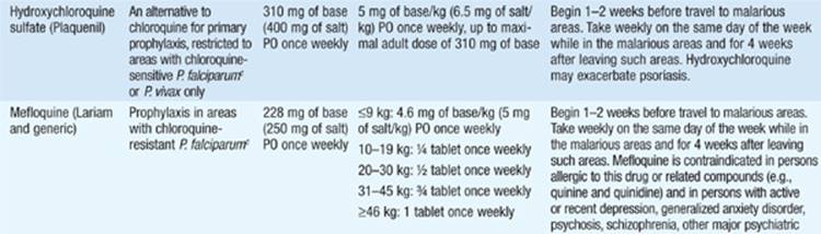
See Table 117-2 for prophylaxis options.
TABLE 117-2 DRUGS USED IN THE PROPHYLAXIS OF MALARIA


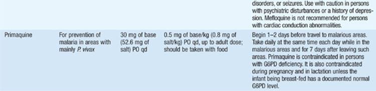

• Mefloquine is the only drug advised for pregnant women traveling to areas with drug-resistant malaria and is generally considered safe in the second and third trimesters; data regarding use in the first trimester, although limited, are reassuring.
BABESIOSIS
Microbiology
Babesiosis is caused by intraerythrocytic protozoa of the genus Babesia. B. microti is the etiologic agent in the northeastern United States, and B. duncani is responsible for disease on the West Coast. B. divergens causes disease in Europe. The deer tick (Ixodes scapularis) transmits B. microti; the vectors of transmission for the other Babesia species are unknown.
Epidemiology
In the United States, infections occur most frequently along the northeastern coast. In 2009, >700 cases were reported in the U.S.; this number is probably an underestimate, given that most pts experience a mild and self-limiting disease and may not seek medical attention.
Clinical Manifestations
Most pts develop a mild illness, but immunosuppressed pts may have more severe disease.
• After an incubation period of 1–6 weeks, pts gradually develop fevers, fatigue, and weakness. Other symptoms may include chills, sweats, myalgias, arthralgias, headache, and—less often—neck stiffness, shortness of breath, and abdominal pain.
• Severe babesiosis is associated with parasitemia levels of >4%.
– Risk factors include an age of >50 years, male gender, asplenia, HIV infection/AIDS, malignancy, and immunosuppression.
– Complications include respiratory failure, DIC, CHF, and renal failure.
– The fatality rate is 5% among all hospitalized pts and 20% among immunocompromised pts.
Diagnosis
Giemsa-stained thin smears identify intraerythrocytic Babesia parasites, which appear round or pear-shaped.
• Ring forms resembling P. falciparum but without pigment are most common.
• Tetrads (“Maltese crosses”)—formed by four budding merozoites—are pathognomonic for B. microti and other small Babesia species.
• PCR and serology can also be used for diagnostic purposes.
TREATMENT Babesiosis
• Mild illness should be treated with atovaquone (750 mg PO q12h) plus azithromycin (500–1000 mg/d PO on day 1 followed by 250 mg/d PO) for 7–10 days.
– Clindamycin plus quinine is equally effective but not as well tolerated.
– Treatment should be given only if Babesia is detected on blood smear, regardless of serology or PCR results.
• Severe disease should be treated with clindamycin (300–600 mg q6h IV or 600 mg q8h PO) plus quinine (650 mg q6–8h PO) for 7–10 days.
– Consider exchange transfusion in cases with high-level parasitemia (>10%); hemoglobin levels of ≤10 g/dL; or pulmonary, hepatic, or renal compromise.
– Immunocompromised pts generally need longer courses of treatment (e.g., 6 weeks), with at least 2 weeks of therapy after parasites are no longer detected on blood smear.
• B. duncani and B. divergens infections can be treated with IV clindamycin and quinine for 7–10 days.
LEISHMANIASIS
Microbiology
Leishmania species are extracellular, flagellated promastigotes while dwelling in their sandfly vector, but are obligate intracellular, nonflagellated amastigotes while living in vertebrate hosts, including humans.
• Organisms of the L. donovani complex usually cause visceral leishmaniasis and are present in Asia, the Middle East, the horn of Africa, the Mediterranean, and Central and South America.
• L. tropica, L. major, and L. aethiopica cause Old World cutaneous leishmaniasis and are present in Asia and in northern and sub-Saharan Africa.
• The L. mexicana complex causes New World cutaneous leishmaniasis and is present in Central America and northern South America.
Epidemiology
Roughly 2 million cases of leishmaniasis occur annually worldwide, of which 1–1.5 million are cutaneous and 500,000 are visceral.
Clinical Manifestations
Visceral Leishmaniasis (kala-azar): Pts most commonly present with an abrupt onset of moderate- to high-grade fever associated with rigor and chills.
• Splenomegaly, hepatomegaly, and (except in the Indian subcontinent) lymphadenopathy are common.
• Leukopenia, anemia, thrombocytopenia, a polyclonal increase in serum immunoglobulins, and hepatic transaminitis are common.
• Up to 50% of pts in India, East Africa, and the Sudan may develop hypopigmented skin lesions (post–kala-azar dermal leishmaniasis) concurrent with or after cure of visceral leishmaniasis. In some cases, these pts may require unusually long treatment courses.
Cutaneous Leishmaniasis: After an incubation period of days or weeks, papular lesions progress to nodules that ulcerate over weeks or months. Lesions usually heal spontaneously after 2–15 months.
• The margins of the ulcer are raised and indurated, and the base of the ulcer is usually painless.
• Disease due to L. tropica may involve leishmaniasis recidivans: development of new scaly, erythematous papules in the area of a healed sore.
Mucosal Leishmaniasis: This disfiguring sequela of New World cutaneous leishmaniasis results from dissemination of parasites from the skin to the naso-oropharyngeal mucosa.
• Disease may occur 1–5 years after the initial cutaneous episode.
• Persistent nasal congestion and bleeding are followed by progressive ulcerative destruction.
• These lesions do not resolve spontaneously.
Diagnosis
• Visceral leishmaniasis: Identification of amastigotes in smears of tissue aspirates is the gold standard for diagnosis.
– The sensitivity of splenic smears is >95%, but splenic aspiration may be very dangerous; smears of bone marrow and lymph node aspirates have sensitivities of 60–85% and 50%, respectively.
– Several serologic techniques, including a rapid test, are available and offer good sensitivity.
• Cutaneous and mucosal leishmaniasis: Diagnosis is made by microscopy, culture, or PCR examination of aspirates and biopsy specimens from skin lesions and lymph nodes.
TREATMENT Leishmaniasis
• Visceral leishmaniasis: The pentavalent antimonial (SbV) compounds sodium stibogluconate and meglumine antimoniate (20 mg/kg per day IV or IM for 28–30 days) are the first-line therapeutic agents.
– Amphotericin B (AmB; either deoxycholate or a lipid formulation) is recommended in areas with SbV resistance (e.g., northeastern India) or if initial SbV therapy fails.
– Paromomycin and the oral agent miltefosine have been approved for treatment of visceral leishmaniasis in India.
– Liposomal AmB is the drug of choice for HIV-infected pts.
• Cutaneous leishmaniasis: Although lesions generally self-resolve, treatment may be needed if lesions spread or persist.
– Topical agents can be effective for a few small lesions. Systemic treatment is needed for multiple lesions; lesions on the face, hands, or joints; and lesions of New World cutaneous leishmaniasis.
– Administration of SbV (20 mg/kg daily for 20 days) constitutes the most effective treatment. Exceptions include disease due to L. guyanensis (pentamidine isethionate preferred) or L. aethiopica(paromomycin preferred).
• Mucosal leishmaniasis: SbV (20 mg/kg for 30 days) is recommended.
– Pts require long-term follow-up, and neither relapse nor failure of therapy is uncommon.
– AmB and potentially miltefosine can be used in cases of relapse or therapy failure.
TRYPANOSOMIASIS
CHAGAS’ DISEASE
Microbiology and Pathology
Trypanosoma cruzi causes Chagas’ disease (American trypanosomiasis) and is transmitted among mammalian hosts by hematophagous reduviid bugs. Organisms disseminate through the lymphatics and the bloodstream, often parasitizing muscles particularly heavily.
Epidemiology
T. cruzi is found exclusively in the Americas and causes disease mostly among the poor in rural areas of Mexico and Central and South America. An estimated 8 million people are chronically infected, with 14,000 deaths annually.
Clinical Manifestations
An indurated area of erythema and swelling (the chagoma) with local lymphadenopathy generally precedes malaise, fever, anorexia, and edema of the face and lower extremities.
• Romaña’s sign—unilateral painless edema of the palpebrae and periocular tissues—occurs when the conjunctiva is the portal of entry.
• Acute disease resolves spontaneously within 4–8 weeks, and pts enter an asymptomatic phase of chronic infection.
• Symptomatic chronic disease becomes apparent years or even decades after the initial infection.
– Cardiac symptoms are common and include rhythm disturbances, segmental or dilated cardiomyopathy, and thromboembolism.
– Pts can develop megaesophagus and suffer from dysphagia, odynophagia, chest pain, and regurgitation.
– Megacolon may develop, leading to abdominal pain, chronic constipation, fecaloma formation, obstruction, and volvulus.
Diagnosis
Microscopic examination of fresh anticoagulated blood, the buffy coat, or blood smears may reveal organisms in cases of acute Chagas’ disease. Serology has no diagnostic role in acute disease. Chronic Chagas’ disease is diagnosed by detection of specific IgG antibodies. Given the frequency of false-positive results, a positive result should be confirmed by at least two assays.
TREATMENT Chagas’ Disease
• Only two drugs—nifurtimox and benznidazole—are available to treat Chagas’ disease; neither is entirely effective.
– Nifurtimox (8–10 mg/kg qd in 4 divided oral doses for 90–120 days) reduces symptom duration, parasitemia level, and mortality rate but offers a parasitologic cure in only ~70% of cases.
– Benznidazole (5 mg/kg qd in 2 or 3 divided doses for 60 days) is the drug of choice in Latin America and may provide parasitologic cure rates as high as 90%.
– Both drugs have a number of side effects.
• Treatment of chronic Chagas’ disease is controversial; no adequate studies demonstrate efficacy. However, a panel of experts convened by the CDC recommends that pts <50 years old with presumably longstanding T. cruziinfection be offered treatment.
SLEEPING SICKNESS
Microbiology and Epidemiology
Sleeping sickness (human African trypanosomiasis, HAT) is caused by parasites of the T. brucei complex and is transmitted via tsetse flies.
• T. b. rhodesiense causes the East African form and T. b. gambiense the West African form; these two forms are epidemiologically and clinically distinct illnesses.
• Humans are the only reservoir for T. b. gambiense; infection occurs primarily in rural populations and rarely develops in tourists. T. b. rhodesiense has reservoirs in antelope and cattle; tourists can be infected when visiting areas where infected game and vectors are present.
• HAT was nearly eradicated in the mid-1960s but resurged in the 1990s. There were an estimated 50,000–70,000 new cases in 2004.
Clinical Manifestations
A trypanosomal chancre develops ~1 week after the bite of an infected tsetse fly. A systemic febrile illness without involvement of the CNS (stage I disease) then evolves as the parasites disseminate through the bloodstream and lymphatics.
• Bouts of high-grade fever lasting several days are separated by afebrile periods. Malaise, headache, arthralgias, hepatosplenomegaly, and other nonspecific manifestations can develop.
• Lymphadenopathy with discrete, rubbery, nontender nodes is prominent in T. b. gambiense disease. Enlargement of nodes of the posterior cervical triangle (Winterbottom’s sign) is a classic manifestation.
• With CNS invasion (stage II disease), pts develop progressive indifference and daytime somnolence, a state that sometimes alternates with restlessness and insomnia. Extrapyramidal signs may include choreiform movements, tremors, and fasciculations; ataxia is common.
• Disease due to T. b. rhodesiense is more acute and, if untreated, can lead to death in weeks to months; in contrast, disease due to T. b. gambiense can smolder for months or years.
Diagnosis
Examination of fluid from the chancre, thin or thick blood smears, buffy coats, lymph node aspirates, bone marrow biopsy specimens, or CSF samples can reveal the parasite.
• Parasitemia is more likely in stage I disease than in stage II disease and in pts infected with T. b. rhodesiense rather than T. b. gambiense.
• CSF should be examined whenever the diagnosis is being considered. Increased opening pressure, increased protein level, and increased mononuclear cell counts are common.
TREATMENT Sleeping Sickness
Stage I disease
• T. b. rhodesiense: suramin (a test dose of 100–200 mg followed by 20 mg/kg IV on days 1, 5, 12, 18, and 26)
– Hypersensitivity reactions and renal damage are the most important side effects.
– A urinalysis should be done before each dose. Treatment should be discontinued if there is hematuria or increasing proteinuria or if casts are present in the sediment.
• T. b. gambiense: pentamidine (4 mg/kg daily IM or IV for 10 days)
– Serious adverse reactions include nephrotoxicity, abnormal liver function, neutropenia, hypoglycemia, and sterile abscesses.
– Suramin is an alternative agent.
Stage II disease
• T. b. rhodesiense: melarsoprol (2–3.6 mg/kg daily in 3 divided doses for 3 days; 1 week later, 3.6 mg/kg per day in 3 divided doses for 3 days; 1 week later, the latter course repeated). To reduce melarsoprol-induced encephalopathy, administer prednisolone (1 mg/kg up to 40 mg daily, starting 1–2 days before the first dose of melarsoprol and continuing through the last dose).
• T. b. gambiense: Eflornithine (100 mg/kg IV qid for 2 weeks) is the first-line agent. Melarsoprol (2.2 mg/kg qd IV for 10 days) is an alternative.
TOXOPLASMOSIS
Microbiology and Epidemiology
Toxoplasmosis is caused by the intracellular parasite Toxoplasma gondii; cats and their prey are the definitive hosts. The primary route of transmission to humans is ingestion of tissue cysts from contaminated soil, food (e.g., undercooked meat), or water.
• Roughly one-third of women who contract T. gondii during pregnancy transmit the parasite to the fetus, with a 65% risk of transmission if the maternal infection is acquired during the third trimester.
• In the United States and most European countries, seroconversion rates increase with age and exposure; 10–67% of persons >50 years old are seropositive.
Pathogenesis
Both humoral and cellular immunity are important, but subclinical infection commonly persists for the pt’s lifetime. Immunocompromised hosts lack factors required to control infection; the consequences are progressive focal destruction and organ failure.
Clinical Manifestations
Disease in immunocompetent hosts is usually asymptomatic (80–90% of cases) and self-limited and does not require therapy. In contrast, immunocompromised pts, including newborns, can develop severe infections typically involving the CNS.
• In the minority of immunocompetent pts who develop symptoms of acute infection, cervical lymphadenopathy is the most common finding; nodes are nontender and discrete. Generalized lymphadenopathy, fever <40°C, headache, malaise, and fatigue occur in 20–40% of pts. Clinical disease usually resolves within several weeks, although lymphadenopathy may persist for several months.
• Immunocompromised pts develop acute toxoplasmosis through reactivation of latent infection in 95% of cases; the remainder of cases are due to new acquisition of parasites.
– CNS findings include encephalopathy, meningoencephalitis, and mass lesions. Pts may develop changes in mental status (75%), fever (10–72%), seizures (33%), headaches (56%), and focal neurologic findings (60%). The brainstem, basal ganglia, pituitary gland, and corticomedullary junction are most often involved.
– Multiple organs (e.g., lungs, GI tract, skin, eyes, heart, liver) can be affected. Toxoplasma pneumonia is often confused with Pneumocystis pneumonia because of an overlapping pt population and similar clinical presentations (i.e., fever, dyspnea, and nonproductive cough rapidly progressing to respiratory failure).
• Congenital infection, which affects 400–4000 infants each year in the U.S., may initially be asymptomatic but can result in reactivation and clinical disease (e.g., chorioretinitis) decades later.
• Toxoplasma causes ~35% of all cases of chorioretinitis in the U.S. and Europe, most of which are thought to be due to congenital infection. Blurred vision, macular involvement with loss of central vision, scotoma, photophobia, and eye pain are manifestations of infection. On examination, yellow-white cotton-like patches with indistinct margins of hyperemia are seen. Older lesions appear as white plaques with distinct borders and black spots.
Diagnosis
Culture of the parasite is difficult and can be done only at specialized laboratories. However, serology is the primary method of diagnosis.
• Results of IgM, IgG, and antibody avidity levels can be combined to help determine when infection may have occurred. (Of note, IgM can persist for >1 year.) These tests, along with a more extensive panel of serologic tests, can be performed at the Toxoplasma reference lab at the Palo Alto Medical Foundation (www.pamf.org/serology/clinicianguide.html).
• In immunocompromised pts, a presumptive clinical diagnosis can be based on clinical presentation, history of exposure (e.g., a positive IgG result), and radiologic evaluation. Radiologic studies demonstrate bilateral contrast-enhancing lesions, typically in the basal ganglia and corticomedullary junction. These lesions can be difficult to distinguish from CNS lymphoma, although the latter more frequently consists of only a single lesion. A brain biopsy may be required for definitive diagnosis.
• Congenital toxoplasmosis is diagnosed by PCR of amniotic fluid (to detect the B1 gene of the parasite) and by the persistence of IgG antibody or a positive IgM titer after the first week of life; IgG antibody determinations should be repeated every 2 months.
• Ocular toxoplasmosis is diagnosed by the detection of typical lesions on ophthalmologic examination and the demonstration of a positive IgG titer in serum or ocular fluids.
TREATMENT Toxoplasmosis
• Immunocompetent pts with only lymphadenopathy do not require treatment unless they have persistent, severe symptoms.
• Immunocompromised pts should receive pyrimethamine plus sulfa-diazine. In resource-poor settings, trimethoprim-sulfamethoxazole (TMP-SMX; one double-strength tablet daily) is an effective alternative.
– Either dapsone plus pyrimethamine or atovaquone with or without pyrimethamine is an alternative for pts who cannot take TMP-SMX.
• Congenital infection is treated daily for 1 year with oral pyrimethamine (1 mg/kg), sulfadiazine (100 mg/kg), and folinic acid.
• Ocular toxoplasmosis is treated with pyrimethamine and either sulfadiazine or clindamycin for 1 month.
Chemoprophylaxis
The risk of disease is very high among AIDS pts who are seropositive for T. gondii and have a  lymphocyte count of <100/μL. TMP-SMX (one double-strength tablet daily) should be given to these pts as prophylaxis against both Pneumocystis pneumonia and toxoplasmosis. Primary or secondary prophylaxis can be stopped if, after institution of antiretroviral treatment, the CD4+ T lymphocyte count is >200/μL for 3 months.
Personal Protection Measures
Toxoplasma infection can be prevented by the avoidance of undercooked meats and oocyst-contaminated materials (e.g., a cat’s litter box).

For a more detailed discussion, see Reed SL, Davis CE: Laboratory Diagnosis of Parasitic Infections, Chap. e25; Moore TA: Pharmacology of Agents Used to Treat Parasitic Infections, Chap. e26; and White NJ, Breman JG: Atlas of Blood Smears of Malaria and Babesiosis, Chap. e27; Moore TA: Agents Used to Treat Parasitic Infections, Chap. 208, p. 1675; White NJ, Breman JG: Malaria, Chap. 210, p. 1688; Vannier E, Gelfand JA: Babesiosis, Chap. 211, p. 1706; Sundar S: Leishmaniasis, Chap. 212, p. 1709; Kirchhoff LV, Rassi A Jr: Trypanosomiasis, Chap. 213, p. 1716; and Kim K, Kasper LH: ToxoplasmaInfections, Chap. 214, p. 1722, in HPIM-18.