
Spinal cord disorders can be devastating, but many are treatable if recognized early (Table 200-1). Knowledge of relevant spinal cord anatomy is often the key to correct diagnosis (Fig. 200-1).
TABLE 200-1 TREATABLE SPINAL CORD DISORDERS

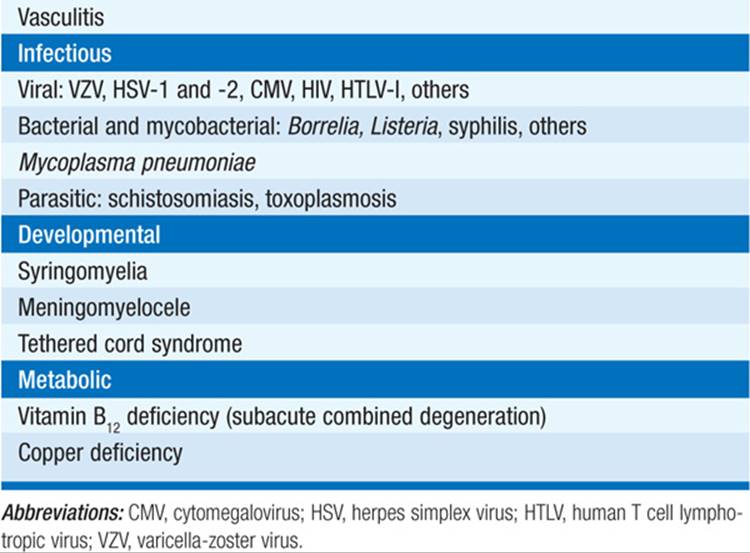
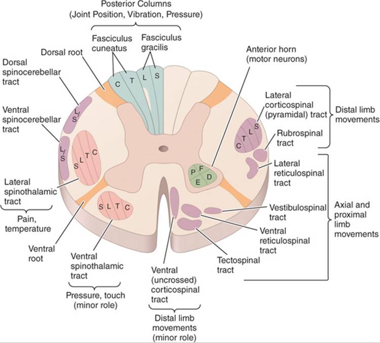
FIGURE 200-1 Transverse section through the spinal cord, composite representation, illustrating the principal ascending (left) and descending (right) pathways. The lateral and ventral spinothalamic tracts ascend contralateral to the side of the body that is innervated. C, cervical; D, distal; E, extensors; F, flexors; L, lumbar; P, proximal; S, sacral; T, thoracic.
SYMPTOMS AND SIGNS
Sensory symptoms often include paresthesias; may begin in one or both feet and ascend. Sensory level to pin sensation or vibration often correlates well with location of transverse lesions. May have isolated pain/temperature sensation loss over the shoulders (“cape” or “syringomyelic” pattern) or loss of sensation to vibration/position on one side of the body and pain/temperature loss on the other (Brown-Séquard hemicord syndrome).
Motor symptoms are caused by disruption of corticospinal tracts that leads to quadriplegia or paraplegia with increased muscle tone, hyperactive deep tendon reflexes, and extensor plantar responses. With acute severe lesions there may be initial flaccidity and areflexia (spinal shock).
Autonomic dysfunction includes primarily urinary retention; should raise suspicion of spinal cord disease when associated with back or neck pain, weakness, and/or a sensory level.
Pain may be present. Midline back pain is of localizing value; interscapular pain may be first sign of midthoracic cord compression; radicular pain may mark site of more laterally placed spinal lesion; pain from lower cord (conus medullaris) lesion may be referred to low back.
SPECIFIC SIGNS BY SPINAL CORD LEVEL
Approximate indicators of level of lesion include the location of a sensory level, a band of hyperalgesia/hyperpathia at the upper end of the sensory disturbance, identification of isolated atrophy or fasciculations, or lost tendon reflex at a specific spinal cord segment.
Lesions Near the Foramen Magnum
Weakness of the ipsilateral shoulder and arm, followed by weakness of ipsilateral leg, then contralateral leg, then contralateral arm, with respiratory paralysis.
Cervical Cord
Best localized by noting pattern of motor weakness and areflexia; shoulder (C5), biceps (C5–6), brachioradialis (C6), triceps/finger and wrist extensors (C7), finger and wrist flexors (C8).
Thoracic Cord
Localized by identification of a sensory level on the trunk. Useful markers are the nipples (T4) and umbilicus (T10).
Lumbar Cord
Upper lumbar cord lesions paralyze hip flexion and knee extension and abolish the patella reflex, whereas lower lumbar lesions affect foot and ankle movements, knee flexion, and thigh extension, and abolish the ankle jerks.
Sacral Cord (Conus Medullaris)
Saddle anesthesia, early bladder/bowel dysfunction, impotence; muscle strength is largely preserved.
Cauda Equina (Cluster of Nerve Roots Derived from Lower Cord)
Lesions below spinal cord termination at the L1 vertebral level produce a flaccid, areflexic, asymmetric paraparesis with bladder/bowel dysfunction and sensory loss below L1; pain is common and projected to perineum or thighs.
INTRAMEDULLARY AND EXTRAMEDULLARY SYNDROMES
Spinal cord disorders may be intramedullary (arising from within the substance of the cord) or extramedullary (compressing the cord or its blood supply). Extramedullary lesions often produce radicular pain, early corticospinal signs, and sacral sensory loss. Intramedullary lesions produce poorly localized burning pain, less prominent corticospinal signs, and often spare perineal/sacral sensation.
ACUTE AND SUBACUTE SPINAL CORD DISEASES (SEE CHAP. 21)
Neoplastic spinal cord compression (Chap. 21): Most are epidural in origin, resulting from metastases to the adjacent spinal bones (Fig. 200-2). Almost any tumor can be responsible: breast, lung, prostate, lymphoma, and plasma cell dyscrasias most frequent. Thoracic cord most commonly involved. Initial symptom is usually back pain, worse when recumbent, with local tenderness preceding other symptoms by many weeks. Spinal cord compression due to metastases is a medical emergency; in general, therapy will not reverse paralysis of >48 h duration.

FIGURE 200-2 Epidural spinal cord compression due to breast carcinoma. Sagittal T1-weighted A. and T2-weighted B. MRI scans through the cervicothoracic junction reveal an infiltrated and collapsed second thoracic vertebral body with posterior displacement and compression of the upper thoracic spinal cord. The low-intensity bone marrow signal in (A) signifies replacement by tumor.
Spinal epidural abscess: Triad of fever, localized midline dorsal spinal pain, and progressive limb weakness; once neurologic signs appear, cord compression rapidly progresses.
Spinal epidural hematoma: Presents as focal or radicular pain followed by variable signs of a spinal cord or conus medullaris disorder.
Acute disk herniation: Cervical and thoracic disk herniations are less common than lumbar.
Spinal cord infarction: Anterior spinal artery infarction produces paraplegia or quadriplegia, sensory loss affecting pain/temperature but sparing vibration/position sensation (supplied by posterior spinal arteries), and loss of sphincter control. Onset sudden or evolving over minutes or a few hours. Associated conditions: aortic atherosclerosis, dissecting aortic aneurysm, vertebral artery occlusion or dissection in the neck, aortic surgery, or profound hypotension. Therapy is directed at the predisposing condition.
Immune-mediated myelopathies: Acute transverse myelopathy (ATM) occurs in 1% of pts with SLE; associated with antiphospholipid antibodies. Sjögren’s and Behçet’s syndromes, mixed connective tissue disease, and p-ANCA vasculitis are other causes. Sarcoid can produce ATM with large edematous swelling of the spinal cord. Demyelinating diseases, either neuromyelitis optica (NMO) or multiple sclerosis, also can present as ATM; glucocorticoids, consisting of IV methylprednisolone followed by oral prednisone, are indicated for moderate to severe symptoms and refractory cases may respond to plasma exchange (Chap. 202). Treatment with mycopheno-late mofetil (250 mg bid gradually increasing to 1000 mg bid) or anti-CD20 monoclonal antibody may protect against relapses in NMO. Other cases of ATM are idiopathic.
Infectious myelopathies: Herpes zoster is the most common viral agent, but herpes simplex virus types 1 and 2, EBV, CMV, and rabies virus are also well described; in cases of suspected viral myelitis, antivirals may be appropriately started pending laboratory confirmation. Bacterial and mycobacterial causes are less common. Schistosomiasis is an important cause worldwide.
CHRONIC MYELOPATHIES
Spondylitic myelopathies: One of the most common causes of gait difficulty in the elderly. Presents as neck and shoulder pain with stiffness, radicular arm pain, and progressive spastic paraparesis with paresthesias and loss of vibration sense; in advanced cases, urinary incontinence may occur. A tendon reflex in the arms is often diminished at some level. Diagnosis is best made by MRI. Treatment is surgical (Chap. 54).
Vascular malformations: An important treatable cause of progressive or episodic myelopathy. May occur at any level; diagnosis is often suggested by contrast-enhanced MRI (Fig. 200-3), but is confirmed by selective spinal angiography. Treatment is embolization with occlusion of the major feeding vessels.

FIGURE 200-3 Arteriovenous malformation. Sagittal MR scans of the thoracic spinal cord: T2 fast spin-echo technique (left) and T1 post-contrast image (right). On the T2-weighted image (left), abnormally high signal intensity is noted in the central aspect of the spinal cord (arrowheads). Numerous punctate flow voids indent the dorsal and ventral spinal cord (arrow). These represent the abnormally dilated venous plexus supplied by the dural arteriovenous fistula. After contrast administration (right), multiple, serpentine, enhancing veins (arrows) on the ventral and dorsal aspect of the thoracic spinal cord are visualized, diagnostic of arteriovenous malformation. This pt was a 54-year-old man with a 4-year history of progressive paraparesis.
Retrovirus-associated myelopathies: Infection with HTLV-I may produce a slowly progressive spastic paraparesis with variable pain, sensory loss, and bladder disturbance; diagnosis is made by demonstration of specific serum antibody. Treatment is symptomatic. A progressive vacuolar myelopathy may also result from HIV infection.
Syringomyelia: Cavitary expansion of the spinal cord resulting in progressive myelopathy; may be an isolated finding or associated with protrusion of cerebellar tonsils into cervical spinal canal (Chiari type 1). Classic presentation is loss of pain/temperature sensation in the neck, shoulders, forearms, or hands with areflexic weakness in the upper limbs and progressive spastic paraparesis; cough headache, facial numbness, or thoracic kyphoscoliosis may occur. Diagnosis is made by MRI; treatment is surgical and often unsatisfactory.
Multiple sclerosis: Spinal cord involvement is common and is a major cause of disability in progressive forms of MS (Chap. 202).
Subacute combined degeneration (vitamin B12 deficiency): Paresthesias in hands and feet, early loss of vibration/position sense, progressive spastic/ataxic weakness, and areflexia due to associated peripheral neuropathy; mental changes (“megaloblastic madness”) and optic atrophy may be present along with a serum macrocytic anemia. Diagnosis is confirmed by a low serum B12 level, elevated levels of homocysteine and methylmalonic acid. Treatment is vitamin replacement beginning with 1 mg of IM vitamin B12 repeated at regular intervals or by subsequent oral treatment.
Hypocupric myelopathy: Clinically nearly identical to subacute combined degeneration (above). Low levels of serum copper and usually ceruloplasmin make the diagnosis. Some cases idiopathic and others follow GI procedures that hinder absorption. Treatment is oral copper supplementation.
Tabes dorsalis (tertiary syphilis): May present as lancinating pains, gait ataxia, bladder disturbances, and visceral crises. Cardinal signs are areflexia in the legs, impaired vibration/position sense, Romberg sign, and Argyll Robertson pupils, which fail to constrict to light but accommodate.
Familial spastic paraplegia: Progressive spasticity and weakness in the legs occurring on a familial basis; may be autosomal dominant, recessive, or X-linked. More than 20 different loci identified.
Adrenomyeloneuropathy: X-linked disorder that is a variant of adrenoleukodystrophy. Usually affected males have a history of adrenal insufficiency and then develop a progressive spastic paraparesis. Female heterozygotes may develop a slower progressive myelopathy without adrenal insufficiency. Diagnosis made by elevated very long chain fatty acids in serum. No therapy is clearly effective although bone marrow transplantation and nutritional supplements have been tried.
COMPLICATIONS
Bladder dysfunction with risk of urinary tract infection; bowel dysmotility; pressure sores; in high cervical cord lesions, mechanical respiratory failure; paroxysmal hypertension or hypotension with volume changes; severe hypertension and bradycardia in response to noxious stimuli or bladder or bowel distention; venous thrombosis and pulmonary embolism.

For a more detailed discussion, see Hauser SL, Ropper AH: Diseases of the Spinal Cord, Chap. 377, p. 3366, in HPIM-18.