
APPROACH TO THE PATIENT Tumor of the Nervous System
Clinical Presentation: Brain tumors of any type can present with general and/or focal symptoms and signs. General nonspecific symptoms include headache, cognitive difficulties, personality change, and gait disorder. The classic headache associated with a brain tumor is most evident in the morning and improves during the day, but this pattern is actually seen in only a minority of pts. Papilledema may suggest elevated intracranial pressure. Focal symptoms and signs include hemiparesis, aphasia, or visual field deficit that are typically subacute and progressive. Seizures are a common presentation, occurring in about 25% of pts with brain metastases or malignant glioma.
Evaluation: Primary brain tumors have no serologic features of malignancy such as an elevated ESR or tumor-specific antigens, unlike metastases. Cranial MRI with contrast is the preferred diagnostic test for any pt suspected of having a brain tumor; CT should be reserved for those pts unable to undergo MRI. Malignant brain tumors typically enhance with contrast and may have central areas of necrosis; they are characteristically surrounded by edema of the neighboring white matter. Low-grade gliomas typically do not enhance. Additional testing such as cerebral angiogram, EEG, or lumbar puncture is rarely indicated or helpful.
TREATMENT Tumors of the Nervous System
SYMPTOMATIC TREATMENT
• Glucocorticoids (dexamethasone 12–16 mg/d in divided doses PO or IV) to temporarily reduce edema
• Anticonvulsants (levetiracetam, topiramate, lamotrigine, valproic acid, or lacosamide) for pts who present with seizures; there is no role for prophylactic anticonvulsant drugs
• Low-dose SC heparin for immobile pts
DEFINITIVE TREATMENT
• Based on the specific tumor types and includes surgery, radiotherapy (RT), and chemotherapy
PRIMARY INTRACRANIAL TUMORS
Astrocytomas
Infiltrative tumors with a presumptive glial cell of origin. Most common primary intracranial neoplasm. Only known risk factors are ionizing radiation and uncommon hereditary syndromes (neurofibromatosis, tuberous sclerosis). Infiltration along white matter pathways often prevents total resection. Imaging studies (Fig. 201-1) fail to indicate full tumor extent. Grade I tumors (pilocytic astrocytomas) are the most common tumor of childhood, typically in the cerebellum; can be cured if completely resected. Grade II astrocytomas usually present with seizures in young adults; if feasible should be surgically resected. RT and chemotherapeutic agents such as temozolomide are increasingly used and may be helpful. Grade III (anaplastic astrocytoma) and grade IV (glioblastoma) astrocytomas are treated similarly with maximal safe surgical resection followed by RT with concurrent and adjuvant temozolomide or with RT and adjuvant temozolomide alone. Median survival in glioblastoma is 12–15 months. Glioblastomas invariably recur and treatment options include reoperation, carmustine wafer implantation, and chemotherapeutic regimens including bevacizumab. The most important adverse prognostic factors in high-grade astrocytomas are older age, histologic features of glioblastoma, poor performance status, and unresectable tumor.
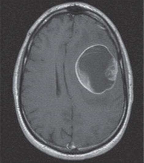
FIGURE 201-1 Postgadolinium T1 MRI of a large cystic left frontal glioblastoma.
Oligodendrogliomas
Generally more responsive to therapy and have a better prognosis than pure astrocytic tumors. Usually nonenhancing; often partially calcified. Treated with surgery and, if necessary, RT and chemotherapy. Median survival in excess of 10 years. Chemotherapy response improved when deletions of chromosomes 1p and 19q present.
Ependymomas
Derived from ependymal cells; highly cellular. Location—spinal canal more than intracranial in adults. If total excision possible, may be curable. Partially resected tumors will recur and require irradiation.
Primary CNS Lymphomas
B cell malignancy; most occur in immunosuppressed pts (organ transplantation, AIDS). May present as a single mass lesion or as multiple mass lesions or meningeal disease. Dramatic, transient responses occur with glucocorticoids; therefore whenever possible steroids should be withheld until after biopsy has been obtained. Pts should be tested for HIV and the extent of disease assessed by performing PET or CT of the body, MRI of the spine, CSF analysis, and slit-lamp examination of the eye. In immuno-competent pts, high-dose methotrexate therapy produces median survival up to 50 months, which may be increased with concurrent whole-brain RT and additional combinations of other chemotherapeutic agents such as cytarabine or rituximab. In immunocompromised pts, prognosis is worse and treatment is with high-dose methotrexate, whole-brain RT, and, in HIV, antiretroviral therapy.
Medulloblastomas
Most common malignant brain tumor of childhood. Half in posterior fossa; highly cellular; derived from neural precursor cells. Treat with surgery, RT, and chemotherapy. Approximately 70% of pts have long-term survival but usually at the cost of significant neurocognitive impairment.
Meningiomas
The most common primary brain tumor. Extraaxial mass attached to dura; dense and uniform contrast enhancement is diagnostic (Fig. 201-2). Total surgical resection of large, symptomatic benign meningiomas is curative. With subtotal resection, local RT reduces recurrence. Small, asymptomatic meningiomas may be followed radiologically without surgery. Treat rare aggressive meningiomas with excision and RT.
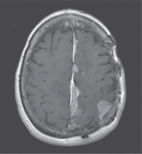
FIGURE 201-2 Postgadolinium T1 MRI demonstrating multiple meningiomas along the falx and left parietal cortex.
Schwannomas
Vestibular schwannomas present as progressive, unexplained unilateral hearing loss. MRI reveals dense, uniformly enhancing tumor at the cerebellopontine angle. Surgical excision may preserve hearing.
TUMORS METASTATIC TO THE NERVOUS SYSTEM
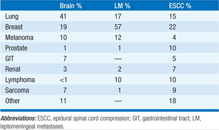
Hematogenous spread most common. Skull metastases rarely invade CNS; may compress adjacent brain or cranial nerves or obstruct intracranial venous sinuses. Primary tumors that commonly metastasize to the nervous system are listed in Table 201-1. Brain metastases are well demarcated by MRI and enhance with gadolinium. Ring enhancement is nonspecific; differential diagnosis includes brain abscess, radiation necrosis, toxoplasmosis, granulomas, tuberculosis, sarcoidosis, demyelinating lesions, primary brain tumors, CNS lymphoma, stroke, hemorrhage, and trauma. Screen for occult cancer: examine skin and thyroid gland; blood carcinoembryonic antigen (CEA) and liver function tests; CT of chest, abdomen, and pelvis. In approximately 10% of pts, a systemic cancer may present with brain metastases; biopsy of primary tumor or accessible brain metastasis is needed to plan treatment. Treatment with glucocorticoids, anticonvulsants, RT, or surgery. Whole-brain RT is often given because multiple microscopic tumor deposits are likely throughout the brain; stereotaxic radiosurgery is of benefit in pts with three or fewer metastases demonstrated by MRI. If a single metastasis is found, it may be surgically excised followed by whole-brain RT. Systemic chemotherapy may produce dramatic responses in rare cases of a highly chemosensitive tumor type such as germ cell tumors.
TABLE 201-1 FREQUENCY OF NERVOUS SYSTEM METASTASES BY COMMON PRIMARY TUMORS
Leptomeningeal Metastases
Presents as headache, encephalopathy, cranial nerve or polyradicular symptoms. Diagnosis by CSF cytology, MRI (nodular meningeal tumor deposits or diffuse meningeal enhancement), or meningeal biopsy. Associated with hydrocephalus due to CSF pathway obstruction. Treatment is palliative, often with RT or chemotherapy.
Spinal Cord Compression from Metastases
(See Chap. 21) Expansion of vertebral body metastasis posteriorly into epidural space compresses cord. Most common primary tumors are lung, breast, or prostate primary. Back pain (>90%) precedes development of weakness, sensory level, or incontinence. Medical emergency; early recognition of impending spinal cord compression essential to avoid devastating sequelae. Diagnosis is by spine MRI.
COMPLICATIONS OF RADIATION THERAPY
Three patterns of radiation injury after CNS RT:
1. Acute—headache, sleepiness, worse neurologic deficits during or immediately after RT. Rarely seen with current protocols. Can be both prevented and treated with glucocorticoids.
2. Early delayed—somnolence (children), Lhermitte’s sign; within weeks to months of RT. Increased T2 signal and sometimes enhancement on MRI. Self-limited and improves with glucocorticoids.
3. Late delayed—dementia or other progressive neurologic deficits; typically months to years after RT. White matter abnormalities on MRI (leukoencephalopathy) or ring-enhancing mass (radiation necrosis). PET can distinguish delayed necrosis from tumor recurrence. Progressive radiation necrosis is best treated palliatively with surgical resection unless it can be managed with glucocorticoids. Radiation injury of large arteries accelerates the development of atherosclerosis, increasing the risk of stroke years after RT. Endocrine dysfunction due to hypothalamus or pituitary gland injury can be due to delayed effects of RT. Development of a second neoplasm after RT also is a risk years after exposure.

For a more detailed discussion, see DeAngelis LM, Wen PY: Primary and Metastatic Tumors of the Nervous System, Chap. 379, p. 3382, in HPIM-18.