
ESOPHAGEAL CARCINOMA
In 2012 in the United States, 17,460 cases and 15,070 deaths; less frequent in women than men. Highest incidence in focal regions of China, Iran, Afghanistan, Siberia, Mongolia. In the United States, blacks more frequently affected than whites; usually presents sixth decade or later; 5-year survival <5% because most pts present with advanced disease.
Pathology
60% squamous cell carcinoma, most commonly in upper two-thirds; <40% adenocarcinoma, usually in distal third, arising in region of columnar metaplasia (Barrett’s esophagus), glandular tissue, or as direct extension of proximal gastric adenocarcinoma; lymphoma and melanoma rare. 10% of all esophageal cancers occur in the upper third, 35% in the middle third, and 55% in the lower third.
Risk Factors
Major risk factors for squamous cell carcinoma: ethanol abuse, smoking (combination is synergistic); other risks: lye ingestion and esophageal stricture, radiation exposure, head and neck cancer, achalasia, smoked opiates, Plummer-Vinson syndrome, tylosis, chronic ingestion of extremely hot tea, deficiency of vitamin A, zinc, molybdenum. Barrett’s esophagus is a risk for adenocarcinoma.
Clinical Features
Progressive dysphagia (first with solids, then liquids), rapid weight loss common, chest pain (from mediastinal spread), odynophagia, pulmonary aspiration (obstruction, tracheoesophageal fistula), hoarseness (laryngeal nerve palsy), hypercalcemia (parathyroid hormone–related peptide hyper-secretion by squamous carcinomas); bleeding infrequent, occasionally severe; examination often unremarkable.
Diagnosis
Double-contrast barium swallow useful as initial test in dysphagia; flexible esophagogastroscopy most sensitive and specific test; pathologic confirmation by combining endoscopic biopsy and cytologic examination of mucosal brushings (neither alone sufficiently sensitive); CT and endoscopic ultrasonography valuable to assess local and nodal spread.
TREATMENT Esophageal Carcinoma
Surgical resection feasible in only 40% of pts; associated with high complication rate (fistula, abscess, aspiration). Squamous cell carcinoma: Surgical resection after chemotherapy [5-fluorouracil (5FU), cisplatin] plus radiation therapy prolongs survival and may provide improved cure rate. Adenocarcinoma: Curative resection rarely possible; <20% of pts with resectable tumors survive 5 years. Palliative measures include laser ablation, mechanical dilatation, radiotherapy, and a luminal prosthesis to bypass the tumor. Gastrostomy or jejunostomy are frequently required for nutritional support. Preoperative chemotherapy with concurrent radiation therapy is somewhat more effective but more toxic therapy.
GASTRIC CARCINOMA
Highest incidence in Japan, China, Chile, Ireland; incidence decreasing worldwide, eightfold in the United States over past 60 years; in 2012, 21,320 new cases and 10,540 deaths. Male:female = 2:1; peak incidence sixth and seventh decades; overall 5-year survival <15%.
Risk Factors
Increased incidence in lower socioeconomic groups; environmental component is suggested by studies of migrants and their offspring. Several dietary factors correlated with increased incidence: nitrates, smoked foods, heavily salted foods; genetic component suggested by increased incidence in first-degree relatives of affected pts; other risk factors: atrophic gastritis, Helicobacter pylori infection, Billroth II gastrectomy, gastrojejunostomy, adenomatous gastric polyps, pernicious anemia, hyperplastic gastric polyps (latter two associated with atrophic gastritis), Ménétrier’s disease, slight increased risk with blood group A.
Pathology
Adenocarcinoma in 85%; usually focal (polypoid, ulcerative), two-thirds arising in antrum or lesser curvature, frequently ulcerative (“intestinal type”); less commonly diffuse infiltrative (linitis plastica) or superficial spreading (diffuse lesions more prevalent in younger pts; exhibit less geographic variation; have extremely poor prognosis); spreads primarily to local nodes, liver, peritoneum; systemic spread uncommon; lymphoma accounts for 15% (most frequent extranodal site in immunocompetent pts), either low-grade tumor of mucosa-associated lymphoid tissue (MALT) or aggressive diffuse large B cell lymphoma; leiomyosarcoma or gastrointestinal stromal tumor (GIST) is rare.
Clinical Features
Most commonly presents with progressive upper abdominal discomfort, frequently with weight loss, anorexia, nausea; acute or chronic GI bleeding (mucosal ulceration) common; dysphagia (location in cardia); vomiting (pyloric and widespread disease); early satiety; examination often unrevealing early in course; later, abdominal tenderness, pallor, and cachexia most common signs; palpable mass uncommon; metastatic spread may be manifest by hepatomegaly, ascites, left supraclavicular or scalene adenopathy, periumbilical, ovarian, or prerectal mass (Blumer’s shelf), low-grade fever, skin abnormalities (nodules, dermatomyositis, acanthosis nigricans, or multiple seborrheic keratoses). Laboratory findings: iron-deficiency anemia in two-thirds of pts; fecal occult blood in 80%; rarely associated with pancytopenia and microangiopathic hemolytic anemia (from marrow infiltration), leukemoid reaction, migratory thrombophlebitis, or acanthosis nigricans.
Diagnosis
Double-contrast barium swallow useful; gastroscopy most sensitive and specific test; pathologic confirmation by biopsy and cytologic examination of mucosal brushings; superficial biopsies less sensitive for lymphomas (frequently submucosal); important to differentiate benign from malignant gastric ulcers with multiple biopsies and follow-up examinations to demonstrate ulcer healing.
TREATMENT Gastric Carcinoma
Adenocarcinoma: Gastrectomy offers only chance of cure (only possible in less than one-third); the rare tumors limited to mucosa are resectable for cure in 80%; deeper invasion, nodal metastases decrease 5-year survival to 20% of pts with resectable tumors in absence of obvious meta-static spread (Table 78-1); CT and endoscopic ultrasonography may aid in determining tumor resectability. Subtotal gastrectomy has similar efficacy to total gastrectomy for distal stomach lesions, but with less morbidity; no clear benefit for resection of spleen and a portion of the pancreas, or for radical lymph node removal. Adjuvant chemotherapy (5FU/leucovorin) plus radiation therapy following primary surgery leads to a 7-month increase in median survival. Neoadjuvant chemotherapy with epirubicin, cisplatin, and 5FU may downstage tumors and increase the efficacy of surgery. Palliative therapy for pain, obstruction, and bleeding includes surgery, endoscopic dilatation, radiation therapy, chemotherapy.
TABLE 78-1 STAGING SYSTEM FOR GASTRIC CARCINOMA


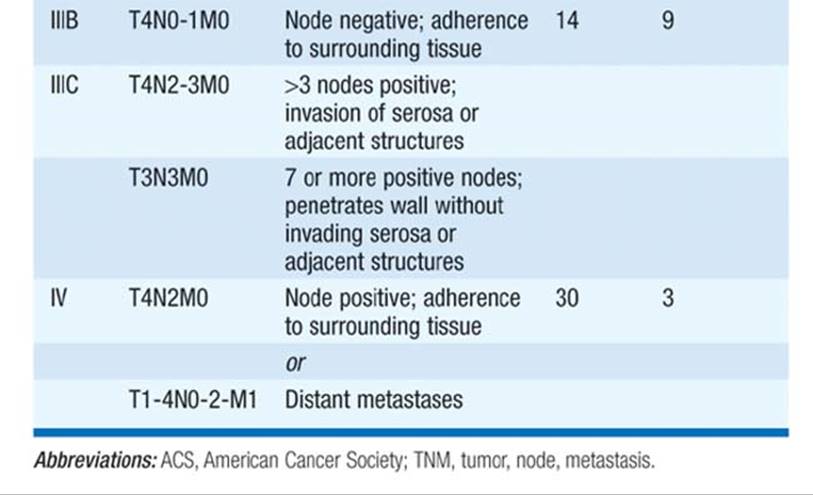
Lymphoma: Low-grade MALT lymphoma is caused by H. pylori infection, and eradication of the infection causes complete remissions in 50% of pts; rest are responsive to combination chemotherapy including cyclophosphamide, doxorubicin, vincristine, prednisone (CHOP) plus rituximab. Diffuse large B cell lymphoma may be treated with either CHOP plus rituximab or subtotal gastrectomy followed by chemotherapy; 50–60% 5-year survival.
Leiomyosarcoma: Surgical resection curative in most pts. Tumors expressing the c-kit tyrosine kinase (CD117)—GIST—respond to imatinib mesylate in a substantial fraction of cases.
BENIGN GASTRIC TUMORS
Much less common than malignant gastric tumors; hyperplastic polyps most common, with adenomas, hamartomas, and leiomyomas rare; 30% of adenomas and occasional hyperplastic polyps are associated with gastric malignancy; polyposis syndromes include Peutz-Jeghers and familial polyposis (hamartomas and adenomas), Gardner’s (adenomas), and Cronkhite-Canada (cystic polyps). See “Colonic Polyps,” below.
Clinical Features
Usually asymptomatic; occasionally present with bleeding or vague epigastric discomfort.
TREATMENT Benign Gastric Tumors
Endoscopic or surgical excision.
SMALL-BOWEL TUMORS
Clinical Features
Uncommon tumors (~5% of all GI neoplasms); usually present with bleeding, abdominal pain, weight loss, fever, or intestinal obstruction (intermittent or fixed); increased incidence of lymphomas in pts with gluten-sensitive enteropathy, Crohn’s disease involving small bowel, AIDS, prior organ transplantation, autoimmune disorders.
Pathology
Usually benign; most common are adenomas (usually duodenal), leiomyomas (intramural), and lipomas (usually ileal); 50% of malignant tumors are adenocarcinoma, usually in duodenum (at or near ampulla of Vater) or proximal jejunum, commonly coexisting with benign adenomas; primary intestinal lymphomas (non-Hodgkin’s) account for 25% and occur as focal mass (Western type), which is usually a T cell lymphoma associated with prior celiac disease, or diffuse infiltration (Mediterranean type), which is usually immunoproliferative small-intestinal disease (IPSID; α-heavy chain disease), a B-cell MALT lymphoma associated with Campylobacter jejuni infection, which can present as intestinal malabsorption; carcinoid tumors (usually asymptomatic) occasionally produce bleeding or intussusception (see below).
Diagnosis
Endoscopy and biopsy most useful for tumors of duodenum and proximal jejunum; otherwise barium x-ray examination best diagnostic test; direct small-bowel instillation of contrast (enteroclysis) occasionally reveals tumors not seen with routine small-bowel radiography; angiography (to detect plexus of tumor vessels) or laparotomy often required for diagnosis; CT useful to evaluate extent of tumor (esp. lymphomas).
TREATMENT Small-Bowel Tumors
Surgical excision; adjuvant chemotherapy appears helpful for focal lymphoma; IPSID appears to be curable with combination chemotherapy used in aggressive lymphoma plus oral antibiotics (e.g., tetracycline); no proven role for chemotherapy or radiation therapy for other small-bowel tumors.
COLONIC POLYPS
TUBULAR ADENOMAS
Present in ~30% of adults; pedunculated or sessile; usually asymptomatic; ~5% cause occult blood in stool; may cause obstruction; overall risk of malignant degeneration correlates with size (<2% if <1.5 cm diam; >10% if >2.5 cm diam) and is higher in sessile polyps; 65% found in rectosigmoid colon; diagnosis by barium enema, sigmoidoscopy, or colonoscopy. Treatment: Full colonoscopy to detect synchronous lesions (present in 30%); endoscopic resection (surgery if polyp large or inaccessible by colonoscopy); follow-up surveillance by colonoscopy every 2–3 years.
VILLOUS ADENOMAS
Generally larger than tubular adenomas at diagnosis; often sessile; high risk of malignancy (up to 30% when >2 cm); more prevalent in left colon; occasionally associated with potassium-rich secretory diarrhea. Treatment: As for tubular adenomas.
HYPERPLASTIC POLYPS
Asymptomatic; usually incidental finding at colonoscopy; rarely >5 mm; no malignant potential. No treatment required.
HEREDITARY POLYPOSIS SYNDROMES
See Table 78-2.
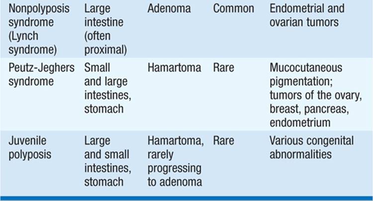
TABLE 78-2 HEREDITABLE (AUTOSOMAL DOMINANT)
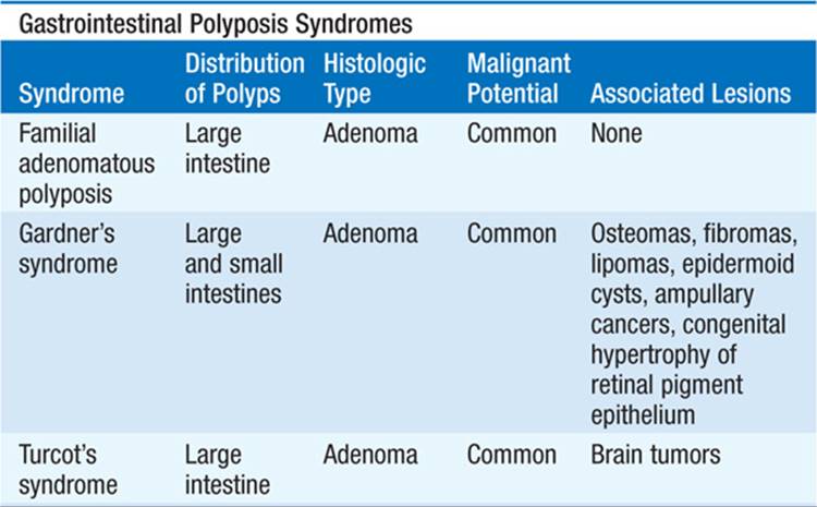
1. Familial polyposis coli (FPC): Diffuse pancolonic adenomatous polyposis (up to several thousand polyps); autosomal dominant inheritance associated with deletion in adenomatous polyposis coli (APC) gene on chromosome 5; colon carcinoma from malignant degeneration of polyp in 100% by age 40. Treatment: Prophylactic total colectomy or subtotal colectomy with ileoproctostomy before age 30; subtotal resection avoids ileostomy but necessitates frequent proctoscopic surveillance; periodic colonoscopic or annual radiologic screening of siblings and offspring of pts with FPC until age 35; sulindac and other nonsteroidal antiinflammatory drugs (NSAIDs) cause regression of polyps and inhibit their development.
2. Gardner’s syndrome: Variant of FPC with associated soft tissue tumors (epidermoid cysts, osteomas, lipomas, fibromas, desmoids); higher incidence of gastroduodenal polyps, ampullary adenocarcinoma. Treatment: As for FPC; surveillance for small-bowel disease with fecal occult blood testing after colectomy.
3. Turcot’s syndrome: Rare variant of FPC with associated malignant brain tumors. Treatment: As for FPC.
4. Nonpolyposis syndrome: Familial syndrome with up to 50% risk of colon carcinoma; peak incidence in fifth decade; associated with multiple primary cancers (esp. endometrial); autosomal dominant; due to defective DNA mismatch repair.
5. Juvenile polyposis: Multiple benign colonic and small-bowel hamartomas; intestinal bleeding common. Other symptoms: abdominal pain, diarrhea; occasional intussusception. Rarely recur after excision; low risk of colon cancer from malignant degeneration of interspersed adenomatous polyps. Prophylactic colectomy controversial.
6. Peutz-Jeghers syndrome: Numerous hamartomatous polyps of entire GI tract, though denser in small bowel than colon; GI bleeding common; somewhat increased risk for the development of cancer at GI and non-GI sites. Prophylactic surgery not recommended.
COLORECTAL CANCER
Second most common internal cancer in humans; accounts for 10% of cancer-related deaths in United States; incidence increases dramatically above age 50, nearly equal in men and women. In 2012, 143,460 new cases, 51,690 deaths.
Etiology and Risk Factors
Most colon cancers arise from adenomatous polyps. Genetic steps from polyp to dysplasia to carcinoma in situ to invasive cancer have been defined, including point mutation in K-ras proto-oncogene, hypomethylation of DNA leading to enhanced gene expression, allelic loss at the APC gene (a tumor suppressor), allelic loss at the DCC (deleted in colon cancer) gene on chromosome 18, and loss and mutation of p53 on chromosome 17. Hereditary nonpolyposis colon cancer arises from mutations in the DNA mismatch repair genes, hMSH2 gene on chromosome 2 and hMLH1 gene on chromosome 3. Mutations lead to colon and other cancers. Diagnosis requires three or more relatives with colon cancer, one of whom is a first-degree relative; one or more cases diagnosed before age 50; and involvement of at least two generations. Environmental factors also play a role; increased prevalence in developed countries, urban areas, advantaged socioeconomic groups; increased risk in pts with hypercholesterolemia, coronary artery disease; correlation of risk with low-fiber, high-animal-fat diets, although direct effect of diet remains unproven; decreased risk with long-term dietary calcium supplementation and, possibly, daily aspirin ingestion. Risk increased in first-degree relatives of pts; families with increased prevalence of cancer; and pts with history of breast or gynecologic cancer, familial polyposis syndromes, >10-year history of ulcerative colitis or Crohn’s colitis, >15-year history of ureterosigmoidostomy. Tumors in pts with strong family history of malignancy are frequently located in right colon and commonly present before age 50; high prevalence in pts with Streptococcus bovis bacteremia.
Pathology
Nearly always adenocarcinoma; 75% located distal to the splenic flexure (except in association with polyposis or hereditary cancer syndromes); may be polypoid, sessile, fungating, or constricting; subtype and degree of differentiation do not correlate with course. Degree of invasiveness at surgery (Dukes’ classification) is single best predictor of prognosis (Fig. 78-1). Rectosigmoid tumors may spread to lungs early because of systemic paravertebral venous drainage of this area. Other predictors of poor prognosis: preoperative serum carcinoembryonic antigen (CEA) >5 ng/mL (>5 μg/L), poorly differentiated histology, bowel perforation, venous invasion, adherence to adjacent organs, aneuploidy, specific deletions in chromosomes 5, 17, 18, and mutation of ras proto-oncogene. 15% have defects in DNA repair.

FIGURE 78-1 Staging and prognosis for pts with colorectal cancer.
Clinical Features
Left-sided colon cancers present most commonly with rectal bleeding, altered bowel habits (narrowing, constipation, intermittent diarrhea, tenesmus), and abdominal or back pain; cecal and ascending colon cancers more frequently present with symptoms of anemia, occult blood in stool, or weight loss; other complications: perforation, fistula, volvulus, inguinal hernia; laboratory findings: anemia in 50% of right-sided lesions.
Diagnosis
Early diagnosis aided by screening asymptomatic persons with fecal occult blood testing (see below); >50% of all colon cancers are within reach of a 60-cm flexible sigmoidoscope; air-contrast barium enema will diagnose ~85% of colon cancers not within reach of sigmoidoscope; colonoscopy most sensitive and specific, permits tumor biopsy and removal of synchronous polyps (thus preventing neoplastic conversion), but is more expensive. Radiographic or virtual colonoscopy has not been shown to be a better diagnostic method than colonoscopy.
TREATMENT Colorectal Cancer
Local disease: Surgical resection of colonic segment containing tumor; preoperative evaluation to assess prognosis and surgical approach includes full colonoscopy, chest films, biochemical liver tests, plasma CEA level, and possible abdominal CT. Resection of isolated hepatic metastases possible in selected cases. Adjuvant radiation therapy to pelvis (with or without concomitant 5FU chemotherapy) decreases local recurrence rate of rectal carcinoma (no apparent effect on survival); radiation therapy without benefit on colon tumors; preoperative radiation therapy may improve resectability and local control in pts with rectal cancer. Total mesorectal excision is more effective than conventional anteroposterior resection in rectal cancer. Adjuvant chemotherapy (5FU/leucovorin plus oxaliplatin, or FOLFOX plus bevacizumab, or 5FU/leucovorin plus irinotecan, or FOLFIRI) decreases recurrence rate and improves survival of stage C (III); survival benefit from adjuvant therapy is not so clear in stage B (II) tumors; periodic determination of serum CEA level useful to follow therapy and assess recurrence. Follow-up after curative resection: Yearly liver tests, complete blood count, follow-up radiologic or colonoscopic evaluation at 1 year—if normal, repeat every 3 years, with routine screening interim (see below); if polyps detected, repeat 1 year after resection. Advanced tumor (locally unresectable or metastatic): Systemic chemotherapy (5FU/leucovorin plus oxaliplatin plus bevacizumab), irinotecan usually used in second treatment; antibodies to the EGF receptor (cetuximab, panitumumab) appear to enhance the effect of chemotherapy; intraarterial chemotherapy [floxuridine (FUDR)] and/or radiation therapy may palliate symptoms from hepatic metastases.
Prevention
Early detection of colon carcinoma may be facilitated by routine screening of stool for occult blood (Hemoccult II, ColonCare, Hemosure); however, sensitivity only ~50% for carcinoma; specificity for tumor or polyp ~25–40%. False positives: ingestion of red meat, iron, aspirin; upper GI bleeding. False negatives: vitamin C ingestion, intermittent bleeding. Annual digital rectal exam and fecal occult blood testing recommended for pts over age 40, screening by flexible sigmoidoscopy every 3 years after age 50, earlier in pts at increased risk (see above); careful evaluation of all pts with positive fecal occult blood tests (flexible sigmoidoscopy and air-contrast barium enema or colonoscopy alone) reveals polyps in 20–40% and carcinoma in ~5%; screening of asymptomatic persons allows earlier detection of colon cancer (i.e., earlier Dukes’ stage) and achieves greater resectability rate; decreased overall mortality from colon carcinoma seen only after 13 years of follow-up. More intensive evaluation of first-degree relatives of pts with colon carcinoma frequently includes screening air-contrast barium enema or colonoscopy after age 40. NSAIDs and cyclooxygenase 2 inhibitors appear to prevent polyp development and induce regression in high-risk groups, but have not been recommended for average-risk pts at this time.
ANAL CANCER
Accounts for 1–2% of large-bowel cancer, 6230 cases and 780 deaths in 2012; associated with chronic irritation, e.g., from condyloma acuminata, perianal fissures/fistulas, chronic hemorrhoids, leukoplakia, trauma from anal intercourse. Women are more commonly affected than men. Homosexual men are at increased risk. Human papillomavirus is etiologic. Presents with bleeding, pain, and perianal mass. Radiation therapy plus chemotherapy (5FU and mitomycin) leads to complete response in 80% when the primary lesion is <3 cm. Abdominoperineal resection with permanent colostomy is reserved for those with large lesions or whose disease recurs after chemoradiotherapy.
BENIGN LIVER TUMORS
Hepatocellular adenomas occur most commonly in women in the third or fourth decades who take birth control pills. Most are found incidentally but may cause pain; intratumoral hemorrhage may cause circulatory collapse. 10% may become malignant. Women with these adenomas should stop taking birth control pills. Large tumors near the liver surface may be resected. Focal nodular hyperplasia is also more common in women but seems not to be caused by birth control pills. Lesions are vascular on angiography and have septae and are usually asymptomatic.
HEPATOCELLULAR CARCINOMA
About 28,720 cases in the United States in 2012, but worldwide this may be the most common tumor; 20,550 deaths in 2012 in US. Male:female = 4:1; tumor usually develops in cirrhotic liver in persons in fifth or sixth decade. High incidence in Asia and Africa is related to etiologic relationship between this cancer and hepatitis B and C infections. Aflatoxin exposure contributes to etiology and leaves a molecular signature, a mutation in codon 249 of the gene for p53.
Modes of Presentation
A pt with known liver disease develops an abnormality on ultrasound or rising α fetoprotein (AFP) or des-gamma-carboxy prothrombin (DCP) due to absence of vitamin K; abnormal liver function tests; cachexia, abdominal pain, fever.
Physical Findings
Jaundice, asthenia, itching, tremors, disorientation, hepatomegaly, splenomegaly, ascites, peripheral edema.
TREATMENT Hepatocellular Carcinoma
Surgical resection or liver transplantation is therapeutic option but rarely successful. Radiofrequency ablation can cause regression of small tumors. Sorafenib may produce partial responses lasting a few months.
Screening and Prevention
Screening populations at risk has given conflicting results. Hepatitis B vaccine prevents the disease. Interferon α (IFN-α) may prevent liver cancer in persons with chronic active hepatitis C disease and possibly in those with hepatitis B. Ribavirin ± IFN-α is most effective treatment of chronic hepatitis C. Teleprevir, a protease inhibitor, is also active.
PANCREATIC CANCER
In 2012 in the United States, about 43,920 new cases and 27,390 deaths. The incidence is decreasing somewhat, but nearly all diagnosed cases are fatal. The tumors are ductal adenocarcinomas and are not usually detected until the disease has spread. About 70% of tumors are in the pancreatic head, 20% in the body, and 10% in the tail. Mutations in K-ras have been found in 85% of tumors, and the p16 cyclin-dependent kinase inhibitor on chromosome 9 may also be implicated. Long-standing diabetes, chronic pancreatitis, and smoking increase the risk; coffee-drinking, alcoholism, and cholelithiasis do not. Pts present with pain and weight loss, the pain often relieved by bending forward. Jaundice commonly complicates tumors of the head, due to biliary obstruction. Curative surgical resections are feasible in about 10%. Adjuvant chemotherapy (5FU) may benefit some pts after resection. Gemcitabine plus erlotinib or capecitabine may palliate symptoms in pts with advanced disease.
ENDOCRINE TUMORS OF THE GI TRACT AND PANCREAS
CARCINOID TUMOR
Carcinoid tumor accounts for 75% of GI endocrine tumors; incidence is about 15 cases per million population. 90% originate in Kulchitsky cells of the GI tract, most commonly the appendix, ileum, and rectum. Carcinoid tumors of the small bowel and bronchus have a more malignant course than tumors of other sites. About 5% of pts with carcinoid tumors develop symptoms of the carcinoid syndrome, the classic triad being cutaneous flushing, diarrhea, and valvular heart disease. For tumors of GI tract origin, symptoms imply metastases to liver.
Diagnosis can be made by detecting the site of tumor or documenting production of >15 mg/d of the serotonin metabolite 5-hydroxyindoleacetic acid (5-HIAA) in the urine. Octreotide scintigraphy identifies sites of primary and metastatic tumor in about two-thirds of cases.
TREATMENT Carcinoid Tumor
Surgical resection where feasible. Symptoms may be controlled with histamine blockers and octreotide, 150–1500 mg/d in three doses. Hepatic artery embolization and chemotherapy (5FU plus streptozocin or doxorubicin) have been used for metastatic disease. IFN-α at 3–10 million units SC three times a week may relieve symptoms. Prognosis ranges from 95% 5-year survival for localized disease to 20% 5-year survival for those with liver metastases. Median survival of pts with carcinoid syndrome is 2.5 years from the first episode of flushing.
PANCREATIC ISLET-CELL TUMORS
Gastrinoma, insulinoma, VIPoma, glucagonoma, and somatostatinoma account for the vast majority of pancreatic islet-cell tumors; their characteristics are shown in Table 78-3. The tumors are named for the dominant hormone they produce. They are generally slow-growing and produce symptoms related to hormone production. Gastrinomas and peptic ulcer disease constitute the Zollinger-Ellison syndrome. Gastrinomas are rare (4 cases per 10 million population), and in 25–50%, the tumor is a component of a multiple endocrine neoplasia type 1 (MEN 1) syndrome (Chap. 186).
TABLE 78-3 GASTROINTESTINAL ENDOCRINE TUMOR SYNDROMES

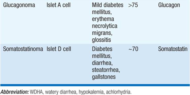
Insulinoma may present with Whipple’s triad: fasting hypoglycemia, symptoms of hypoglycemia, and relief after IV glucose. Normal or elevated serum insulin levels in the presence of fasting hypoglycemia are diagnostic. Insulinomas may also be associated with MEN 1.
Verner and Morrison described a syndrome of watery diarrhea, hypokalemia, achlorhydria, and renal failure associated with pancreatic islet tumors that produce vasoactive intestinal polypeptide (VIP). VIPomas are rare (1 case per 10 million) but often grow to a large size before producing symptoms.
Glucagonoma is associated with diabetes mellitus and necrolytic migratory erythema, a characteristic red, raised, scaly rash usually located on the face, abdomen, perineum, and distal extremities. Glucagon levels >1000 ng/L not suppressed by glucose are diagnostic.
The classic triad of somatostatinoma is diabetes mellitus, steatorrhea, and cholelithiasis.
Provocative tests may facilitate diagnosis of functional endocrine tumors: tolbutamide enhances somatostatin secretion by somatostatinomas; pentagastrin enhances calcitonin secretion from medullary thyroid (C cell) tumors; secretin enhances gastrin secretion from gastrinomas. If imaging techniques fail to detect tumor masses, angiography or selective venous sampling for hormone determination may reveal the site of tumor. Metastases to nodes and liver should be sought by CT or MRI.
TREATMENT Pancreatic Islet-Cell Tumors
Tumor is surgically removed, if possible. Everolimus 10 mg PO qd or sunitinib 37.5 mg PO qd may produce meaningful delay (~12 months) in progressive disease and prolong survival in pts with metastatic disease. Octreotide inhibits hormone secretion in the majority of cases. IFN-α may reduce symptoms. Streptozotocin plus doxorubicin combination chemotherapy may produce responses in 60–90% of cases. Embolization or chemoembolization of hepatic metastases may be palliative.

For a more detailed discussion, see Mayer RJ: Gastrointestinal Tract Cancer, Chap. 91, p. 764; Carr BI: Tumors of the Liver and Biliary Tree, Chap. 92, p. 777; Chong I, Cunningham D: Pancreatic Cancer, Chap. 93, p. 786; and Jensen RT: Endocrine Tumors of the Gastrointestinal Tract and Pancreas, Chap. 350, p. 3056, in HPIM-18.