
MICROBIOLOGY
Staphylococci are gram-positive cocci that form grapelike clusters on Gram’s stain; they are catalase positive (unlike streptococci), nonmotile, aerobic, and facultatively anaerobic. Staphylococcus aureus, which is distinguished from other staphylococci by its production of coagulase, is the most virulent species.
S. AUREUS INFECTIONS
Epidemiology
S. aureus is an important cause of community-acquired infections and is the leading cause of nosocomial infections.
• S. aureus is a component of the normal human flora, most frequently colonizing the anterior nares but also colonizing the skin (particularly damaged skin), vagina, axilla, perineum, and oropharynx. These sites of colonization are reservoirs for future infection.
• Of healthy persons, 25–50% may be persistently or transiently colonized with S. aureus, and the rate is especially high among insulin-dependent diabetic pts, HIV-infected persons, injection drug users, hemodialysis pts, and pts with skin damage.
• Methicillin-resistant S. aureus (MRSA) is common in hospitals, and its prevalence is increasing dramatically in community settings among individuals without prior medical exposure.
– In the United States, strain USA300 (defined by pulsed-field gel electrophoresis) causes most community-acquired MRSA (CA-MRSA) infections and can cause severe disease in immunocompetent pts.
– Outbreaks of CA-MRSA infection occur among diverse groups, including prisoners, athletes, and drug users.
Pathogenesis
S. aureus is a pyogenic pathogen known for its capacity to induce abscess formation.
• Invasive Disease For invasive S. aureus infection to occur, some or all of the following steps are necessary:
– Colonization/inoculation: Bacteria colonize tissue surfaces or are inoculated directly into tissue—e.g., as a result of minor abrasions or via IV access catheters.
– Invasion: Bacteria replicate at the site of infection and elaborate enzymes that facilitate survival and local spread. CA-MRSA isolates that produce the Panton-Valentine leukocidin toxin have been linked to more serious infections.
– Evasion of host defense mechanisms: S. aureus possesses an antiphagocytic polysaccharide microcapsule that facilitates evasion of host defenses and plays a role in abscess formation. Organisms can survive in a quiescent state in various tissues and then cause recrudescent infections when conditions are suitable.
– Metastatic spread: S. aureus can survive in PMNs and may use these cells to spread to and seed other tissue sites.
• Toxin-Mediated Disease S. aureus produces three types of toxin: cytotoxins, pyrogenic toxin superantigens, and exfoliative toxins.
– Antitoxin antibodies are protective against toxin-mediated staphylococcal illness.
– Enterotoxins and toxic shock syndrome toxin 1 (TSST-1) act as “super-antigens” or T cell mitogens and cause the release of large amounts of inflammatory mediators, producing multisystem disease that includes fever, rash, and hypotension.
Diagnosis
S. aureus infections are readily diagnosed by Gram’s stain and microscopic examination of infected tissue.
• Routine cultures of infected material usually yield positive results, and blood cultures are sometimes positive even when infections are localized to extravascular sites.
• PCR assays have been developed for rapid testing and are increasingly being used.
Clinical Syndromes
Skin and Soft Tissue Infections S. aureus causes a variety of cutaneous infections characterized by pus-containing blisters, many of which can also be caused by group A streptococci and other streptococcal species. Predisposing factors include skin disease (e.g., eczema), skin damage (e.g., minor trauma), injections, and poor personal hygiene.
• Infections can be superficial (e.g., folliculitis, cellulitis, impetigo) or deep and painful (e.g., furuncles, carbuncles, hidradenitis suppurativa).
– Carbuncles (often located in the lower neck) are more severe and painful than furuncles (boils that extend from hair follicles) and are due to coalesced lesions extending to deeper SC tissue.
– Mastitis in lactating women can range from superficial cellulitis to abscess.
Musculoskeletal Infections See Chap. 93 for additional details.
• S. aureus is the most common cause of osteomyelitis arising from either hematogenous dissemination or contiguous spread from a soft tissue site (e.g., diabetic or vascular ulcers).
– Hematogenous osteomyelitis in adults is often vertebral and occurs in pts with endocarditis, pts undergoing hemodialysis, injection drug users, or diabetics. Intense back pain and fever can occur, but infections may also be clinically occult.
– Epidural abscess is a serious complication that can present as trouble voiding or walking or as radicular pain in addition to symptoms of osteomyelitis; neurologic compromise can develop in the absence of timely treatment, which often requires surgical intervention.
– Osteomyelitis from contiguous soft tissue infections is suggested by exposure of bone, a draining fistulous tract, failure to heal, or continued drainage.
• S. aureus is the most common cause of septic arthritis in native joints of both adults and children. S. aureus septic arthritis in adults may result from trauma, surgery, or hematogenous dissemination.
– The joints most commonly affected are the knees, shoulders, hips, and phalanges.
– Examination of synovial fluid reveals >50,000 PMNs/μL and gram-positive cocci in clusters on Gram’s stain.
• Pyomyositis, an infection of skeletal muscles that is seen in tropical climates and in seriously compromised pts (including HIV-infected pts), causes fever, swelling, and pain overlying involved muscle and is usually due to S. aureus.
Respiratory Tract Infections
• Newborns and infants can develop serious infections characterized by fever, dyspnea, and respiratory failure; pneumatoceles (shaggy, thin-walled cavities), pneumothorax, and empyema are known complications.
• Community-acquired pneumonia usually follows viral infections (e.g., after influenza) and manifests as fever, bloody sputum production, and midlung-field pneumatoceles or multiple patchy pulmonary infiltrates.
• Nosocomial pneumonia is commonly seen in intubated pts.
– The clinical presentation resembles that of pneumonia of other bacterial etiologies.
– Pts produce an increased volume of purulent sputum and develop fever, new pulmonary infiltrates, and respiratory distress.
Bacteremia and Sepsis The incidence of metastatic seeding during bacteremia has been estimated to be as high as 31%.
• Bones, joints, kidneys, and lungs are most commonly infected.
• Diabetes, HIV infection, and renal insufficiency are often seen in association with S. aureus bacteremia and increase the risk of complications.
Infective Endocarditis (See also Chap. 89)
S. aureus is the leading cause of endocarditis worldwide and accounts for 25–35% of cases.
• The incidence is increasing as a result of injection drug use, hemodialysis, intravascular prosthetic devices, and immunosuppression.
• Mortality rates range from 20% to 40% despite the availability of effective antibiotics.
• The four clinical settings in which S. aureus endocarditis is encountered are (1) right-sided endocarditis in association with injection drug use, (2) left-sided native-valve endocarditis, (3) prosthetic-valve endocarditis, and (4) nosocomial endocarditis.
Urinary Tract Infections UTIs due to S. aureus are uncommon and suggest hematogenous dissemination.
Prosthetic Device–Related Infections Compared with coagulase-negative staphylococci, S. aureus causes more acute disease, with localized and systemic manifestations that tend to be rapidly progressive. Successful treatment usually involves removal of the prosthetic device.
CA-MRSA Infections While the skin and soft tissues are the most common sites of infection associated with CA-MRSA, 5–10% of these infections are invasive and potentially life-threatening (e.g., necrotizing fasciitis, necrotic pneumonia, sepsis, purpura fulminans).
Toxin-Mediated Disease Each class of toxin produced by S. aureus results in a characteristic syndrome.
• Food poisoning: results from inoculation of toxin-producing S. aureus into food by colonized food handlers, with subsequent toxin elaboration in growth-promoting foods (e.g., custard, potato salad, processed meat).
– The heat-stable toxin is not destroyed even if heating kills the bacteria.
– Because the disease is caused by preformed toxins, its onset is rapid and explosive, occurring within 1–6 h of ingestion of contaminated food.
– The chief signs and symptoms are nausea and vomiting, but diarrhea, hypotension, and dehydration may occur. Fever is absent.
– Symptoms resolve within 8–10 h; treatment is entirely supportive.
• Toxic shock syndrome (TSS): results from elaboration of an enterotoxin (many nonmenstrual TSS cases) or TSST-1 (some nonmenstrual cases and >90% of menstrual cases).
– Although the specific toxin may differ, the clinical presentation is similar in menstrual and nonmenstrual cases.
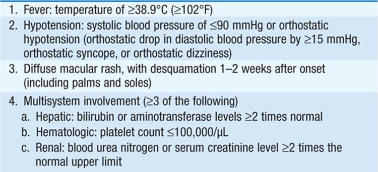
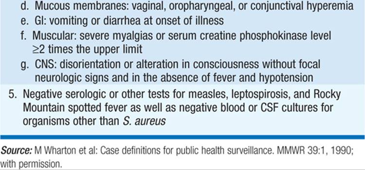
– Diagnosis is based on a constellation of clinical findings. Table 95-1 summarizes the case definition for staphylococcal TSS.
TABLE 95-1 CASE DEFINITION OF S. AUREUS TOXIC SHOCK SYNDROME
– Menstrual cases occur 2–3 days after menses begin.
– Illness occurs only in people who lack antibody to the toxin.
• Staphylococcal scalded-skin syndrome (SSSS): most often affects newborns and children. Fragility of the skin, with tender, thick-walled, fluid-filled bullae, can lead to exfoliation of most of the skin surface. Nikolsky’s sign is positive when gentle pressure on bullae causes rupture of lesions and leaves denuded underlying skin.
Prevention
Hand washing and careful attention to appropriate isolation procedures prevent the spread of S. aureus infection. Elimination of nasal carriage of S. aureus (e.g., with mupirocin) has reduced the incidence of postsurgical infection and the rates of infection among hemodialysis and peritoneal dialysis pts.
INFECTIONS CAUSED BY COAGULASE-NEGATIVE STAPHYLOCOCCI (CoNS)
Microbiology
CoNS are generally less virulent than S. aureus but are important and common causes of prosthetic-device infections.
• Of CoNS species, S. epidermidis most often causes disease. This organism is a normal component of the skin, oropharyngeal, and vaginal flora.
• S. saprophyticus is a cause of UTIs.
• S. lugdunensis and S. schleiferi are more virulent than other CoNS species and cause serious infections, possibly because they apparently share more virulence determinants with S. aureus than do other CoNS species.
Pathogenesis
CoNS are uniquely adapted to cause prosthetic-device infections because they can elaborate an extracellular polysaccharide (glycocalyx or slime) that forms a biofilm on the device surface, protecting bacteria from host defenses as well as from antibiotic treatment while allowing bacterial survival.
Clinical Syndromes
CoNS cause diverse prosthetic device–related infections. Signs of localized infection are usually subtle, disease progression is slow, and systemic findings are limited. Fever and mild leukocytosis may be documented. Infections not associated with prosthetic devices are infrequent, but up to 5% of native-valve endocarditis cases have been due to CoNS in some series.
Diagnosis
CoNS are readily detected by standard methods, but distinguishing infection from colonization is often problematic because CoNS are common contaminants of cultures of blood and other sites. Only 10–25% of blood cultures positive for CoNS reflect true bacteremia.
TREATMENT Staphylococcal Infections
• Suppurative collections should be surgically drained. The emergence of CA-MRSA has increased the importance of culturing material from all collections to identify the pathogen and determine its antimicrobial susceptibility.
• In most cases of prosthetic-device infection, the device should be removed, although some CoNS infections can be managed medically.
• Antibiotic therapy for S. aureus infection is generally prolonged (i.e., 4–8 weeks), particularly if blood cultures remain positive 48–96 h after initiation of therapy, if the infection was acquired in the community, if a removable focus of infection is not removed, or if cutaneous or embolic manifestations of infection occur. For immunocompetent pts in whom shorter therapy is planned, a transesophageal echocardiogram to rule out endocarditis is warranted.
• Antimicrobial therapy for serious staphylococcal infections is summarized in Table 95-2.
TABLE 95-2 ANTIMICROBIAL THERAPY FOR STAPHYLOCOCCAL INFECTIONSa


– Penicillinase-resistant β-lactams, such as nafcillin, oxacillin, and cephalosporins, are highly effective against penicillin-resistant strains.
– The incidence of MRSA is high in hospital settings, and strains intermediately or fully resistant to vancomycin have been described. In general, vancomycin is less reliably bactericidal than the β-lactams and should be used only when absolutely indicated.
– Among newer antistaphylococcal agents, daptomycin is bactericidal but is not effective in pulmonary infections; quinupristin/dalfopristin is typically bactericidal but is only bacteriostatic against isolates resistant to erythromycin or clindamycin; line-zolid is bacteriostatic and offers similar bioavailability after oral or parenteral administration; and telavancin—a derivative of vancomycin—is active against strains with reduced susceptibility to vancomycin (VISA). Tigecycline, a broad-spectrum minocycline analogue, is bacteriostatic against MRSA.
• Other alternatives include the quinolones, but resistance to these drugs is increasing, especially among MRSA strains.
• Trimethoprim-sulfamethoxazole (TMP-SMX) and minocycline have been used successfully to treat MRSA infections in cases of vancomycin toxicity or intolerance.
• Although some drugs have been used in combination (e.g., rifampin, aminoglycosides, fusidic acid), clinical studies have not demonstrated a therapeutic benefit.
Special considerations for treatment include:
• Uncomplicated skin and soft tissue infections: Oral agents are usually adequate.
• Native-valve endocarditis: A β-lactam is recommended for MSSA and vancomycin (1 g q12h) for MRSA. Treatment should continue for 4–6 weeks.
• Prosthetic-valve endocarditis: Surgery is often needed in addition to antibiotics. A β-lactam drug (or vancomycin if MRSA is involved) with gentamicin and rifampin is indicated.
• Hematogenous osteomyelitis or septic arthritis: A 4-week treatment course is adequate for children, but adults require longer courses. Joint infections require repeated aspiration or arthroscopy to prevent damage from inflammatory cells.
• Chronic osteomyelitis: Surgical debridement—in addition to antibiotic therapy—is needed in most cases.
• Prosthetic-joint infections: Ciprofloxacin and rifampin have been used successfully in combination, particularly when the prosthesis cannot be removed.
• TSS: Supportive therapy and removal of tampons or other packing material or debridement of an infected site are most important. The role of antibiotics is less clear, but a clindamycin/semisynthetic penicillin combination is recommended.
– Clindamycin is recommended because it is a protein synthesis inhibitor and has been shown to decrease toxin synthesis in vitro; its efficacy in vivo is less clear.
– IV immunoglobulin may be helpful.

For a more detailed discussion, see Lowy FD: Staphylococcal Infections, Chap. 135, p. 1160, in HPIM-18.