
STREPTOCOCCAL AND ENTEROCOCCAL INFECTIONS
MICROBIOLOGY
Streptococci and enterococci are gram-positive cocci that form chains when grown in liquid media.
• Culture on blood agar reveals three hemolytic patterns:
– α-Hemolysis results in partial hemolysis that imparts a greenish appearance to agar. This pattern is seen with S. pneumoniae and viridans streptococci.
– β-Hemolysis results in complete hemolysis around a colony. This pattern is seen with streptococci of Lancefield groups A, B, C, and G. Lancefield grouping is based on cell-wall carbohydrate antigens.
– γ-Hemolysis describes the absence of hemolytic ability. This pattern is typical of enterococci, nonenterococcal group D streptococci, and anaerobic streptococci.
• Streptococci and enterococci colonize the respiratory, GI, and genitourinary tracts as part of the normal flora. Several of these species are also important causes of human diseases.
GROUP A STREPTOCOCCUS (GAS)
Epidemiology and Pathogenesis
GAS (S. pyogenes) causes suppurative infections and is associated with postinfectious syndromes such as acute rheumatic fever (ARF) and post-streptococcal glomerulonephritis (PSGN).
• Up to 20% of people may have asymptomatic pharyngeal colonization with GAS.
– Pharyngitis due to GAS is one of the most common bacterial infections of childhood.
– GAS accounts for 20–40% of all cases of exudative pharyngitis in children >3 years of age.
• The incidence of all GAS infections is ~10-fold higher in low-income than in high-income countries. Worldwide, GAS contributes to ~500,000 deaths per year.
• The major surface protein (M protein) and the hyaluronic acid poly-saccharide capsule protect GAS against phagocytic ingestion and killing.
• GAS makes a large number of extracellular products that may contribute to local and systemic toxicity; these include streptolysins S and O, streptokinase, DNases, and the pyrogenic exotoxins that cause the rash of scarlet fever and contribute to the pathogenesis of toxic shock syndrome (TSS) and necrotizing fasciitis.
• Respiratory droplets provide the usual route of transmission, although other mechanisms have been described.
Clinical Manifestations
Pharyngitis After an incubation period of 1–4 days, pts develop sore throat, fever, chills, malaise, and GI manifestations.
• Examination may reveal an erythematous and swollen pharyngeal mucosa, purulent exudates over the posterior pharynx and tonsillar pillars, and tender anterior cervical adenopathy.
• Viral pharyngitis is the more likely diagnosis when pts have coryza, hoarseness, conjunctivitis, or mucosal ulcers.
• Throat culture is the gold standard for diagnosis.
– Latex agglutination or enzyme immunoassay is highly specific (>95%) and can be relied on for a rapid, definitive diagnosis.
– Given a variable sensitivity of 55–90%, a negative rapid-assay result should be confirmed with a throat culture.
TREATMENT GAS Pharyngitis
• See Table 96-1 for recommended treatments.
TABLE 96-1 TREATMENT OF GROUP A STREPTOCOCCAL INFECTIONS
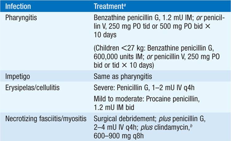

– The primary goal of treatment is to prevent suppurative complications (e.g., lymphadenitis, abscess, sinusitis, bacteremia, pneumonia) and ARF; therapy does not seem to reduce the duration of symptoms or prevent PSGN.
– Follow-up cultures after completion of therapy are not routinely recommended.
• Asymptomatic pharyngeal GAS carriage usually is not treated; however, when the pt is a potential source of infection in others (e.g., health care workers), penicillin V (500 mg PO qid for 10 days) with rifampin (600 mg PO bid for the final 4 days) is used.
Scarlet Fever Scarlet fever is the designation for GAS infection—usually pharyngitis—associated with a characteristic rash. It is much less common now than in the past.
• The rash typically appears in the first 2 days of illness over the upper trunk and spreads to the extremities but not to the palms and soles. The skin has a sandpaper feel.
• Other findings include strawberry tongue (enlarged papillae on a coated tongue) and Pastia’s lines (accentuation of rash in skin folds).
• Rash improves in 6–9 days, with desquamation on palms and soles.
Skin and Soft Tissue Infections See Chap. 93 for further discussion of clinical manifestations and treatment.
• Impetigo: A superficial skin infection, impetigo is most often seen in young children in warmer months or climates and under poor hygienic conditions.
– Red papular lesions evolve into pustules that ultimately form characteristic honeycomb-like crusts, usually affecting the facial areas around the nose and mouth and the legs. Pts are usually afebrile.
– GAS impetigo is associated with PSGN.
– For treatment, see Table 96-1. Given an increasing incidence of impetigo due to Staphylococcus aureus, empirical antibiotic therapy should cover GAS and S. aureus.
• Thus dicloxacillin or cephalexin (250 mg PO qid for 10 days) is used.
• Topical mupirocin ointment is also effective.
• Cellulitis: GAS cellulitis develops at anatomic sites where normal lymphatic drainage has been disrupted (e.g., due to surgery or prior cellulitis). When skin integrity is breached, organisms may enter at sites distant from the area of cellulitis.
– GAS may cause rapidly developing postoperative wound infections with a thin exudate.
– Erysipelas is a form of cellulitis characterized by pain, fever, and acute onset of bright red swelling that is sharply demarcated from normal skin.
• It usually involves the malar facial area or the lower extremities and is caused almost exclusively by GAS.
• The skin often has a peau d’orange texture, and blebs or bullae may form after 2 or 3 days.
– For treatment of erysipelas or cellulitis known to be due to GAS, see Table 96-1; empirical treatment should be directed against GAS and S. aureus.
• Necrotizing fasciitis: See Chap. 93 for details. GAS causes ~60% of cases of necrotizing fasciitis. For treatment, see Table 96-1.
Pneumonia and Empyema GAS is an occasional cause of pneumonia in previously healthy pts.
• Pts have pleuritic chest pain, fever, chills, and dyspnea; ~50% have accompanying pleural effusions that—unlike the sterile parapneumonic effusions of pneumococcal pneumonia—are almost always infected and should be drained quickly to avoid loculation.
• For treatment, see Table 96-1.
Bacteremia In most cases of GAS bacteremia, a focus is readily identifiable. Bacteremia occurs occasionally with cellulitis and frequently with necrotizing fasciitis.
• If no focus is immediately evident, a diagnosis of endocarditis, occult abscess, or osteomyelitis should be considered.
Toxic Shock Syndrome Unlike those with TSS due to S. aureus, pts with streptococcal TSS generally lack a rash, have bacteremia, and have an associated soft-tissue infection (cellulitis, necrotizing fasciitis, or myositis).
• Table 96-2 presents a proposed case definition for streptococcal TSS.
TABLE 96-2 PROPOSED CASE DEFINITION FOR STREPTOCOCCAL TOXIC SHOCK SYNDROMEa
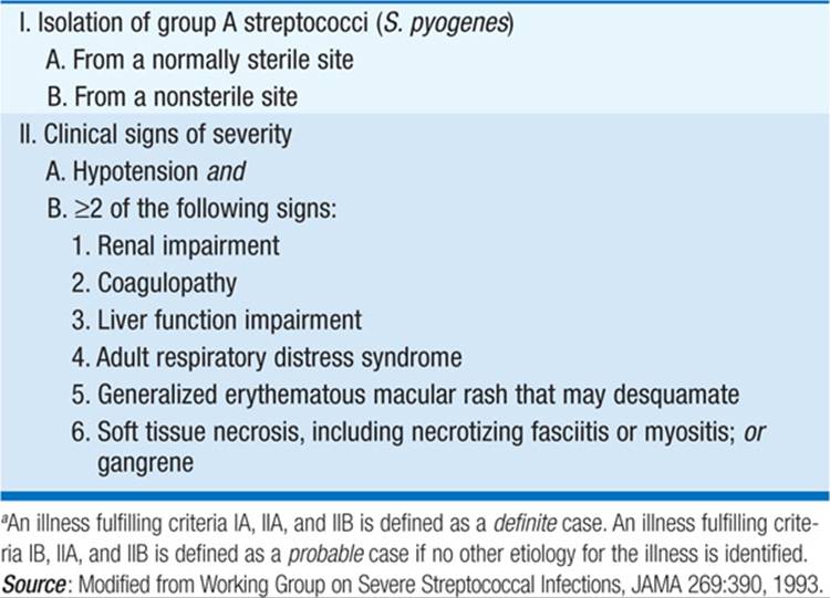
• The mortality rate for streptococcal TSS is ~30%, with most deaths due to shock and respiratory failure.
• For treatment, see Table 96-1.
Prevention
Although household contacts of individuals with invasive GAS infection are at increased risk of infection, the attack rate is low enough that antibiotic prophylaxis is not routinely recommended.
STREPTOCOCCI OF GROUPS C AND G
• Streptococci of groups C and G cause infections similar to those caused by GAS, including cellulitis, bacteremia (particularly in elderly or chronically ill pts), pneumonia, and soft tissue infections.
• Strains that form small colonies (<0.5 mm) on blood agar are generally of the S. milleri group (S. intermedius, S. anginosus); large-colony groups C and G streptococci are now considered a single species (S. dysgalactiae subsp. equisimilis).
• Treatment is the same as for similar syndromes due to GAS.
– Although it has not been shown to be superior, the addition of gentamicin (1 mg/kg IV q8h) is recommended by some experts for endocarditis or septic arthritis due to group C or G streptococci because of a poor clinical response to penicillin alone.
– Joint infections can require repeated aspiration or open drainage for cure.
GROUP B STREPTOCOCCUS (GBS)
• GBS is a major cause of meningitis and sepsis in neonates and a common cause of peripartum fever in women.
– About half of the infants delivered vaginally to mothers colonized with GBS (5–40% of women) become colonized, but only 1–2% develop infection.
– With maternal colonization, the risk of neonatal GBS infection is high if delivery is preterm or if the mother has an early rupture of membranes (>24 h before delivery), prolonged labor, fever, or chorioamnionitis.
• Widespread prenatal screening for GBS has reduced the incidence of neonatal infection to 0.8 cases per 1000 live births; adults now account for a larger proportion of invasive GBS infections than do newborns.
Neonatal Infections
• Early-onset infection occurs within the first week of life (median age, 20 h). The infection is acquired within the maternal genital tract during birth.
– Neonates typically have respiratory distress, lethargy, and hypotension.
– Bacteremia is noted in ~100% of cases, pneumonia in one-third to one-half, and meningitis in one-third.
• Late-onset infection develops in infants >1 week old and generally ≤3 months of age (mean age, 3–4 weeks). The organism is acquired during delivery or during later contact with a source.
– Meningitis is the most common manifestation.
– Infants present with lethargy, fever, irritability, poor feeding, and occasionally seizures.
TREATMENT GBS Infections in Neonates
• Penicillin is the agent of choice for all GBS infections.
– Empirical therapy for suspected bacterial sepsis consists of ampicillin and gentamicin while cultures are pending.
– Many physicians continue to give gentamicin until the pt improves clinically.
Prevention
Identification of high-risk mothers and prophylactic administration of ampicillin or penicillin during delivery reduce the risk of neonatal infection.
• Maternal screening for anogenital colonization with GBS at 35–37 weeks of pregnancy is currently recommended.
• Women who have previously given birth to an infant with GBS disease, who have a history of GBS bacteriuria during pregnancy, or who have an unknown culture status but risk factors noted above should receive intrapartum prophylaxis (usually 5 mU of penicillin G followed by 2.5 mU q4h until delivery).
– Cefazolin can be used for pts with a penicillin allergy who are at low risk for anaphylaxis.
– If the mother is at risk for anaphylaxis and the GBS isolate is known to be susceptible, clindamycin or erythromycin can be used; otherwise, vancomycin is indicated.
Infections in Adults
Most GBS infections in adults are related to pregnancy and parturition. Other GBS infections are seen in the elderly, especially pts with underlying conditions such as diabetes mellitus or cancer.
• Cellulitis and soft tissue infection, urinary tract infection (UTI), pneumonia, endocarditis, and septic arthritis are most common.
• Penicillin (12 mU/d for localized infections and 18–24 mU/d for endocarditis or meningitis, in divided doses) is recommended. Vancomycin is an acceptable alternative for penicillin-allergic pts.
• Relapse or recurrent invasive infection occurs in ~4% of cases.
NONENTEROCOCCAL GROUP D STREPTOCOCCI
The main nonenterococcal group D streptococci that cause human infections are S. gallolyticus, S. pasteurianus, S. infantarius, and S. lutetiensis (previously classified together as S. bovis).
• These organisms have been associated with GI malignancies and other bowel lesions, which are found in ≥60% of pts presenting with group D streptococcal endocarditis.
• Unlike enterococcal endocarditis, group D streptococcal endocarditis can be adequately treated with penicillin alone.
VIRIDANS STREPTOCOCCI
• Many viridans streptococcal species are part of the normal oral flora, residing in close association with the teeth and gingiva. Minor trauma such as flossing or tooth-brushing can cause transient bacteremia.
• Viridans streptococci have a predilection to cause endocarditis. Moreover, they are often part of a mixed flora in sinus infections and brain and liver abscesses.
• Bacteremia is common in neutropenic pts, who can develop a sepsis syndrome with high fever and shock. Risk factors in these pts include chemotherapy with high-dose cytosine arabinoside, prior treatment with trimethoprim-sulfamethoxazole (TMP-SMX) or a fluoroquinolone, mucositis, or therapy with antacids or histamine antagonists.
• The S. milleri group (including S. intermedius, S. anginosus, and S. constellatus) differs from other viridans streptococci in both hemolytic pattern (i.e., they may be α-, β-, or γ-hemolytic) and clinical syndromes. These organisms commonly cause suppurative infections, especially abscesses of brain and viscera, as well as respiratory tract infections such as pneumonia, empyema, and lung abscess.
• Neutropenic pts should receive vancomycin pending susceptibility testing; other pts may be treated with penicillin.
ABIOTROPHIA AND GRANULICATELLA SPECIES (NUTRITIONALLY VARIANT STREPTOCOCCI)
• The organisms formerly known as nutritionally variant streptococci are now classified as Abiotrophia defectiva and three species within the genus Granulicatella. These fastidious organisms require media that are enriched (e.g., with vitamin B6) for growth.
• These organisms are more frequently associated with treatment failure and relapse in cases of endocarditis than are viridans streptococci. Thus gentamicin (1 mg/kg q8h) must be added to the penicillin regimen.
ENTEROCOCCAL INFECTIONS
Microbiology
Enterococci are gram-positive cocci that are observed as single cells, diplococci, or short chains.
• Enterococci share many morphologic and phenotypic characteristics with streptococci and thus were previously classified as the latter.
• Enterococci are generally nonhemolytic when cultured on blood agar plates.
• Enterococci are inherently resistant to a variety of commonly used antibiotics. E. faecium is the most resistant species, with >80% of U.S. isolates resistant to vancomycin (VRE) and >90% resistant to ampicillin. In contrast, only ~7% of E. faecalis isolates are resistant to vancomycin and ~4% to ampicillin.
Epidemiology
Although 18 enterococcal species have been isolated from human infections, E. faecalis and E. faecium cause the overwhelming majority of enterococcal infections.
• Enterococci are the second most common cause of nosocomial infection (after staphylococci), with roughly equal numbers of cases caused by E. faecalis and E. faecium.
• Colonization with VRE (as opposed to antibiotic-susceptible strains) predisposes to enterococcal infection. Risk factors for VRE colonization include prolonged hospitalization; long antibiotic courses; hospitalization in long-term-care facilities, surgical units, and/or ICUs; organ transplantation; renal failure; high APACHE scores; and physical proximity to pts colonized with VRE.
Clinical Manifestations
Enterococci cause UTIs, especially in pts who have undergone instrumentation; chronic prostatitis; bacteremia related to intravascular catheters; bacterial endocarditis of both native and prosthetic valves (usually with a subacute presentation); meningitis, particularly in pts who have undergone neurosurgery; soft tissue infections, particularly involving surgical wounds; and neonatal infections. These organisms can also be a component of mixed intraabdominal infections.
TREATMENT Enterococcal Infections
• Given low cure rates with β-lactam monotherapy, combination therapy with a β-lactam plus gentamicin or streptomycin is recommended for serious enterococcal infections. High-level resistance to aminoglycosides (i.e., minimal inhibitory concentrations of >500 and >2000 μg/mL for gentamicin and streptomycin, respectively) abolishes the synergism otherwise obtained by adding an aminoglycoside to a cell wall–active agent. This phenotype must be assessed in isolates from serious infections.
• For E. faecium isolates resistant to ampicillin:
– Daptomycin, quinupristin/dalfopristin, or linezolid plus another active agent (doxycycline with rifampin, tigecycline, or a fluoroquinolone) may be used.
– If daptomycin is used and if high-level resistance is not noted, an aminoglycoside should be added to the regimen.
• If high-level aminoglycoside resistance is present, two other active agents should be used.
CORYNEBACTERIAL AND RELATED INFECTIONS
CORYNEBACTERIUM DIPHTHERIAE
Microbiology
C. diphtheriae, the causative agent of the nasopharyngeal and skin infection known as diphtheria, is a club-shaped, gram-positive, unencapsulated, nonmotile, nonsporulating rod.
• The bacteria often form clusters of parallel arrays (palisades) in culture, referred to as Chinese characters.
• Some strains produce diphtheria toxin, which can cause myocarditis, polyneuropathy, and other systemic toxicities and is associated with the formation of pseudomembranes in the pharynx during respiratory infection.
Epidemiology and Pathogenesis
As a result of routine immunization, fewer than five cases of diphtheria are diagnosed per year in the United States.
• Low-income countries in Africa and Asia continue to have significant outbreaks; globally, there were ~7000 cases of diphtheria in 2008 and ~5000 deaths related to diphtheria in 2004.
• C. diphtheriae is transmitted via the aerosol route, primarily during close contact.
• Diphtheria toxin—the primary virulence factor—irreversibly inhibits protein synthesis, thereby causing the death of the cell.
Clinical Manifestations
• Respiratory diphtheria: Upper respiratory tract illness due to C. diphtheriae typically has a 2- to 5-day incubation period and is diagnosed on the basis of a constellation of sore throat; low-grade fever; and a tonsillar, pharyngeal, or nasal pseudomembrane.
– Unlike that of GAS pharyngitis, the pseudomembrane of diphtheria is tightly adherent; dislodging the membrane usually causes bleeding.
– Massive swelling of the tonsils and “bull-neck” diphtheria resulting from submandibular and paratracheal edema can develop. This illness is further characterized by foul breath, thick speech, and stridorous breathing.
– Respiratory tract obstruction due to swelling and sloughing of the pseudomembrane can be fatal.
– Neurologic manifestations may appear during the first 2 weeks of illness, beginning with dysphagia and nasal dysarthria and progressing to cranial nerve involvement (e.g., weakness of the tongue, facial numbness, blurred vision due to ciliary paralysis).
• Several weeks later, a generalized sensorimotor polyneuropathy with prominent autonomic dysfunction (including hypotension) may occur.
• Pts who survive the acute phase gradually improve.
• Cutaneous diphtheria: This variable dermatosis is generally characterized by punched-out ulcerative lesions with necrotic sloughing or pseudo-membrane formation. Pts typically present to medical care because of nonhealing or enlarging ulcers; the lesions rarely exceed 5 cm in diameter.
Diagnosis
A definitive diagnosis is based on compatible clinical findings and detection of C. diphtheriae or toxigenic C. ulcerans (by isolation or histologic identification) in local lesions.
• The laboratory should be notified that diphtheria is being considered, and appropriate selective media must be used.
• In the U.S., respiratory diphtheria is a notifiable disease; cutaneous diphtheria is not.
TREATMENT Diphtheria
• Diphtheria antitoxin is the most important component of treatment and should be given as soon as possible. To obtain antitoxin, contact the Emergency Operations Center at the CDC (770-488-7100). See www.cdc.gov/vaccines/vpd-vac/diphtheria/dat/dat-main.htm for further information.
• Antibiotic therapy is administered for 14 days to prevent transmission to contacts. The recommended options are (1) procaine penicillin G (600,000 U IM q12h in adults; 12,500–25,000 U/kg IM q12h in children) until the pt can take oral penicillin V (125–250 mg qid); or (2) erythromycin (500 mg IV q6h in adults; 40–50 mg/kg per day IV in 2–4 divided doses in children) until the pt can take oral erythromycin (500 mg qid).
– Rifampin and clindamycin are other options for pts who cannot tolerate penicillin or erythromycin.
– Cultures should document eradication of the organism 1 and 14 days after completion of antibiotic therapy. If the organism is not eradicated after 2 weeks of therapy, an additional 10-day course followed by repeat cultures is recommended.
• Respiratory isolation and close monitoring of cardiac and respiratory functions should be instituted.
Prognosis
Risk factors for death include a long interval between onset of local disease and antitoxin administration; bull-neck diphtheria; myocarditis with ventricular tachycardia; atrial fibrillation; complete heart block; an age of >60 years or <6 months; alcoholism; extensive pseudomembrane elongation; and laryngeal, tracheal, or bronchial involvement.
Prevention
DTaP (diphtheria and tetanus toxoids and acellular pertussis vaccine adsorbed) is recommended for primary immunization of children up to age 7 years; Tdap (tetanus toxoid, reduced diphtheria toxoid, and acellular pertussis) is recommended as the booster vaccine for children 11–12 years old and as the catch-up vaccine for children 7–10 and 13–18 years old.
• Td (tetanus and diphtheria toxoids) is recommended for routine booster use in adults at 10-year intervals or for tetanus-prone wounds. When >10 years have elapsed since the last Td dose, adults 19–64 years old should receive a single dose of Tdap.
• Close contacts of pts with respiratory diphtheria should have throat specimens cultured for C. diphtheriae, should receive a 7- to 10-day course of oral erythromycin or one dose of benzathine penicillin (1.2 mU for persons ≥6 years old; 600,000 U for children <6 years old), and should be vaccinated if immunization status is uncertain.
INFECTIONS WITH OTHER CORYNEBACTERIA AND RELATED ORGANISMS
Nondiphtherial Corynebacterium species and related organisms are common components of the normal human flora. Although frequently considered contaminants, these bacteria are associated with invasive disease in immunocompromised hosts.
• C. ulcerans infection is a zoonosis that causes diphtheria-like illness and requires similar treatment.
• C. jeikeium infects pts with cancer or severe immunodeficiency and can cause severe sepsis, endocarditis, device-related infections, pneumonia, and soft tissue infections. Treatment consists of removal of the source of infection and administration of vancomycin.
• C. urealyticum is a cause of sepsis and nosocomial UTI, including alkaline-encrusted cystitis (a chronic inflammatory bladder infection associated with deposition of ammonium magnesium phosphate on the surface and walls of ulcerating lesions in the bladder). Vancomycin is an effective therapeutic agent.
• Rhodococcus species appear as spherical to long, curved, clubbed gram-positive rods that are often acid-fast. The most common presentation—nodular cavitary pneumonia of the upper lobe (similar to tuberculosis and nocardiosis) in an immunocompromised host—often occurs in conjunction with HIV infection. Vancomycin is the drug of choice, but macrolides, clindamycin, rifampin, and TMP-SMX have also been used to treat these infections.
• Arcanobacterium haemolyticum can cause pharyngitis and chronic skin ulcers, often in association with a scarlatiniform rash similar to that caused by GAS. The organism is susceptible to β-lactam agents, macrolides, fluoroquinolones, clindamycin, vancomycin, and doxycycline. Penicillin resistance has been reported.

For a more detailed discussion, see Wessels MR: Streptococcal Infections, Chap. 136, p. 1171; Arias CA, Murray BE: Enterococcal Infections, Chap. 137, p. 1180; and Bishai WR, Murphy JR: Diphtheria and Other Infections Caused by Corynebacteria and Related Species, Chap. 138, p. 1188, in HPIM-18.