
Timur S. Durrani, MD, MPH, MBA
Robert J. Harrison, MD, MPH
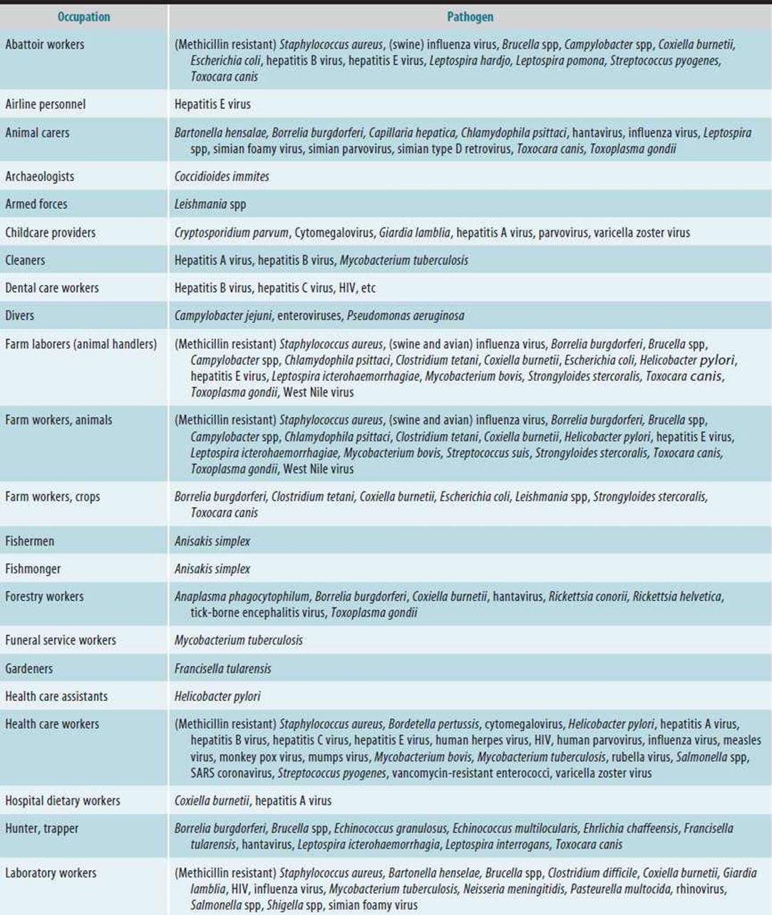
Occupational infections are human diseases caused by work-associated exposure to microbial agents, including bacteria, viruses, fungi, and parasites. It has been hypothesized that certain infectious diseases such as malaria, dengue fever, and cholera will become more prevalent with climate change. An infection is distinguished as occupational by some aspect of the work that involves contact with a biologically active organism. Occupational infection can occur following contact with infected persons or surfaces, as in the case of health care workers; with infected animal or human tissue, secretions, or excretions, as in laboratory workers; with asymptomatic or unknown contagious humans, as happens during business travel; or with infected animals, as in agriculture. See Table 20–1 for work-related pathogens by specific job title.
Table 20–1. Work-related pathogens by specific job title or broader occupational groups.

The etiology, pathogenesis, clinical findings, diagnosis, and treatment of occupational, nonoccupational, and bioterrorism infections are essentially the same except for practical differences related to identification of the source of exposure, epidemiologic control, and prevention. This chapter focuses on the occupational aspects of microbial exposures and relevant strategies for prevention. This chapter highlights Middle East Respiratory Syndrome (MERS) and influenza, tuberculosis, hepatitis B and C, acquired immunodeficiency syndrome (AIDS), and travel-related infections as examples of different types of exposure.
NEWLY EMERGING INFECTIOUS DISEASES
 Middle East Respiratory Syndrome (MERS)
Middle East Respiratory Syndrome is a viral respiratory illness first reported in Saudi Arabia in 2012. Similar to the Severe Acute Respiratory Syndrome (SARS) epidemic which peaked in 2003, MERS is caused by a coronavirus. By the end of the epidemic, health care workers accounted for 21% of SARS cases, demonstrating the vulnerability of health care workers to occupational infections. Most people who have been confirmed to have MERS-CoV infection developed severe acute respiratory illness. They had fever, cough, and shortness of breath. About half of these people died. All the cases have been linked to countries in or near the Arabian Peninsula, these countries include Bahrain, Iraq, Iran, Israel, Jordan, Kuwait, Lebanon, Oman, Palestinian territories, Qatar, Saudi Arabia, Syria, the United Arab Emirates (U.A.E.), and Yemen.
The 2003 SARS epidemic evoked a global public health emergency response as the disease, in the absence of control measures, spread rapidly on a global scale. SARS was clearly an occupational health hazard because health care workers who cared for patients accounted for 21% of all cases (>50% in some settings). Here health care workers were not only affected but also propagated the outbreaks in hospitals and into the community. Other groups of workers at risk include wildlife traders, food handlers, laboratory workers, traveling workers, and flight attendants.
Corona viruses such as MERS and SARS are transmitted primarily by respiratory droplets from coughing and sneezing. Droplets can be propelled up to 6 ft and deposited on mouth, nose, and eyes (hence the need for masks and goggles). Droplets also can contaminate surfaces, which become fomites. Another possible mode is airborne transmission which occurs by dissemination of either airborne droplet nuclei or small particles in the respirable size range containing infectious agents that remain infective over time and distance. Microorganisms carried in this manner may be dispersed over long distances by air currents and may be inhaled by susceptible individuals who have not had face-to-face contact with (or been in the same room with) the infectious individual
Response in Health Care Facilities
The Centers for Disease Control and Prevention (CDC) recommends airborne precautions for the management of hospitalized patients suspected of a MERS infection. This includes basic hygiene measures incorporating respiratory hygiene/cough etiquette, use of personal protective equipment (PPE), in particular N95 masks, use of airborne infection isolation rooms (AIIR), and procedural protocols for the use of ventilators, nebulizers, endotracheal intubation, and other droplet-and aerosol-generating devices and procedures.
Occupational Issues
In terms of occupational health, some health care workers may be placed on “working quarantine (allowed to travel only between home and health care facility)” to ensure sufficient staffing levels. In relation to the corporate setting, “snow day” measures may be warranted involving closure of businesses and cancelation of public gatherings. The International Labor Office (ILO) documented a working paper on practical and administrative responses to SARS in the workplace in relation to preexisting ILO standards.
INFLUENZA
Influenza, or flu, is a respiratory infection caused by several flu viruses.
Seasonal flu is the term used to refer to the flu outbreaks that occur annually, mainly in the late fall and winter. Pandemic flu refers to particularly virulent strains of flu that spread rapidly from person to person to create a worldwide epidemic (pandemic).
Flu viruses are classified as types A, B, and C. Flu A viruses are divided into subtypes based on two proteins on the surface of the virus, that is, hemagglutinin (H; 15 subtypes: H1–H15) and neuraminidase (N; 9 subtypes: N1–N9). Wild birds are the primary natural reservoir for all subtypes of flu A viruses, and while shedding the viruses they mostly remain healthy hosts. When domesticated birds (ie, chickens, ducks, turkeys, etc) become infected, the infection spreads quickly and widely and leads to a bird flu outbreak in poultry. If the virus is of a highly pathogenic type, the epidemic is highly lethal. Viruses can be transmitted from farm to farm by contaminated equipment, vehicles, feed, cages, or clothing. Thus, the standard control measures in poultry are quarantine and depopulation (or culling) and surveillance around affected flocks. Although the risk of infection to humans from bird flu is generally low, people should avoid contact with infected birds or contaminated surfaces and should be careful when handling and cooking poultry.
Flu A viruses genetically are labile and well adapted to escape host defenses. Consequently, small changes constantly occur in the antigenic composition (antigenic drift). Alternatively, an abrupt major change (antigenic shift) may occur if viruses from two different species infect the same host (human or animal) and mix together. The new virus may acquire most genes from a human virus, but a hemagglutinin and/or neuraminidase from an avian virus, and as such likely will become capable of efficient spread from person to person.
An outbreak of human infections with a new avian influenza A (H7N9) virus was first reported in China by the World Health Organization (WHO) on April 1, 2013. The virus was detected in poultry in China as well. During the outbreak, more than 130 human infections with H7N9 were reported, the vast majority during the month of April. Many of the people infected with H7N9 reported contact with poultry. The working assumption is that human infections occurred after exposure to infected poultry or contaminated environments. While some mild illness in human cases was seen, most patients had severe respiratory illness and 43 people died. Close contacts of confirmed H7N9 patients were followed to determine whether any human-to-human spread of H7N9 was occurring. No evidence of sustained person-to-person spread of the H7N9 virus was found. No cases of H7N9 outside of China have been reported.
In the twentieth century, the great influenza pandemic of 1918–1919 (“Spanish flu” [A(H1N1)]) caused an estimated 40–50 million deaths worldwide and was followed by pandemics in 1957–1958 (“Asian flu” [A(H2N2)]) and 1968–1969 (“Hong Kong flu” [A(H3N2)]). Although the origin of the 1918–1919 pandemic virus is not clear, the latter two pandemics were caused by viruses containing a combination of genes from a human flu virus and an avian flu virus. Influenza pandemics may occur when new virus subtypes emerge and are readily transmitted from person to person. WHO asserts that another influenza pandemic is inevitable but unpredictable. Influenza pandemics are distinct from the more usual and smaller-scale influenza epidemics occurring almost every winter caused by type A or B flu viruses (the only other type of flu virus, type C, causes mild respiratory illness and does not cause epidemics).
INFLUENZA IN THE WORKPLACE
While each workplace is unique, and a worker’s risk for occupational exposure to influenza can vary widely depending on the nature of their job, the CDC encourages workplaces to promote influenza vaccination among workers, encourage proper hand and respiratory hygiene practices, and educate workers on influenza signs and symptoms. Because health care workers and first responders will be at high risk of infection in a pandemic situation, preparedness strategies with a strong occupational health component are highly warranted.
Guidance for screening, prevention and treatment of influenza are updated regularly.
INFECTIONS CAUSED BY THE WORKERS ENVIRONMENT
Coccidioidomycosis
Coccidioidomycosis, also known as Valley fever, is a recognized occupational illness that has been reported in workers engaged in soil-disrupting activities, including agricultural workers, military personnel, anthropologists, and archaeologists working in endemic areas. In nonendemic areas, work-related cases of disease have been reported in various occupations, including laboratory and hospital personnel.
Coccidioidomycosis is caused by the inhalation of airborne fungal spores following soil disruption from either of the two soil-dwelling Coccidioides species: C immitis, native to California, and C posadasii, found outside California. The fungus is endemic to certain semiarid areas of California, Arizona, New Mexico, Nevada, and Texas, and of Central and South Americas. Coccidioidomycosis can be a severe illness and result in disability due to pulmonary involvement and disseminated disease; however, most infections are asymptomatic. Influenza-like illness is the most common clinical presentation. Infection generally imparts immunity to reinfection, although rare cases of reinfection have been reported. Groups at increased risk for developing disseminated disease are African Americans and Asians, people of Filipino descent, pregnant women during their third trimester, and immunocompromised patients.
Prevention of occupationally acquired coccidioidomycosis requires a multidisciplinary approach, including engineering controls such as continuous soil wetting of disturbed soil and washing all equipment prior to removal from the worksite, administrative controls such as increasing training on prevention, signs and symptoms of infection, and use of personal protective equipment such as powered air purifying respirators and requiring a change of clothing at the worksite.
Ketoconazole is the only antifungal that has been approved by the Food and Drug Administration (FDA) for use in coccidioidomycosis; however, most experts prefer either fluconazole or itraconazole. Some initial studies have indicated that azole antifungals should be used at a minimum dose of 400 mg daily and that relapses are frequent once therapy has been discontinued. Fluconazole has shown to be the most effective for treating coccidioidal meningitis.
Hantavirus
Hantavirus of the bunyavirus family of viruses is a negative-sensed, single-stranded RNA virus. Animal laboratory workers and persons working in rodent infested buildings are at increased risk for coming in contact with dried droppings, urine, or saliva of mice and rats that carry Hantavirus and developing Hantavirus pulmonary syndrome (HPS). The disease begins as a flu-like illness characterized by fever, chills, and muscle aches, but it can rapidly progress to a life-threatening condition marked by respiratory failure as the lungs fill with fluid.
There is no specific treatment or cure for hantavirus infection. Treatment of patients with HPS remains supportive in nature. Patients should receive appropriate, broad-spectrum antibiotic therapy while awaiting confirmation of a diagnosis of HPS. Care during the initial stages of the disease should include antipyretics and analgesia as needed. If there is a high degree of suspicion of HPS, patients should be immediately transferred to an emergency department or intensive care unit (ICU) for close monitoring and care.
Biosafety Level 2 (BSL-2) facilities and BSL-2 practices are recommended for laboratory handling of sera and tissues from persons potentially infected with the agents of HPS.
Prevention of exposure should rely on sealing holes in workplace structure to prevent entry of rodents, placing traps to reduce rodent population and placing food in sealed containers to eliminate nesting sites. If rodent excreta are found, the CDC recommends cleaning with wetting agents and disinfectants prior to creating an aerosolized dust contaminates.
Tick Borne
Outdoor workers are at risk of exposure to tick-borne diseases if they work at sites with ticks. Worksites with woods, bushes, high grass, or leaf litter are likely to have more ticks. Outdoor workers in most regions of the United States should be extra careful to protect themselves in the spring, summer, and fall when ticks are most active. Ticks may be active all year in some regions with warmer weather. Workers at risk for tick-borne diseases include outdoor workers such as construction workers, painters, roofers, pavers, laborers, mechanics, landscapers, forestry workers, brush clearers, land surveyers, farmers, railroad workers, oil field workers, utility line workers, park or wildlife management workers, entomologists and wildlife biologists. Laboratory, field, and clinical workers who perform necropsies of infected birds or handle infected tissues or fluids are also at risk of infection (particularly West Nile Virus). Other diseases transmitted by tick include Lyme disease, Babesiosis, Ehrlichiosis, Rocky Mountain spotted fever, tick-borne relapsing fever, tularemia, and Q fever.
Ticks are usually more active in the months of April through October and peak in the summer months of June through August. The time of year when ticks are active may vary with the geographic region and climate. Outdoor workers should be extra careful to protect themselves in the late spring and summer when immature ticks are most active. Common symptoms of infection with tick-borne diseases include body/muscle aches, fever, headaches, fatigue, joint pain or rash. Tick-borne diseases are diagnosed based on symptoms and the possibility that the worker has been exposed to infected ticks. Most cases can be successfully treated with antibiotics.
Prevention of tick-borne diseases includes wearing light-colored clothing, including long-sleeved shirts and long pants tucked into boots or socks, use of insect repellents containing 20–30% DEET on exposed skin and clothing to prevent tick bites and application of permethrin to pants, socks, and shoes which typically stays effective through several washings. Workers should be advised check their skin and clothes for ticks every day. The immature forms of these ticks are small and may be hard to see. They should also be advised to shower or bathe as soon as possible after working outdoors to wash off and check for ticks, particularly in the hair, underarms, and groin. Any ticks discovered should be removed immediately using fine-tipped tweezers, and the area cleansed with soap and water. Removing infected ticks within 24 hours reduces the risk of being infected with the Lyme disease bacterium. Finally wash and dry work clothes in a hot dryer to kill any ticks present.
INFECTIONS CAUSED BY EXPOSURE TO INFECTED HUMANS OR THEIR TISSUES
Health care and clinical laboratory workers are also at increased risk of infection by organisms whose natural hosts are humans, as in the case of hepatitis, rubella, AIDS, tuberculosis, and staphylococcal disease. Some infections may be transmitted through close personal contact with infected patients. Exposure and infection caused by almost any of the viruses, bacteria, fungi, and parasites pathogenic for humans can result from direct contact with the organism in culture or in human tissue. Tuberculosis is an example of a relatively common occupational infection resulting from repeated close contact with infected patients, and hepatitis B exemplifies a serious and relatively frequent infection resulting from manipulation of infected human blood and inoculation by infectious virus particles.
Methicillin-Resistant Staphylococcus aureus
Methicillin-resistant Staphylococcus aureus (MRSA) is reported to colonize health care workers at a rate between 1% and 15%, compared to 1% of the general population. Correctional staff (officers and staff working in jails or prisons) also have increased rates of MRSA. Other locations at increased risk of MRSA exposure include schools (including athletic departments), dormitories, military barracks, households, correctional facilities, and daycare centers. Factors that increase risk of transmission of MRSA include crowding of populations, frequent skin-to-skin contact, compromised skin, contaminated items (such as towels or used bandages) and lack of cleanliness. MRSA is transmitted most frequently by direct skin-to-skin contact or contact with shared items or surfaces that have come into contact with someone else’s infection. MRSA infections can vary from simple skin abscesses to life-threatening systemic infections.
Prevention of transmission relies on improved hygiene practices at highest-risk locations. In health care settings this can include ensuring suspected or infected patients be placed in private rooms, if available, and the appropriate use of PPE. Environmental controls should include daily use of commercially available cleaners or detergents on high-touch surfaces (eg, doorknobs, counters, bedside tables, bedrails bathtubs, toilet seats) that may come in contact with bare skin or uncovered infections.
Simple abscesses or boils may be managed with incision and drainage alone. Antibiotics for 5–10 days are recommended for patients who have abscesses associated with severe or extensive disease (eg, multiple sites of infection) or rapid progression in the presence of associated cellulitis; signs and symptoms of systemic illness or associated comorbidities or immunosuppression. Oral antibiotic options for treating skin and soft-tissue infections in patients with community-associated MRSA include clindamycin, trim-ethoprim/sulfamethoxazole, a tetracycline (doxycycline or minocycline), and linezolid.
Decolonization may be considered if a worker develops a recurrent infection despite good personal hygiene and wound care, or if other household members develop infections. Strategies for decolonization include nasal decolonization with mupirocin twice per day for 5–10 days, or nasal decolonization with mupirocin twice per day for 5–10 days plus topical body decolonization with a skin antiseptic solution (eg, chlorhexidine) for 5–14 days.
TUBERCULOSIS
Mycobacterium tuberculosis can cause disseminated disease but is associated most frequently with pulmonary infections. The bacilli are transmitted by airborne route and, depending on host factors, may lead to latent tuberculosis infection (LTBI) or tuberculosis disease (TB). Only people who are ill with TB of the lungs are infectious, whereas people with LTBI are not.
Globally, it is estimated that TB caused 8.7 million incident cases and 1.4 million deaths in 2011. Geographically, the burden of TB is highest in Asia and Africa. India and China combined have almost 40% of the world’s TB cases; the South-East Asia and Western Pacific Regions of which they are a part account for 60%. The African Region has approximately one quarter of the world’s cases, and the highest rates of cases and deaths relative to population. There has been major progress in reducing TB cases and deaths in the past two decades, with TB incidence falling globally for several years and declining at a rate of 2.2% between 2010 and 2011. Globally, the TB mortality rate has fallen by 41% since 1990 and the world is on track to reach the global target of a 50% reduction by 2015.
Medical house staffs have two to three times the tuberculosis infection rate of nonmedical personnel, and laboratory workers exposed to M tuberculosis have three times the incidence of nonexposed workers. Staffs of laboratories and necropsy rooms are estimated to be between 100 and 200 times more likely than the general public to develop tuberculosis. Other high-prevalence work environments include most health care settings (especially hospitals, long-term care facilities, and dialysis centers), refugee/immigration centers, homeless shelters, substance abuse treatment centers, and correctional institutions.
Tubercle bacilli may be present in gastric fluid, cerebro-spinal fluid, urine, sputum, and tissue specimens harboring active lesions. Infectious patients disseminate the organism when coughing, sneezing, or talking by expelling small infectious droplets that may remain suspended in the air for several hours and then be inhaled by susceptible persons. After an incubation period of 4–12 weeks, infection usually remains subclinical and dormant without development of active disease, but the Mantoux tuberculin skin test (TST) will become positive. However, the organism may be activated at any time, resulting in acute severe pulmonary or other systemic disease. The risk of development of clinical disease following infection is higher in selected age groups (infancy, 16–21 years of age), in states of undernutrition, in certain immunopathologic states (eg, AIDS), in certain genetic groups (persons with HLA-Bw15 histocompatibility antigen), and in persons with some coexisting diseases (silicosis, end-stage renal disease, malnutrition, leukemia, lymphoma, upper gastrointestinal tract carcinoma, diabetes).
Tuberculin Skin Test
TST is a chemical fractionation product of tubercle bacilli culture filtrate. Intradermal injection of 5 tuberculin units of TST in a patient with subclinical or clinical tuberculous infection results in a delayed hypersensitivity reaction manifested by induration at the site of injection within 48–72 hours. A minimum of 5 mm of induration is required for a test to be positive or reactive in close contacts of infectious patients, immunosuppressed patients, organ recipients, or persons with known or suspected human immunodeficiency infection (HIV) infection. A reaction of 10 mm or more is considered positive in other high-prevalence (>5%), high-risk occupational groups (above) or high-risk groups such as immigrants from high-prevalence areas, alcoholics, intravenous drug users, and those with the other disease states just mentioned. In persons with no risk factors in areas of low prevalence, induration of 15 mm or more is required for a positive reaction. The TST test may be negative in the presence of overwhelming tuberculosis, measles, Hodgkin disease, sarcoidosis, or immunosuppressive states. If the initial test is negative in individuals with suspected reduced immune response or in those who will be screened annually because of occupational or other risk, it should be repeated.
“Two-Step” Method
Some people infected with M tuberculosis may have a negative reaction to the TST if many years have passed since they became infected. They may have a positive reaction to a subsequent TST because the initial test stimulates their ability to react to the test. This is commonly referred to as the “booster phenomenon” and may incorrectly be interpreted as a skin test conversion (going from negative to positive). For this reason, the “two-step method” is recommended at the time of initial testing for individuals who may be tested periodically (eg, health care workers). If the first TST result in the two-step baseline testing is positive, consider the person infected and evaluate and treat the person accordingly. If the first test result is negative, the TST should be repeated in 1–3 weeks. If the second test result is positive, consider the person infected and evaluate and treat the person accordingly; if both steps are negative, consider the person uninfected and classify the TST as negative at baseline testing (Figure 20–1).

 Figure 20–1. TST skin testing.
TST skin testing is an accepted method for screening high-risk populations for primary infection. Persons having a reactive test are at risk of developing active clinical infection at any time (lifelong) following the primary infection owing to reactivation of the primary infection as long as viable tubercle bacilli remain in the body.
Blood Tests for TB Infection
Interferon-Gamma Release Assays (IGRAs) are whole-blood tests that can aid in diagnosing M tuberculosis infection. They do not differentiate latent tuberculosis infection from tuberculosis disease. The advantages of the IGRAs, when compared to TSTs include requiring only a single patient visit to conduct the test, results can be available within 24 hours, there is no “booster phenomenon” measured by subsequent tests, and prior bacille Calmette-Guérin (BCG) vaccination does not cause a false-positive IGRA test result. Disadvantages include samples must be processed within a specified time (8–30 hours) after collection while white blood cells are still viable, errors in collecting or transporting blood specimens or in running and interpreting the assay can decrease the accuracy of IGRAs, there is limited data on the use of IGRAs to predict who will progress to TB disease in the future and limited data on the use of IGRAs for: children under 5 years of age, persons recently exposed to M tuberculosis, immunocompromised persons and serial testing, and tests may be expensive.
When IGRAs are used for serial testing (such as in health care workers), there is no need for a second test because boosting does not occur.
Control & Treatment
TST testing can identify persons whose tests are reactive, indicating primary infection. Serial testing (biennially or more frequently) can identify recently infected individuals whose tests have become reactive (converters) within the past 2 years. Occupational candidates for periodic TST testing include those having contact with suspected or known infected patients, persons working with potentially infected primates or cattle (eg, veterinarians, zoo keepers, primate handlers), and all others working in the higher-risk environments mentioned earlier.
Recent asymptomatic converters or others recently discovered to be tuberculin-reactive (reactors) whose date of conversion is unknown and who are least likely to develop complications as a consequence of antibiotic therapy should receive drug treatment according to protocols recommended by the CDC or local health departments.
Prophylaxis is recommended for persons found to have a positive PPD who fall into any of the following categories: newly infected persons, including recent converters (within 2 years); household contacts of active cases; persons with an abnormal chest radiograph consistent with clinical tuberculosis and inadequate past antituberculous therapy or prior active disease with inadequate past therapy; persons whose reactivation may have public health consequences (eg, school teachers); patients with AIDS (or persons with antibodies to HIV), silicosis, insulin-dependent diabetes mellitus, hematologic or reticuloendothelial cancer, prior gastrectomy, chronic undernutrition, ileal bypass, renal failure requiring dialysis, or a history of prolonged use of glucocorticoid or immunosuppressive therapy, as well as intravenous drug users; and all reactors younger than 35 years of age who have none of the preceding risk factors.
Before starting prophylaxis, a chest radiograph should be taken on all skin test reactors. Any abnormalities found should be thoroughly evaluated for evidence of clinically active disease. If adequate prior prophylaxis or therapy for active disease has been completed, prophylaxis should not be given.
Current treatment regimens recommended by the American Thoracic Society (ATS)/CDC are based on evidence from clinical trials and are rated by a system using a letter (A, B, C, D, or E) that indicates strength of the recommendation and a roman numeral (I, II, or III) that indicates the quality of evidence supporting the recommendation. There are four recommended regimens for patients with tuberculosis caused by drug-susceptible organisms, each with significant potential toxicity and drug-drug interactions. Each regimen has an initial phase of 2 months followed by a choice of several options for the continuation phase of either 4 or 7 months. Isoniazid and rifampicin are the two most powerful anti-TB drugs included in the regimen in most circumstances. Because of rapidly changing drug resistance patterns, the reader should refer to current recommendations regarding prophylaxis or therapy of active disease.
Bacilli in lungs of patients may develop resistance to anti-TB medicines when the patient fails to complete standard treatment regimens or is given the wrong treatment regimen. A particularly dangerous form of drug-resistant TB is multidrug-resistant TB (MDR-TB), which is defined as the disease caused by TB bacilli resistant to at least isoniazid and rifampicin. High rates of MDR-TB are known in some countries and threaten TB control efforts. For a cost-effective control of TB, WHO and other international organizations advocate a comprehensive strategy centered on direct observed therapy (DOT). Health and community workers or trained volunteers observe patients swallowing the full course of the correct dosage of anti-TB medicines. By doing so, DOT prevents the development of drug resistance.
Persons for whom prophylactic antibiotic therapy is contraindicated should receive surveillance chest radiographs if they become symptomatic. Persons having known contact with an infectious patient for whom PPD status is not previously documented should be PPD tested immediately and then retested 8–12 weeks after the infectious contact. If conversion occurs, physical examination and chest radiography should occur to rule out acute clinical infection.
Attenuated tubercle bacilli—particularly BCG—have been used in many countries as a vaccine. However, BCG has variable efficacy in preventing the adult forms of TB and interferes with skin testing for latent TB infection. Thus, it is not recommended routine for use in the United States.
HEPATITIS B
Prior to the introduction of the hepatitis B vaccine in 1981, hepatitis B infections constituted the most frequent occupational infection among health care, laboratory, and public safety workers following human blood or body fluid exposures in the United States. Hepatitis B virus (HBV) can cause fulminant hepatitis and also can lead to chronic carrier states in up to 10% of those following acute infection. Chronic carriers suffer higher rates of cirrhosis and liver failure as well as liver cancer. The prevalence of HBV infection among health care personnel was 10 times higher than the general population in the decade preceding the HBV vaccine’s release.
Blood contains the highest titers of virus in infected individuals, with lower levels in various other body fluids, including cerebrospinal, synovial, pleural, peritoneal, pericardial, and amniotic fluids, as well as semen and vaginal secretions. Viral titers in urine, feces, tears, and saliva are low enough that these are not felt to be routes of transmission except in cases of human bites that usually involve some blood transmission. Sexual and maternal-child transmissions are alternative modes of contracting HBV in the general population.
The risk for transmission of HBV through needlestick injuries is approximately 30%. However, over 50% of acute infections in adults are asymptomatic. Given that 10% of acute HBV infections lead to chronic infections, a significant number of those with occupational infections become chronic asymptomatic carriers.
HBV can remain viable for at least 1 month on dried surfaces at room temperature. This poses additional opportunities to acquire occupational HBV infections when individuals with open cuts or abraded skin or mucous membranes contact contaminated surfaces. In fact, most occupational infections have no clear percutaneous injury leading to HBV transmission.
Prescreening serologic testing prior to vaccination generally is not recommended because the prevalence of HBV-infected individuals in the United States is low. Some groups have instituted prescreening of all potential vaccine recipients with hepatitis B core antibodies when a high percentage of potential vaccines come from endemic countries. Positive core antibodies indicate past or present HBV infections and should prompt testing for surface antigens to identify chronic carriers and for surface antibodies to identify those with resolved past infections.
While the original hepatitis B vaccine was plasma-derived, studies showed no transmission of infectious agents with this vaccine. The development of a recombinant DNA vaccine in 1986 provided an even more acceptable and highly safe method for mass vaccination of health care personnel. Since 1991, it has been recommended to vaccinate newborn infants at birth even though the prevalence of chronic hepatitis B is less than 0.5% of the populace. That same year, the Bloodborne Pathogens Act was passed, mandating employer-funded vaccination for at-risk health care workers. Since that time, a dramatic reduction in occupational HBV transmission has occurred. However, there are still some workers who have not completed or have refused vaccination and remain vulnerable to infection. There is an additional subset of those vaccinated who do not develop antibodies and who remain susceptible to infection.
Known exposures to HBV-infected blood or blood products in those who were not vaccinated or where antibody protection did not develop require the use of hepatitis B immune globulin (HBIG), which is expensive and needs a second dose 1 month later unless hepatitis B vaccination is administered concomitantly.
The usual schedule for HBV vaccination for health care workers, public safety workers, and staff of facilities for developmentally disabled persons at risk for exposure to blood or body fluids is two doses separated by no less than 4 weeks, and a third dose 4–6 months after the second dose. Those who have received only one or two doses do not need to restart the series: They only need to complete the doses they did not receive (as with most other vaccines requiring multiple dosing). Since only 50–60% of those vaccinated with two doses get immunity, some institutions will consider an accelerated series for those who will be actively working with blood or blood products, giving doses at 0, 1, 2, and 12 months with the Engerix-B (GlaxoSmithKline) vaccine (which has twice as much antigen as the Recombivax (Merck) vaccine and is the only one approved by the FDA for this series). Since this still leaves a window of time before antibody protection is achieved in high-risk workers, an extremely accelerated schedule with doses at 0, 1, and 3 weeks and a final dose at 12 months is used in some cases.
This schedule provides up to 83% protection by week 4 (and this continues to rise without additional doses of vaccine) and is used in over 15 European countries. The final dose, given at 6–12 months depending on the schedule, is critical because this provides long-term protection. Once the three-to four-dose initial series is given, no further doses are necessary assuming that hepatitis B surface antibodies are produced.
In 1997, the Advisory Committee on Immunization Practices began recommending testing for hepatitis B surface antibodies in those with ongoing exposures to blood or blood products. While the committee did not recommend testing those vaccinated prior to December 1997, there are several reasons to verify antibody responses in this population (with positive surface antibodies defined as levels greater than 10 mIU/mL). While the vaccine is highly protective in infants vaccinated at birth, the degree of protection declines with advancing age, with 90% responding to a three-dose series by 40 years of age and 75% by 60 years of age. Moreover, those who develop antibodies lose them over time, although they remain protected. If a positive antibody response is never verified, it will not be possible to differentiate the responder who lost antibodies (who is protected) from the nonresponder who never developed antibodies (who is not protected). Therefore, documenting the development of surface antibody protection at any time following the vaccination series significantly improves postexposure management for hepatitis B.
The current recommendations are to check for surface antibodies 4 weeks to 6 months following the primary series. If the person tests negative for antibodies, the data show that one additional dose of vaccine will induce antibody protection in 15–25% of nonresponders and that three additional doses (for a total of six doses) will induce antibodies in 30–50% of nonresponders. The official recommendation for additional doses is to follow the 0-, 1-, and 6-month schedule. This can be shortened effectively to 0, 1, and 2 months because a 6-month spacing from first to last dose (important for long-term immunity) already has been achieved with the primary vaccination series. Those who do not develop antibodies after six total doses should consider changing positions at work not involving blood or blood products. In cases where this is not possible or feasible, consideration of a three-dose series with 40 μg of antigen with either the Merck Recombivax HB formulated for hemodialysis patients (Note: Routine Recombivax HB contains only 10 μg of antigen) or two doses of the Engerix-B vaccine containing 20 μg of antigen per dose can be attempted. If more than 6 months have elapsed since vaccination and the individual tests negative for surface antibodies, giving one additional dose of vaccine and retesting for antibodies 4 weeks later is warranted because 50% of patients lose antibodies after 7 years. Fewer than 5% of persons receiving six doses of hepatitis B vaccine administered by the appropriate schedule in the deltoid muscle fail to develop detectable anti-HBs antibody. Some persons who are anti-HBs negative following six doses may have a low level of antibody that is not detected by routine serologic testing (“hyporesponder”). However, one reason for persistent nonresponse to hepatitis B vaccine is that the person is chronically infected with HBV.
See Table 20–2 on interpretation of hepatitis B serologic test results.
Table 20–2. Interpretation of hepatitis B serologic test results.

HEPATITIS C
Hepatitis C is a viral infection of the liver caused by the hepatitis C virus (HCV) and now known to be responsible for more than 75% of what was previously termed post-transfusion non-A, non-B hepatitis. In the United States, HCV is more frequently associated with a history of blood transfusion (prior to the introduction of EIA in the late 1980s), parenteral drug use, sexual or household exposures, and in some instances, bloodborne pathogen transmission. Worldwide there are six major genotypes of HCV, with type 1 the most frequent in the United States.
The current estimate for transmission of HCV following a needlestick injury from a positive carrier of HCV is approximately 1.8%. Transmission following mucous membrane exposure is rare, with no apparent transfer following exposures to intact skin.
With the current third-generation enzyme immunoassay (EIA), sensitivity is estimated to be approximately 97% within 6–8 weeks of exposure. However, the presence of antibodies does not correlate with protection because 70–90% of those infected become chronic carriers despite positive antibodies. Chronic carriers have a 20% chance of developing cirrhosis and an increased risk for developing hepatocellular carcinomas. Positive EIA tests usually warrant confirmatory testing with highly sensitive RT-PCR assays for HCV RNA. The recombinant immunoblot assay (RIBA) still can be used in cases where the EIA is positive with a negative HCV RNA to determine whether the EIA is a false-positive result.
Major advances in the treatment of chronic carriers have occurred recently with the introduction of parenteral peginterferon-alpha combined with oral ribavirin. Sustained virologic response (SVR) is defined as no measurable virus 6 months following completion of treatment. Unfortunately, type 1 genotypes (most frequent in the United States), are less responsive to this regimen (42–46% SVR) and require 12 months of treatment, unlike other genotypes (76–82% SVR) that usually respond after 6 months.
Following exposures to known HCV-positive blood or blood products, HCV RNA testing is often considered 2–4 weeks after exposure as a sensitive diagnostic tool to detect early disease. The issues of postexposure prophylaxis are currently less clear. However, some studies now seem to indicate that treatment of early seroconversions with inter-feron-alpha possibly with ribavirin may prevent chronic carrier states. There is no indication for the use of immune globulin in postexposure management of these cases.
HUMAN IMMUNODEFICIENCY VIRUS
The advent of the human immunodeficiency virus (HIV) has lead to devastating effects in the world, particularly affecting the poorest countries least equipped to handle this infectious disease. The symptomatic phase of HIV, manifest with opportunistic infections and Kaposi sarcoma, is the AIDS. With the evolution of highly active antiretroviral therapy (HAART), greater control of the HIV-infected populations in the United States and other nations able to afford such treatment has led to a marked drop in the numbers of deaths attributable to this disease.
HIV transmission occurs via blood and sexual contact. Fortunately, occupationally acquired infection has been a relatively infrequent (albeit serious) occurrence. The body fluids other than blood that are considered higher risk for HIV transmission include semen and vaginal secretions and cerebrospinal, synovial, pleural, peritoneal, pericardial, and amniotic fluids. Nasal secretions, saliva, sputum, sweat, tears, urine, and vomitus are not considered potentially infectious unless they are visibly bloody.
The established rate of transmission following a positive HIV exposure from a needlestick injury is approximately 0.3%, making it approximately 10-fold less transmissible as HCV and 100-fold less transmissible as HBV. Moreover, the incidence of occupational HIV transmission appears to have declined substantially in recent years. There are several factors that may account for this including the widespread use of antiretroviral agents in HIV-infected individuals leading to lower viral loads as well as broader use of antiretroviral treatment following HIV exposures.
However, the growing number of HIV-resistant strains has required a greater understanding of the various treatment options available when these high-risk injuries occur. Similarly, the problems encountered with drug toxicity make it imperative that postexposure prophylaxis be used only in high-risk injuries.
Postexposure antiretroviral medications now include reverse-transcriptase inhibitors, nonnucleoside reverse-transcriptase inhibitors, protease inhibitors, and the newest class of agents, the fusion inhibitors. Two or three drug regimens are now considered following HIV exposures, with multiple drugs used when injuries involve larger amounts of HIV-infected blood (eg, large-bore needles, deep punctures, and visible blood on devices or needles that were used in patients’ arteries or veins) or when higher concentrations of virus are suspected (eg, AIDS patients, acute seroconversions, high viral loads, and concentrated virus in special laboratory situations).
It is important to understand that in situations where the source is unknown or has an unknown HIV status, postexposure prophylaxis generally is not warranted. Expert advice on the need and choice of antiretroviral agents can be obtained 24/7 by calling the University of California, San Francisco National Clinician’s Hotline.
HIV postexposure prophylaxis usually constitutes a 4-week course of treatment. Close monitoring for drug side effects should be conducted within the first 3 days. Monitoring throughout the 4-week course of treatment is highly advisable because many people on these regimens experience side effects that lead to discontinuation of treatment. Close follow-up monitors for potential side effects and ensures a complete course of treatment. Baseline testing for preexisting infection at the time of the exposure always should be established, with follow-up testing at 6 weeks and 3 and 6 months. Prolonged testing up to 12 months can be considered for situations where source patients are coinfected with HIV and HCV or where the exposed individual is HCV-positive.
Counseling and support of the exposed individual (and partner) are imperative because these injuries are psychologically traumatic and include recommendations for sexual abstinence or the use of condoms if postexposure prophylaxis is warranted.
New guidelines for managing these injuries were published in the Morbidity and Mortality Weekly Report (CDC publication) on September 30, 2005. It is imperative that all clinicians regard these types of occupational exposures as urgent issues because early initiation of treatment can prevent occupational transmission.
TRAVEL
The ever-expanding global marketplace has continued to have an impact on the need for international business travel. Travel to areas of the world with suboptimal public health systems and/or tropical diseases warrant special considerations because there are many vaccine-or medication-preventable diseases that can have significant morbidity and mortality in healthy adults. Moreover, illnesses contracted during travel that are specific to the destination, such as malaria or hepatitis A, are covered under workers’ compensation. Even more significant is the loss of productivity when an employee becomes ill and cannot function in activities that warranted the trip. Unfortunately, many businesses and physicians fail to prepare traveling employees adequately with proper vaccinations and pretravel preparations.
Few vaccinations are currently required for entry into some countries. What is often not recognized is the larger number of vaccine-preventable diseases for which vaccinations are not required but where their administration could prevent significant illness, such as hepatitis A. Another area often ignored is the repeat business traveler returning multiple times to a foreign subsidiary where multiple trips will add up to many months of travel. In light of this, vaccinations considered for longer durations of travel, such as hepatitis B, Japanese encephalitis, and in some cases, preexposure rabies need to be considered. These and other vaccines also should be considered when preparing families for long-term foreign assignments.
Useful guidelines for determining which vaccines would be appropriate for a particular country can be found on the CDC’s Travelers Health Web site or by referring to the CDC’s Health Information for International Travel (also known as the “Yellow Book”). However, these sources list all recommended vaccines and do not differentiate between short-or long-term or urban or rural travel, which are important factors when deciding which vaccines should be used. Additionally, military deployments present unique challenges. Further information immunization of military personnel can be obtained at the U.S. Department of Defense Military Vaccine Agency Web site.
REQUIRED TRAVEL VACCINATIONS
As stated previously, required vaccinations are those that certain countries require proof of in order to enter the country. Often they do not include the many diseases that are endemic to that country that can be contracted by travelers from nonendemic locations.
Yellow Fever
Yellow fever is an acute viral hemorrhagic disease transmitted through mosquitoes that occurs in tropical areas of Africa, South America, and parts of Panama. Yellow fever vaccination may be required when entering a country (even if only in transit) when travel has occurred through another country, where yellow fever is known or thought to be present. It is also recommended when travel into endemic zones occurs.
Vaccination must be obtained from a certified yellow fever vaccination center, where the International Certificate of Vaccination (or “yellow card” as it is commonly referred to) is stamped and signed. These centers can be located by checking the CDC Web site (http://wwwnc.cdc.gov/travel/yellow-fever-vaccination-clinics/search). The Certificate of Vaccination must be presented at customs in order to enter and is valid 10 days following vaccination and for the next 10 years (the duration of protection from vaccination).
Since yellow fever vaccine is a live-virus vaccination, it should not be given to immunosuppressed individuals and is relatively contraindicated in pregnancy (although it can be given if travel to high-risk areas is unavoidable). It is contraindicated in those with severe allergies to eggs or when a severe allergic reaction has occurred with previous doses. There have been rare cases of yellow fever vaccine–associated neurotropic disease (YEL-AND) with encephalitis, primarily in infants but also in a few adults along with autoimmune neurologic disease (eg, Guillain-Barré syndrome), estimated to occur in 4–6 persons per million. Since 1996, cases of vaccine-associated viscerotropic disease (YEL-AVD) with febrile multiorgan-system failure have been reported in 26 individuals in the world (3–5 cases per million doses administered). This seems to occur slightly more frequently in those over 60 years of age (19 cases per million doses). Recent studies have shown a high association among those with thymic disorders (eg, myasthenia gravis), which now are a contraindication to vaccination. Both YEL-AND and YEL-AVD have occurred with primary vaccination and do not appear to be problems for those needing booster doses.
It is also important to remember that live-virus vaccines (yellow fever, MMR, and varicella) must be given simultaneously or be separated by at least 4 weeks.
Travel Prophylaxis
Malaria
Malaria is a significant protozoal disease transmitted by infected female Anopheles mosquitoes, which bite in the evening hours. Malaria infects up to 300 million people around the world each year. Moreover, those coming from nonendemic countries such as the United States have a greater chance for developing severe illness or developing symptoms many months after returning from malarious regions when the diagnosis is more likely to be missed. There are four types of malaria that infect humans: Plasmodium falciparum, P vivax, P ovale, and P malariae. P falciparum is the most serious form and has developed resistance in many areas of the world.
In nonresistant areas for P falciparum and other forms (generally in Central America and the Middle East), chloroquine or hydroxychloroquine can be used. This is taken weekly beginning 1 week before entering, weekly during travel, and for 4 weeks after leaving the malarious area.
In areas of chloroquine resistance (Asia, Southeast Asia, India, Africa, and South America), other forms of malaria prophylaxis must be used. These include mefloquine, doxycycline, and atovaquone-proguanil. Mefloquine has been associated with bad dreams, anxiety, depression, psychosis, a lowered seizure threshold, and cardiac conduction abnormalities. This drug is taken once a week in a schedule similar to chloroquine. Doxycycline, as a form of tetracycline, has been associated with photosensitivity, gastrointestinal disorders, rash, and diarrhea. This is taken once a day 1–2 days before entering and daily continuing up to 4 weeks after leaving the malarious area. Atovaquone-proguanil is the most recent addition to the antimalarial agents and has relatively few adverse effects that include abdominal pain, nausea, vomiting, diarrhea, headache, elevated transaminases, and pruritus. This medication is taken daily 1 day before entering and continuing up to 7 days after leaving the malarious region.
Since antimalarial medications are highly but not completely protective, additional measures to reduce mosquito bites are important. These should include the use of an effective DEET-containing repellent on exposed skin (avoiding the eyes and mouth), use of mosquito netting if sleeping in nonprotected areas, treatment of clothing and mosquito netting with permethrin, and avoidance of outdoor activity during the evening hours.
Traveler’s Diarrhea
Traveler’s diarrhea (TD) is a common problem for travel to areas where food and sanitation are less than optimal. This problem affects up to 30–70% of traveler’s during the first 2 weeks of travel. While TD can be due to noninfectious causes such as jet lag and changes in diet, the infectious causes can include a host of organisms, such as enterotoxigenic Escherichia coli (ETEC), Campylobacter, Salmonella, Shigella, enteroaggregative E coli, and many other bacterial agents. Viral agents include norovirus (affecting many cruise ships) as well as rotaviruses. Protozoal infections are less likely, although they often lead to more chronic diarrheal states.
Prevention by eating piping-hot foods, avoiding foods handled by hand and not thoroughly cooked, and avoiding contaminated water (including ice) can be useful, although often difficult to adhere to. Standby treatment with quinolone antibiotics (given as single dose of 750–1000 mg for uncomplicated diarrhea or as a 3-day course for more severe forms) or rifaximin (a nonabsorbable antibiotic given 200 mg tid for 3 days) can be used often in conjunction with loperamide as long as fever or bloody diarrhea is absent. With the advent of quinolone-resistant Campylobacter in Thailand and India, use of azithromycin as a backup should be considered.
INFECTIONS TRANSMITTED FROM ANIMALS TO HUMANS: ZOONOSES
Zoonoses are defined as any disease and/or infection that is naturally transmissible from vertebrate animals to humans. Occupations involving contact with infected animals and/or their infected secretions or tissues or contact with arthropod vectors from infected animals can result in work-related zoonotic disease. Zoonoses involve different types of agents: bacteria (eg, salmonellosis and campylobacteriosis), parasites (eg, cysticercosis/taeniasis), Rickettsia(eg, Q-fever), viruses (eg, rabies and avian influenza), and unconventional agents (eg, bovine spongiform encephalopathy [BSE] as a cause of variant Creutzfeldt-Jakob disease). Avian flu, BSE and Nipah virus are examples of “emerging” zoonoses, defined by WHO/FAO/OIE.
BRUCELLOSIS
Brucellosis is an infectious disease caused by the bacteria of the genus Brucella. The species varies with the animal host as follows: B abortus, cattle; B melitensis, goats and sheep; B suis, swine; and B canis, dogs. US cattle herds had nearly been rid of B. abortus infection by 2003. The CDC asserts that the risk of contracting brucellosis through occupational exposure to livestock in the United States or consumption of domestically produced dairy products is minimal. The majority of US cases of brucellosis occur among returned travelers or recent immigrants from endemic areas.
Pathogenesis & Clinical Findings
Occupational brucellosis occurs as a result of mucous membrane or skin contact with infected animal tissues. Aborted placental and fetal membrane tissues from cattle, swine, sheep, and goats are well-documented sources of human exposure. The incubation period is from 1–6 weeks. The onset is insidious, with fever, sweats, malaise, aches, and weakness. The fever has a characteristic pattern, often rising in the afternoon and falling during the night (undulant fever). The infection is systemic and may result in gastric, intestinal, neurologic, hepatic, or musculoskeletal involvement. There is usually an initial septicemic phase, following which a more chronic stage may develop characterized by low-grade fever, malaise, and in some cases, psychoneurotic symptoms.
Diagnosis & Treatment
Brucellosis is diagnosed by finding Brucella organisms in samples of blood or bone marrow or by detecting antibodies. Treatment will vary with organism sensitivity, but brucellae are often sensitive to tetracyclines or ampicillin. More resistant species may require combined therapy with streptomycin and trimethoprim-sulfamethoxazole. Prolonged treatment often is necessary.
Prevention
Identification and treatment or slaughter of infected animals combined with effective immunization of susceptible animals can eliminate disease in livestock populations. Personal hygiene and protective precautions should be observed in handling potentially infected animal tissues or secretions, particularly those resulting from abortion. Immunization of humans is still experimental.
Q FEVER
Q Fever is a zoonosis caused by Coxiella burnetii, an intracellular bacterium that infects mononuclear phagocytes but can infect other cell types as well. Infection in humans usually occurs by inhalation of bacteria from air that is contaminated by excreta of infected animals.
Q fever is an occupational disease in persons whose work involves contact with animals, such as slaughterhouse workers, veterinarians, and farmers, although infection is not limited to these groups. Q fever outbreaks have been reported among workers in animal research facilities, military units, and, rarely, hospitals and diagnostic laboratories. Urban outbreaks and cases with no known exposure or close proximity to livestock have been reported, as have nonoccupational exposures such as through a hobby farm. Approximately 200 cases of acute Q fever were reported in US military personnel who had been deployed to Iraq since 2003. Investigations of these cases linked illness to tick bites, sleeping in barns, and living near helicopter zones with environmental exposure resulting from helicopter-generated aerosols.
Acute Q fever symptoms vary, the condition typically is characterized by a nonspecific febrile illness, hepatitis, or pneumonia. Asymptomatic infections followed by serocon-version have been reported in up to 60% of cases identified during outbreak investigations. Onset of symptoms usually occurs within 2–3 weeks of exposure, and symptomatic patients might be ill for weeks or months if untreated. Chronic Q fever can manifest within a few months or several years after acute infection and can follow symptomatic or asymptomatic infections. Chronic disease is rare and typically is characterized by endocarditis in patients with preexisting risk factors such as valvular or vascular defects. Unlike acute Q fever, which has a low mortality rate, chronic Q fever endocarditis is always fatal if untreated. Routine blood cultures are negative in patients with chronic Q fever endocarditis. Diagnosis of chronic Q fever endocarditis can be extremely difficult because vegetative lesions are visualized by echocardiography in approximately 12% of patients
Diagnosis
Acute Clinical Features
A prolonged fever greater than 10 days with a normal leukocyte count, thrombocytopenia, and increased liver enzymes is suggestive of acute Q fever infection.
Women infected with Q fever during pregnancy are at increased risk for miscarriage and preterm delivery. Women of child-bearing age who receive a diagnosis of Q fever can benefit from pregnancy screening and counseling to guide health care management decisions.
Chronic Clinical Features
Conditions that increase the risk for development of chronic Q fever include preexisting valvular heart disease, vascular grafts, or arterial aneurysms. Infection during pregnancy and immunosuppression (eg, from chemotherapy) are both conditions that have been linked to chronic Q fever development. Endocarditis and infections of aneurysms or vascular prostheses are the most common forms of chronic Q fever and generally are fatal if untreated.
Laboratory Analysis
Polymerase chain reaction (PCR) of whole blood or serum provides rapid results and can be used to diagnose acute Q fever in approximately the first 2 weeks after symptom onset but before antibiotic administration. A fourfold increase in phase II immunoglobulin G (IgG) antibody titer by immunofluorescent assay (IFA) of paired acute and convalescent specimens is the diagnostic gold standard to confirm diagnosis of acute Q fever. A negative acute titer does not rule out Q fever because an IFA is negative during the first stages of acute illness. Most patients seroconvert by the third week of illness. A single convalescent sample can be tested using IFA in patients past the acute stage of illness; however, a demonstrated fourfold rise between acute and convalescent samples has much higher sensitivity and specificity than a single elevated, convalescent titer. Diagnosis of chronic Q fever requires demonstration of an increased phase I IgG antibody (≥1:1024) and an identifiable persistent infection (eg, endocarditis). PCR, immunohistochemistry, or culture of affected tissue can provide definitive confirmation of infection by C burnetii.
Treatment & Management
Because of the delay in seroconversion often necessary to confirm diagnosis, antibiotic treatment should never be withheld pending laboratory tests or discontinued on the basis of a negative acute specimen. In contrast, treatment of chronic Q fever should be initiated only after diagnostic confirmation. Treatment for acute or chronic Q fever should only be given in clinically compatible cases and not based on elevated serologic titers alone. Doxycycline is the drug of choice, and 2 weeks of treatment is recommended for adults. Women who are pregnant when acute Q fever is diagnosed should be treated with trimethoprim/sulfamethoxazole throughout the duration of pregnancy. Serologic monitoring is recommended following acute Q fever infection to assess possible progression to chronic infection. The recommended schedule for monitoring is based on the patient’s risk for chronic infection.
Prevention
Educational efforts should describe groups vulnerable to development of chronic Q fever, such as workers who have preexisting valvulopathy, a prosthetic heart valve, a vascular prosthesis, an aneurysm, are pregnant or might become pregnant, or are immunosuppressed, because these employees have a higher risk for a severe outcome or death if infected. Although protection for at-risk workers can be provided by Q fever vaccination, a licensed vaccine for humans is only commercially available in Australia.
Management of Occupational Exposure
The majority of occupationally related Q fever outbreaks in the United States have occurred among biomedical research facility workers exposed to infected pregnant ewes. Workplaces with employees at high risk for C burnetiiexposure (eg, laboratories that experiment with C burnetii and animal research facilities) should institute a Q fever medical surveillance and health education monitoring program. Engineering controls, administrative controls, and use of PPE are recommended when appropriate. The use of standard precautions by health care providers is sufficient to prevent Q fever transmission during routine care. Additional precautions should be used during aerosol-generating procedures. The use of postexposure prophylaxis is not recommended for workers after a known or potential exposure; any acute febrile illness that occurs within 6 weeks of exposure warrants immediate treatment and medical evaluation.
B VIRUS
B virus infection is caused by Macacine herpesvirus 1, an alphaherpesvirus closely related to herpes simplex virus. B virus is also commonly referred to as herpes B, monkey B virus, herpesvirus simiae, and herpesvirus B.
The virus is commonly found among macaque monkeys, including rhesus macaques, pig-tailed macaques, and cynomolgus monkeys any of which can harbor latent B virus infection and appear to be natural hosts for the virus. Monkeys infected with B virus usually have no or only mild symptoms. In addition, rabbits, guinea pigs, and mice can be experimentally infected with B virus.
Infection with B virus is extremely rare in humans; however, when it does occur, the infection can result in severe neurologic impairment or fatal encephalomyelitis if the patient was not treated soon after exposure.
Reported cases of infection in humans are very rare; since the identification of the virus in 1932, there have only been 31 documented human infections by B virus, 21 of which were fatal. Most of these infections have resulted from animal bites or scratches or from percutaneous inoculation with infectious materials. However, in 1997 a researcher died from B virus infection following a mucosal splash exposure.
Initial treatment of workers exposed to B virus infection should include cleaning of the exposed area by thoroughly washing and scrubbing the area or wound with soap, concentrated solution of detergent, povidone-iodine, or chlorhexidine and water, and irrigate the washed area with running water for 15–20 minutes. A specimen for testing should not be obtained from the wound area prior to washing the site because it could force virus more deeply into the wound, reducing the effectiveness of the cleansing protocol. After the site is cleansed, a serum specimen should be obtained from the patient to provide a baseline antibody level. Consideration should be given to prophylaxis with valacyclovir 1 g tid × 14 days or acyclovir 800 mg 5 times/day for 14 days.
The affected worker should be counseled to seek immediate care if they develop skin lesions, flu-like symptoms or neurologic symptoms.
OCCUPATIONAL IMMUNIZATION, PROPHYLAXIS, & BIOLOGIC SURVEILLANCE
Laboratory workers at risk of contact with live organisms and travelers to areas of endemic infection should be considered for appropriate immunization, prophylaxis, or surveillance if the technology is available. Preparations are available for protection against diphtheria, pertussis, tetanus, measles, mumps, rubella, smallpox, yellow fever, poliomyelitis, hepatitis A, hepatitis B, influenza, rabies, cholera, pneumococcal pneumonia, meningococcal disease (certain serotypes), plague, typhoid fever, tuberculosis, Q fever, adenovirus infection, anthrax, pertussis, and Hemophilus influenzae infection. In addition, many unlicensed or experimental vaccines are available through the CDC (eg, for various arthropod-borne viruses).
Skin testing can be useful in surveillance of tuberculosis and some mycoses (eg, coccidioidomycosis, histoplasmosis, and blastomycosis). Skin tests also may detect prior infection with mumps and vaccinia. Serologic testing for evidence of subclinical infection in selected high-risk populations should be considered carefully but may be of value for the following diseases: brucellosis, chlamydial infections, leptospirosis, plague, tularemia, salmonellosis, toxoplasmosis, some parasitic diseases (amebiasis, trichinosis), most occupational viral diseases (hepatitis A and B, herpes simplex, influenza, rabies, infectious mononucleosis), mycoplasmal pneumonia, and some rickettsioses.
As with the administration of any surveillance test or therapeutic agent, disease prevalence, occupational exposure risk, contraindications, and side effects from the prophylactic agent all should be considered before administration of any immunologic agent or use of any biologic surveillance test. Measles-mumps-rubella (MMR) vaccine, for example, should not be given within 3 months before or during pregnancy. Yellow fever and oral polio vaccines should not be given during pregnancy unless there is a substantial risk of exposure.
Exposure Evaluation
Serologic or other clinical microbiologic techniques can be used to investigate human or animal sources of infectious agents. Environmental exposure evaluation associated with inanimate sources such as contaminated ventilation systems or centrifuges is more esoteric. However, technologies exist for collection and measurement of airborne bacteria and viruses. A knowledgeable industrial hygienist can select the appropriate instrumentation and sampling strategy based on the presumed biologic characteristics of the organism, air velocity, sampler efficiency, anticipated concentration, “particle” size, sampler physical requirements, and the study objective.
REFERENCE
Anderson A: Diagnosis and management of Q fever—United States, 2013: recommendations from CDC and the Q Fever Working Group. MMWR Recomm Rep 2013;62:1 [PMID: 23535757]. http://www.cdc.gov/mmwr/PDF/rr/rr6203.pdf.
Das R: Occupational coccidioidomycosis in California: outbreak investigation, respirator recommendations, and surveillance findings. J Occup Environ Med 2012;54:564 [PMID: 22504958].
Haagsma JA: Infectious disease risks associated with occupational exposure. Occup Environ Med 2012;69:140 [PMID: 22006935].
International Labour Office. SARS—Practical and administrative responses to an infectious disease in the workplace. http://www.ilo.org/safework/info/publications/WCMS_108546/lang– en/index.htm.
Liu C: Clinical practice guidelines for the treatment of methicillin-resistant Staphylococcus aureus infections in adults and children. Clin Infect Dis 2011;52:18 [PMID: 21208910].
U.S. Centers for Disease Control and Prevention. Latent Tuberculosis Infection: A Guide for Primary Health Care Providers. http://www.cdc.gov/tb/publications/ltbi/diagnosis.htm.
U.S. Centers for Disease Control and Prevention. Middle East Respiratory Syndrome (MERS). http://www.cdc.gov/coronavirus/mers/index.html.
U.S. Department of Health and Human Services. Checklist to help businesses prepare for a pandemic, http://www.flu.gov/planning-preparedness/business/.
U.S. Public Health Service Guidelines for the Management of Occupational Exposures to HIV and Recommendations for Postexposure Prophylaxis. http://www.cdc.gov/mmwr/preview/mmwrhtml/rr5409a1.htm.
Watson DC: Epidemiology of Hantavirus infections in humans. Crit Rev Microbiol 2014;40:261 [PMID: 23607444].
 SELF-ASSESSMENT QUESTIONS
Select the one correct answer for each question.
Question 1: Middle East Respiratory Syndrome is
a. a bacterial respiratory illness
b. quite unlike Severe Acute Respiratory Syndrome (SARS)
c. caused by a coronavirus
d. of small concern to health care workers
Question 2: Tuberculosis
a. incidence is increasing worldwide because of the emergence of drug-resistant strains
b. is most frequently a gastrointestinal infection
c. is transmitted solely by airborne route
d. is most prevalent in the African region
Question 3: Tuberculin skin test
a. is recommended on a monthly basis for occupational high-risk workers
b. is reliably positive even in the presence of overwhelming tuberculosis
c. is reliably positive even in the presence of measles, Hodgkin disease, sarcoidosis, or immunosuppressive states
d. is considered positive in high-risk occupational groups with a reaction of 10 mm or more
Question 4: Hepatitis C
a. is a bacterial infection of the liver caused by the hepatitis C virus
b. causes an estimated 25% of what was previously termed posttransfusion non-A, non-B hepatitis
c. is infrequently associated with a history of blood transfusion
d. transmission following mucous membrane exposure is rare, with no apparent transfer following exposures to intact skin
Question 5: Hepatitis A
a. is a viral hepatitis transmitted through the fecal-oral route
b. is rarely encountered by travelers
c. always causes pronounced acute symptoms in children
d. has been eradicated in the United States
Question 6: Occupational brucellosis
a. infection is systemic and may result in gastric, intestinal, neurologic, hepatic, or musculoskeletal involvement
b. occurs as a result of inhalation of infected animal tissues
c. has an incubation period from 6 to 12 weeks
d. onset is profound and acute, with fever, sweats, malaise, aches, and weakness