Kazeem B. Salako, MBBS, MRCP
Mahbub M.U. Chowdhury, MBChB, FRCP
Although human skin can withstand many of the assaults of a hostile environment; skin is the most commonly injured organ in industry. Skin disorders comprise more than 35% of all occupationally related diseases, affecting annually approximately one worker per thousand. Reporting remains highly incomplete, however, and the hardship and financial loss to workers and employers alike are substantial. Most occupational skin disease results from contact with a chemical substance, of which there are more than 90,000 in the environment today. Under certain conditions, all of them can irritate the skin, and approximately 2000 substances are now recognized as contact allergens. In addition, workers bring to their work preexisting diseases, which can be aggravated by their work.
Contact dermatitis (CD) of the hands is the most common occupational skin disease and atopy is often an important cofactor. CD can be subdivided into irritant contact dermatitis (ICD) and allergic contact dermatitis (ACD) (Figure 21–1).

![]() Figure 21–1. Types of contact dermatitis.
Figure 21–1. Types of contact dermatitis.
CONTACT DERMATITIS
Irritant Contact Dermatitis
ESSENTIALS OF DIAGNOSIS
Acute and subacute effects
![]() Single exposure to a strong irritant is sufficient
Single exposure to a strong irritant is sufficient
![]() Usually hands involved
Usually hands involved
![]() Raw appearance and erythema of the affected body part
Raw appearance and erythema of the affected body part
![]() Demarcated areas from the normal skin
Demarcated areas from the normal skin
![]() Cracking\chapping of the affected body part
Cracking\chapping of the affected body part
![]() Fissuring
Fissuring
![]() Bleeding
Bleeding
![]() Pustular skin changes
Pustular skin changes
![]() Itching\burning with or without visible skin changes
Itching\burning with or without visible skin changes
Chronic effects
![]() Repeated exposures required
Repeated exposures required
![]() Skin dryness
Skin dryness
![]() Hyperkeratosis
Hyperkeratosis
![]() Skin itching (less than in ACD)
Skin itching (less than in ACD)
![]() Skin wrinkling
Skin wrinkling
![]() Development of allergic contact dermatitis
Development of allergic contact dermatitis
![]() General Considerations
General Considerations
ICD is a spectrum of disease processes with a complex pathophysiology, a varied natural history, and divergent clinical appearance. This contrasts with ACD, in which a specific chemical is the central cause. Many factors can induce irritant reactions, either in isolation or in combination. These include the intrinsic nature of the substance (ie, pH, solubility, physical state, and concentration), environmental factors (ie, temperature, humidity, and pressure), predisposing individual characteristics (ie, age, gender, ethnicity, concurrent and preexisting skin disease, and the skin region exposed), and genetic factors such as filaggrin (FLG) gene mutation. Irritant contact dermatitis is a common form of occupational skin disease and, in the United States, accounts for nearly 80% of all occupational dermatitis.
There are at least 14 biologic entities within the irritant dermatitis syndrome.
Acute irritation/corrosion refers to a single exposure of a material that is so irritant that damage is seen within hours to a day or so. Typically, this is caused by exposure to strong acids and bases (Table 21–1). Many other chemicals produce similar exaggerated effects. The likelihood of a mixture producing such acute irritation frequently can be estimated by high concentrations of chemicals with extremes of pH.
Table 21–1. Examples of contact irritants and allergens.
Irritants
Soaps/detergents
Water
Acids/alkalis
Organic solvents
Metalworking fluids
Allergens
Chromate
Epoxy resins
Biocides
Fragrances
Formaldehyde
Rubber chemicals
Methacrylates
Subjective/sensory irritation is a form of irritation that consists of burn, sting, itch, and other discomfort but without visible signs. The same symptoms can occur with visible dermatitis, but this is not then called subjective/sensory irritation. The syndrome is readily confused with low-dose chemicals that also produce burn, sting, and itch but which, with higher doses, will produce contact urticaria. This must be ruled out in order for the symptoms to be defined as subjective/sensory irritation. Although visible damage does not occur, some individuals are highly annoyed by the symptoms. A classic chemical class that induces this is the pyrethroids.
Irritant reaction refers to a slowly developing redness and chapping of the skin that, with prompt cessation, usually leads to prompt amelioration without therapy. The prototypic situation is the hairdresser trainee who becomes the shampoo person, washing heads many times a day for weeks and months. The erythema and chapping frequently start on the dorsal hand. When discontinued, resolution is rapid. Many (but not all) moisturizers will inhibit the response. Some individuals will go on, with repeated exposure, to a cumulative irritant dermatitis, which may become severe.
Delayed acute irritant dermatitis refers to acute (primary) irritant dermatitis that develops within hours to a day or so. Another form exists in which a single exposure produces irritation as late as 2 and 3 days. This form of irritant dermatitis can be confused with allergic contact dermatitis responses.
Suberythematous irritation is defined as skin discomfort in which there is no visible erythema, induration, or scaling. However, careful examination of the skin with a stratum corneum assay (squamometry) reveals changes in the protein conformation of the stratum corneum. This nonvisible clinical problem is well worth noting by the occupational health care professional because it can be the first sign of early clinical (visible) irritant dermatitis.
Cumulative irritation is often confused with allergic contact dermatitis. This biologic entity refers to the fact that some chemicals (frequently at appropriately low doses) may not produce irritation on multiple exposures until weeks, months, or years of exposure. It is essential, when a visible dermatitis develops after a prolonged period of time, to exclude allergic contact dermatitis with appropriate diagnostic patch testing. If the worker is patch-test-negative, the clinical dermatitis then may be cumulative irritation. Discontinuing the irritant and allowing healing eventually may allow the chemical to be used without clinical difficulty.
Traumative irritant dermatitis refers to an uncommon and little understood clinical phenomenon where a small area of dermatitis heals and then exacerbates. The subsequent dermatitis may be long lasting (weeks to years). Triggering factors include acute irritant dermatitis, occasionally allergic contact dermatitis, and trauma such as cuts.
Pustular and acneiform irritant dermatitis occurs in individuals who develop, on exposure to irritants such as oils, greases, and tars, acne-like lesions such as comedones (Table 21–2 and Table 21–3). They also develop pustules, which the individual frequently identifies, if on the face, as acne.
Table 21–2. Examples of acne in the workplace.

Table 21–3. Chloracne-producing chemicals.
Polyhalogenated naphthalenes
Polyhalogenated biphenyls
Polyhalogenated dibenzofurans
Contaminants of polychlorophenol compounds: herbicide 4, 5-T
Contaminants of 3, 4-dichloroaniline and related herbicides
Dichlorodiphenyltrichloroethane (DDT) (crude trichlorobenzene)
Exsiccation eczematoid dermatitis refers to a chronic low-humidity dermatitis leading to an eczematous morphology. The trigger is low humidity and often frequent changes of air. This is nonimmunologic, and management consists of raising the relative humidity.
Friction occurs in many industries with repetitive exposures of the skin leading to friction. Friction has been studied extensively and can be measured readily with various bioengineering instruments. This form of irritation is not chemically induced.
Nonimmunologic contact urticaria (NICU) is a common event but fortunately is typically of minimal clinical significance. An appropriate dose of a chemical such as sorbic acid or dimethyl sulfoxide (DMSO) will produce at low doses burn, sting, and itch. At higher doses, they will produce erythema, and at still higher doses will produce a frank wheal. Involution is rapid.
Airborne irritant dermatitis refers to irritation (with appropriate negative patch tests and a photopatch test) in a photoexposed area.
Photoirritation (phototoxicity) refers to chemical irritation that requires typically ultraviolet light A to elicit it. It would not occur in the dark. The prototype chemical that has been most studied is bergaptene. Predictive tests to identify chemicals that produce photoirritation are well developed and highly predictive. Management generally requires removing the chemical from the environment.
Tandem irritant dermatitis refers to cases when one irritant may not produce clinical disease, but two irritants may do so. This is not a common phenomenon, and some combinations do not produce tandem irritation.
Other general clinical patterns include repeated rubbing and friction in many individuals producing a thickened, sharply demarcated, scaly plaque resembling psoriasis known as lichen simplex. Excessive sweating, especially under occlusion, and ultraviolet and infrared radiation may cause miliaria. Irritation also may result in hyperpigmentation or hypopigmentation, alopecia, urticaria, and granulomas.
![]() Mechanisms of Action
Mechanisms of Action
ICD is a nonimmunogenic skin reaction to toxic substances either in low or high concentrations. Any substance (including water after long-term exposure) has a potential to cause skin irritation. Skin exposure to irritating toxic substances in minor concentrations over a long period is a predisposing factor, as are atopic skin diathesis and hyperhidrosis.
The exact mechanism of ICD is not well elucidated. Currently, either alone or in combination, two mechanisms have been proposed: damage to the barrier function of the stratum corneum of the skin, and/or the direct effect of the irritant on the skin cells.
ICD results from the denaturation and delipidation of the lipid-rich stratum corneum leading to altered barrier function and transepidermal water loss. This may result in the further penetration of and damage to the deeper epidermal layer containing living keratinocytes.
ICD mechanism is best illustrated by surfactants and emulsifiers (Figure 21–2). Surfactants have hydrophilic and hydrophobic tails; hence can reduce the surface tension of and form micelles in solution. They cause cytoplasm release of proinflammatory cytokines such as IL-1α which further expresses IL-6, IL-8, phospholipase A2 (PLA2) and TNF-α. The process is then followed by the morphological changes and clinical manifestations of ICD.

![]() Figure 21–2. Pathophysiology of irritant contact dermatitis.
Figure 21–2. Pathophysiology of irritant contact dermatitis.
![]() Clinical Findings
Clinical Findings
A. Symptoms and Signs
Clinical features vary and depend on many factors. These include the skin integrity, physical and chemical properties of the substance involved, duration of exposure, surface area of the exposed skin and the location. The commonest predisposing factor to ICD in workplace is atopy, occurring in 15–20% of the population. Dry skin and advancing age are also important predisposing factors. ICD in workplace manifests as erythema, edema, and scaling (Figure 21–3). It usually involves the hands and results from exposures to irritants. Symptoms appear at work, some improvements occur over the weekend and holidays with complete resolution only after a prolonged leave of absence or change of job.

![]() Figure 21–3. Subacute effects of ICD on the palm, erythema, edema, and scaling. (Source: Cardiff & Vale NHS Trust, Cardiff, UK.) (See Color Plate 1.)
Figure 21–3. Subacute effects of ICD on the palm, erythema, edema, and scaling. (Source: Cardiff & Vale NHS Trust, Cardiff, UK.) (See Color Plate 1.)
Anatomic differences in exposure site are important. Irritation usually is greater in areas where the skin is thin, such as dorsa of the hands, between the fingers, volar forearms, inner thighs, and dorsum of the feet. Irritant dermatitis from airborne substances such as dusts and volatile chemicals develops most commonly on regions most heavily exposed, such as the face, hands, and arms.
B. Special Tests
The diagnosis of ICD is often confirmed by exclusion of allergic contact dermatitis. Patch testing is necessary to rule out allergic contact dermatitis, but it should be emphasized that testing should be avoided with irritants unless in nonirritant concentrations.
![]() Specific Types of Cutaneous Irritation
Specific Types of Cutaneous Irritation
A. Phototoxic (Photoirritation) Reactions
A nonimmunologic phototoxic eruption may result from contact with certain chemicals, such as the juice of a plant, with simultaneous exposure to natural or artificial light. Vesicle and bullae formation are characteristic, with sunburn-like erythema, followed by hyperpigmentation. Pseudoporphyria, photo-onycholysis, slate-gray hyperpigmentation, and lichenoid eruptions are less frequent. The degree of phototoxicity is correlated to the dose or concentration of the phototoxic substance. The most common causes are the polycyclic aromatic hydrocarbons in tar and furocoumarins (psoralens) found in certain plants (Table 21–4). Numerous systemic drugs also can cause these reactions. The exact mechanism of photoirritation has not been fully elucidated; ultraviolet absorption (usually UVA spectrum), tissue damage from reactive oxygen species (ROS) generation, photo-dynamic lipid peroxidation and DNA cleavage have been considered to play major roles in the process. Avoiding the offending substance(s) is curative.
Table 21–4. Causes of phototoxic (photoirritant) reactions.
Coal tars
Furocoumarins: Psoralen; 8-methoxypsoralen; 4,5,8-trimethylpsoralen
Aminobenzoic acid derivative: Amyl-ortho-dimethylaminobenzoic acid
Dyes: Disperse blue 35
Drugs: Sulfonamides; phenothiazines; tetracyclines; thiazides
B. Cement Burns
Severe burns can result from contact with wet cement because of its high alkalinity resulting from the presence of calcium oxide and hydroxide. The burns usually result from workers kneeling in wet cement or spilling it into their boots or gloves. Workers frequently delay removing contaminated boots and gloves in order to finish a job before the concrete hardens. Initially, there is burning and erythema, with ulceration delayed for several hours and followed by deep necrosis. Healing is slow, requiring several weeks and leaving disfiguring scars.
The loss of work in these cases is extensive, lasting many weeks. There are numerous cosmetic and functional residual problems. The importance of taking precautionary measures by cement users cannot be overemphasised.
C. Fiberglass Dermatitis
Commercially produced since the 1930s, fiberglass is available in two forms: wool fiberglass and textile fiberglass. The former is used chiefly for insulation, acoustic panels, and ceiling boards in construction. Textile fiberglass is made into yarns or processed into short fibers for reinforcement of plastics, rubber, and paper. Binders are used on wool fiberglass, such as thermosetting phenol formaldehyde-type resins. The sizing agent for textile fiberglass varies, but once the sizing agent is cured, the risk of allergic contact dermatitis is diminished. Almost all fiberglass manufactured has a diameter of more than 4.5 μm, which can readily penetrate the sweat glands and cause irritation.
Contact with fiberglass produces irritation, with itching and prickling of the skin, especially in skin folds and areas where clothing rubs. A maculopapular rash may be present, usually obscured by excoriations. When widespread, the rash can be diagnosed incorrectly as scabies. Application of a piece of cellophane tape to the skin and then to a microscopic slide will disclose the uniform, rodlike fibers of glass (readily visualized with polarization).
The symptoms usually subside after a few days. Allergic sensitization has not been proven, and many workers develop “hardening” and thus are able to return to work and continue without recurrence.
D. Pigmentary Changes
Chemical agents may induce either increased or decreased pigmentation or sometimes both in the same patient. Melanosis denotes hyperpigmentation, whereas leukoderma refers to loss of pigment. Inflammation usually precedes the color change. Repeated trauma, friction, chemical and thermal burns, and exposure to ultraviolet (UV) light can increase pigmentation, especially in dark-skinned persons. Coal tar, pitch, asphalt, creosote, and other tar and petroleum derivatives can induce skin darkening. Psoralens, found in certain plants, induce phytophotodermatitis with contact followed by sun exposure, which can cause hyperpigmentation.
Occupational leukoderma resembles idiopathic vitiligo, and differentiation can be difficult. However, to be considered work-induced, the initial site of leukoderma, usually the hands and forearms, should be the site of repeated contact with a known depigmenting chemical (Table 21–5). With continued contact, depigmentation may spread to distant body sites not in direct contact with the chemical (Figure 21–4).
Table 21–5. Chemicals causing leukoderma.
Hydroquinone
Monobenzylether of hydroquinone
Monomethylether of hydroquinone
para-Tertiary-butylphenol
para-Tertiary-butylcatechol
para-Tertiary-amylphenol
para-Isopropylcatechol

![]() Figure 21–4. Hypopigmentation on hands. (Source: Cardiff & Vale NHS Trust, Cardiff, UK.) (See Color Plate 2.)
Figure 21–4. Hypopigmentation on hands. (Source: Cardiff & Vale NHS Trust, Cardiff, UK.) (See Color Plate 2.)
Chemical leukoderma is reversible if exposure is discontinued soon after onset. If continued exposure occurs, it may be permanent. Topical and oral psoralen and ultraviolet A (PUVA) therapy has been used to induce repigmentation, but acral lesions, especially on the hands, often are refractory to treatment.
![]() Differential Diagnosis
Differential Diagnosis
• Allergic contact dermatitis
• Atopic dermatitis\endogenous eczema
• Lichen simplex chronicus
• Pompholyx
• Palmoplantar pustulosis
• Phytophotodermatitis
• Id reaction
• Dermatitis artefacta—self-induced lesions that are seen occasionally and can be recognized by their bizarre shapes and locations with an inconsistent and suspicious history of occurrence.
• Scabies
• Drug eruptions
• Porphyria cutanea tarda
• Pseudoporphyria
• Bullous diseases of dialysis
ALLERGIC CONTACT DERMATITIS
ESSENTIALS OF DIAGNOSIS
![]() Once allergic sensitization has occurred, the dermatitis begins within 24–48 hours after contact.
Once allergic sensitization has occurred, the dermatitis begins within 24–48 hours after contact.
![]() Pruritus—very prominent feature
Pruritus—very prominent feature
![]() Erythema—usually rapid
Erythema—usually rapid
![]() Papule formation
Papule formation
![]() Vesicles
Vesicles
![]() Blistering
Blistering
![]() General Considerations
General Considerations
Although reportedly occurring less often than irritant contact dermatitis, ACD is of great importance because ordinary protective measures can be ineffective, and many workers have to change jobs or learn a new trade. By contrast, workers with irritant dermatitis often can return to work, provided they use adequate personal protective measures, such as gloves, and if the workplace is made less hazardous.
ACD is an immunologic reaction classified as a delayed type IV or cell-mediated hypersensitivity. This distinguishes it from type I reactions, which are immediate and antibody mediated. See Chapter 17 for a discussion of Immunology.
A. Mechanisms of Action
Development of ACD results from a very complex interplay of inherited risk factors such as polymorphism (genetic variations) and acquired risk factors like include atopic dermatitis, ICD, and venous stasis. The mechanism is not yet fully understood. Atopic skin diathesis remains the single most important risk factor in the occupational settings.
Langerhans cells (LCs), epidermal and dermal dendritic cells (DCs) play vital roles in the sensitisation and elicitation of ACD. During sensitisation, the potential allergens react with DCs via interaction with neighboring keratinocytes, migration to the local draining lymph nodes and the priming of naive T cells. These processes are mediated by inflammatory cytokines, chemokines, and adhesion molecules. When skin is in contact with the same allergens, the allergen-specific effector T cells are then recruited resulting in elicitation. Following their recruitment, these T cells are then activated by antigen-presenting skin cells, including LCs, dermal DCs, and most likely keratinocytes.
Cytotoxic effector T cells in the dermo-epidermal junction will attack (causing cell death) among others, keratinocytes at the suprabasal layer. The interaction of DCs, keratinocytes and the loss of regulatory T (Treg) cell-mediated inhibition will result in the subsequent activation of skin-specific effector cells, that is, cytotoxic T (CD8+ Tc1) cells and T helper (Th) cells 1 and 17 (Figure 21–5).

![]() Figure 21–5. Pathophysiology of allergic contact dermatitis.
Figure 21–5. Pathophysiology of allergic contact dermatitis.
Lymphocyte-mediated immune mechanisms in contact allergy in sensitization phase. The contact allergen interacts with dendritic cells in the skin via “pattern recognition receptors” such as TLRs. Subsequently naive T helper (Th) cells are polarized upon specific recognition of the haptenated allergen by the major histocompatibility complex (MHC), costimulatory signals and cytokines such as IL-12, IL-4, IL-1b, and IL-6. This process is followed by the elicitation phase where hapten-specific cytotoxic CD8 T lymphocytes (CTLs) release inflammatory cytokines and induce disease-specific local skin lesions following reexposure of the skin to the same contact allergen.
Occasionally a more acute dermatitis can occur on reexposure to the allergen or with aggravation by contact with irritating substances. There is considerable variation in the intensity of reaction depending on the body area affected. The mucous membranes usually are not affected, and the hair-bearing scalp usually is much less involved than the adjacent skin. The palms and soles may be less affected than the dorsal and interdigital areas. The eyelids and periorbital skin are especially sensitive, whereas involvement of the vault of the axillae is rare. It is important to consider and address the perception of patients regarding their symptom as this is beneficial in relieving the symptoms on the long term.
Examples of occupational contact allergens include epoxy resins, biocides, chromate, and formaldehyde (see Table 21–1).
![]() Clinical Findings
Clinical Findings
A. Symptoms and Signs
Although most contact allergens produce sensitization in only a small percentage of exposed persons, there is great variation among individuals depending on numerous factors such as the nature of the allergen itself. The allergen in poison ivy or poison oak will sensitize nearly 70% of exposed persons, whereas p-phenylenediamine, the allergen in permanent hair dyes, sensitizes a relatively small percentage of persons who repeatedly come into contact with it.
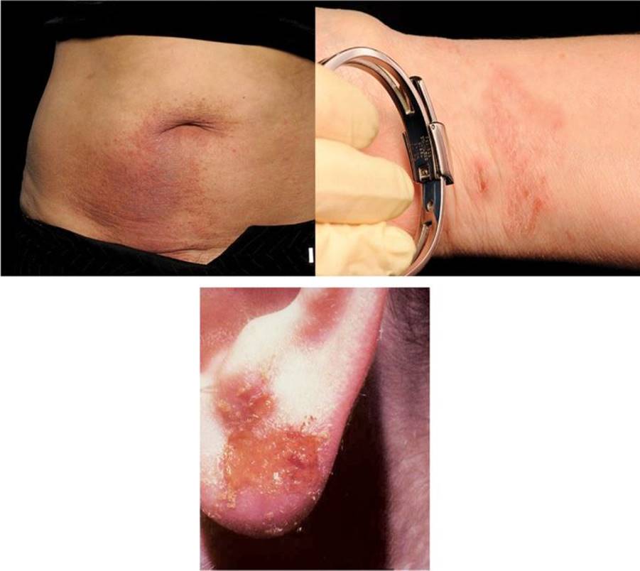
Sensitization requires at least 4 days to develop. Many workers, however, contact an allergen repeatedly in their work for months and even years before developing clinical sensitivity. The precipitating cause of sensitization can be a minor episode of irritant dermatitis or even increased frequency of contact with greater pressure and sweating at the site. After poison oak, nickel is the most common cause of contact dermatitis (Figure 21–6).

![]() Figure 21–6. Nickel ACD resulting from a metal belt buckle, a bracelet, and an ear ring. (Source: Cardiff & Vale NHS Trust, Cardiff, UK.) (See Color Plate 3.)
Figure 21–6. Nickel ACD resulting from a metal belt buckle, a bracelet, and an ear ring. (Source: Cardiff & Vale NHS Trust, Cardiff, UK.) (See Color Plate 3.)
The dermatitis originates at the site of contact with the allergen, but new lesions may appear at distant, seemingly unrelated sites, usually because of inadvertent transfer of the allergen by the hands.
A subacute and chronic stage can evolve. It is characterized by skin thickening, dryness, and fissuring.
B. Patch Testing
The key to diagnosis of allergic contact dermatitis is diagnostic patch testing. The opportunity to select the site of application and the ability to use only a minute concentration of test substance, confining it to a small area, are important features. The organ tested is the same as that affected by the disease and the same mechanism for production of the disease is used; hence, the patch test remains one of the most direct and valuable of all methods of medical testing.
Standardized procedures in patch testing are important, especially the concentration of the allergen and the type and characteristics of the vehicle. During recent decades, attempts to standardize patch testing have occurred.
Two methods are currently in use worldwide. The older method is the Finn chamber, which employs an aluminum cup, 8 mm in diameter, fixed to a strip of Scanpor tape, a finely meshed paper tape with a polyacrylate adhesive. The allergens are applied to the cups, covering more than half the diameter of each cup, and fixed to the skin with Scanpor tape. A newer method, the T.R.U.E. test, manufactured in Denmark, is a convenient, ready-to-use strip of tape on which a measured amount of allergen is incorporated in a thin hydrophilic gel film printed on a polyester patch measuring 9 × 9 mm. The patches contain 24 different allergens, are mounted on strips of acrylic tape protected by a plastic sheet, and are packaged in airtight envelopes. The thin sheet of plastic is removed, and the strips are placed on the skin. On contact with skin moisture, the dry film dissolves into the gel, and the allergen is released onto the skin. This method permits rapid application and avoids the hazard of mistakes in preparation of the application. The T.R.U.E. test system was not designed for occupational use and is now out of date in terms of current knowledge.
The upper back is the favored site for patch testing. Any hair must be removed using an electric rather than a safety razor to minimize damage to the keratin layer. The patches are left on the skin for 48 hours and then removed, and the sites are identified with a fluorescent-inked pen. Reading is done at 72 or 96 hours after application and occasionally at 1 week. When a fluorescent pen has been used to delineate the allergens, a hand-held black light will identify the sites. A single reading at 48 hours misses approximately 35% of positive results. Table 21–6 lists patch test interpretation codes.
Table 21–6. Patch test interpretation codes.

Clinical interpretation is the most difficult aspect of patch testing. Irritant reactions show varied patterns such as fine wrinkling, erythematous follicular papules, petechiae, pustules, and sometimes large bullae. A classic positive patch test reaction consists of erythema, mild oedema, and small, closely set vesicles.
Table 21–7 describes the allergens present in the T.R.U.E. test and additional allergens for detecting vehicle and preservative allergy. Table 21–8 lists other additional occupational series available for patch testing.
Table 21–7. Allergens tested in various standard series: Main uses (European, International, British).

Table 21–8. Additional occupational series for patch testing.
Hairdressing
Bakery
Dental
Epoxy
Fragrance
Isocyanate
Oils and cooling fluid
Methacrylates: dental, nails, printers
Photographic chemicals
Plant
Plastics and glues
Rubber additives
Textile colors and finish
Adverse reactions can occur but are rare. The most common are increased pigmentation at the site of a positive reaction, persistence of a reaction (especially with a positive reaction to gold), mild flare of the original dermatitis with brisk reactions, the development of psoriasis in a positive test site (rare), active sensitization (very rare), and anaphylactoid reactions (exceedingly rare).
Keep in mind that the test is a template of allergic contact sensitization developed over a person’s lifetime. Therefore, clinical relevance of each positive reaction must be determined. This can be accomplished only with extensive knowledge of commercial and industrial materials and their ingredients. Information can be obtained from numerous sources, including standard textbooks, manufacturers, and material safety data sheets. A review of the patient’s clinical history, a workplace visit, chemical analysis of other allergens or cross-reacting substances, and further patch testing may be required (Figure 21–7).

![]() Figure 21–7. Patch test to European standard battery and facial series with positive reactions. (Source: Cardiff & Vale NHS Trust, Cardiff, UK.) (See Color Plate 4.)
Figure 21–7. Patch test to European standard battery and facial series with positive reactions. (Source: Cardiff & Vale NHS Trust, Cardiff, UK.) (See Color Plate 4.)
Other special tests
• Photopatch test
• Skin prick test
• Spot test, for example, nickel, epoxy resin, etc
• Provocative use test (PUT) or a repeat open application test (ROAT)
SPECIFIC TYPES OF ALLERGIC CONTACT DERMATITIS
![]() Epoxy Resin Dermatitis
Epoxy Resin Dermatitis
Epoxy resins are used commonly as adhesives and can be found in paints, cement, and electrical insulation. Most epoxy resins are based on diglycidyl ether of bisphenol A. Epichlorohydrin combined with bisphenol A produce an epoxy resin of varying molecular weights from 340 to larger polymers, which are less sensitizing. However, there are other potential allergens, including pigments, fillers, reactive diluents, and solvents, that are mixed with a curing or hardening agent to polymerize the resin. Once hardened, the sensitizing potential is reduced.
Patch testing to epoxy resins must be thorough because there may be unknown compounds, and testing with the patient’s own resins is essential. Irritant reactions and sensitization on patch testing may occur, particularly to the amine epoxy hardeners. Facial dermatitis may suggest allergy to hardener rather than to the epoxy resin itself because the latter has low volatility. Detection of epoxy resin can be undertaken by a spot test with sulfuric acid or thin-layer chromatography.
Epoxy resin dermatitis can be prevented with exclusion or low concentrations of molecular weight 340 and 624 epoxy oligomers, high-molecular-weight (>1000) reactive diluents, and hardeners that exclude aliphatic amines.
![]() Photoallergic Reactions
Photoallergic Reactions
Photoallergic reactions are immunologically based. They are more uncommon than phototoxic reactions and develop only in individuals previously sensitized by simultaneous exposure to a photosensitizing chemical and appropriate UV radiation. The biologic process is similar to ACD, except that UV converts the chemical to a complete allergen. The radiation is usually in the UVA spectrum, although it may extend into the UVB.
Photoallergic reactions appear suddenly with an acute eczematous eruption, later becoming lichenoid and thickened, on the face, neck, dorsum of the hands, and exposed arms, often extending to other areas. The diagnosis is suggested by the distribution and character of the eruption, but confirmation requires careful questioning and photopatch testing. Sparing of skin under the chin and upper eyelids is strongly suggestive of a photo eruption (Figure 21–8). Table 21–9 lists some causes of photoallergic reactions.

![]() Figure 21–8. Photoallergic reaction. (Source: Cardiff & Vale NHS Trust, Cardiff, UK.) (See Color Plate 5.)
Figure 21–8. Photoallergic reaction. (Source: Cardiff & Vale NHS Trust, Cardiff, UK.) (See Color Plate 5.)
Table 21–9. Causes of photoallergic reactions
Halogenated salicylanilides
Tetrachlorosalicylanilide
3,4,5-tribromosalicylanilide
4,5-dibromosalicylanilide
Phenothiazines: Chlorpromazine, promethazine
Fragrances: Musk ambrette
Optical brighteners (stilbenes)
Sunscreens: PABA esters, Benzophenone 3, butyl methoxydibenzoylmethane Compositaeplants
![]() Operational Definition of Occupational ACD
Operational Definition of Occupational ACD
There are many steps in the full assessment and operational definition or final diagnosis of occupational ACD. A history of occupational exposure and a definite time relationship between exposure and onset of dermatitis is essential. Other factors required are a consistent morphology of the dermatitis and positive diagnostic testing with appropriate vehicle and concentration such as patch testing. Clinical relevance needs to be defined, and this may require a provocative use test (PUT) or a repeat open application test (ROAT) with the suspected allergens. This involves application of the substance onto the inner forearm twice daily for 7–28 days until a red, itchy patch appears, confirming ACD. Serial dilutions of the chemicals tested may be needed to confirm initial findings and suspicions. Control subject testing is essential to confirm nonirritating concentrations. Finally, clearing of the dermatitis once the allergen is removed or exposure is reduced significantly provides further information regarding the relevance of the allergen.
![]() Differential Diagnosis
Differential Diagnosis
• Irritant contact dermatitis
• Atopic dermatitis, psoriasis
• Pustular eruptions of the palms and soles (palmoplantar pustulosis)
• Herpes simplex and zoster
• Idiopathic vesicular reactions secondary to Trichophyton infections of the feet
• Dyshidrotic and nummular eczemas
• Drug eruptions
![]() Prevention of Contact Dermatitis
Prevention of Contact Dermatitis
In addition to the above mentioned treatment strategies, other measures to lower the incidence of contact dermatitis in the workplace include
• Identification of potential irritants and allergens in the workplace
• Chemical substitution or removal to prevent recurrence
• Personal protective measures
• Personal and environmental hygiene
• Education to promote awareness of potential irritants and allergens both at work and at home
• Pre-employment and periodic health screening and
• Engineering controls with automated, closed systems
![]() Treatment of Contact Dermatitis
Treatment of Contact Dermatitis
Treatment of contact dermatitis depends on the stage of the disease. Acute vesicular eruptions are treated with wet dressings for the first 24–36 hours using Burow’s solution or potassium permanganate KMNO4 solutions, followed by a topical corticosteroid; only the most potent topical corticoids (classes 1 and 2) are effective in the acute phase. In addition, based on current evidence, cold compresses have been shown to decrease inflammation in contact dermatitis.
A. Corticosteroids
When the eruption begins to dry, corticosteroid creams can be used, accompanied by oral sedating antihistamines for itching. Oral antibiotic therapy is indicated only when secondary infection is suspected. Topical antibiotic and anti-histamine preparations should be avoided, however, because of risk of sensitization. High-potency topical corticosteroids decrease mild to moderate, but not severe, ACD. Topical corticosteroids are possibly not significantly effective with some irritants such as sodium lauryl sulphate. There are no controlled studies, but oral corticosteroids are effective in severe ACD.
B. Skin Cleaners
These should be readily available and designed for the use intended, for example, heavy-duty cleansers for mechanics and others working with grease and oils and mild bar or liquid soaps for workers in less dirty occupations. Industrial cleansers often contain harsh abrasives and potentially allergenic antibacterial agents.
Waterless hand cleaners remove industrial dirt without water and can be of value in work sites without convenient washing facilities. Most are based on relatively nonirritating detergents and are removed from the skin with towels, waste papers, or rags. When used repeatedly, rags may contain a large number of irritants from the work site.
C. Protective Clothing and Gloves
Protective clothing is available for most work situations and exposures. It must be selected with specific consideration of the type of work and exposure and must be inspected regularly for holes and tears. Remember that certain allergens, such as methyl and ethyl methacrylate, glyceryl monothioglycolate, and paraphenylenediamine, pass readily through rubber gloves. Workers may wear gloves to protect an active dermatitis, but the occlusion can aggravate an existing eruption, and contact with rubber can lead to allergic sensitization to ingredients of the gloves.
D. Barrier Creams
Barrier creams are popularly termed “invisible gloves.” Although the benefit of this physical barrier to penetration is debated widely, barrier creams have reduced allergic and irritant contact dermatitis in both experimental and clinical studies. Barrier creams should be applied to intact skin only and prior to contact with irritants, including application after breaks. High frequency of application with adequate amounts is essential. Barrier creams may induce irritant or allergic contact dermatitis caused by various preservatives, lanolin, and fragrances. Workers should not become lax in other protective measures because of this “invisible glove” provides a sense of false security.
E. Emollients
Emollients and moisturizers are designed to increase the water content of the skin and can be used on irritated skin. They play an important role in treating and preventing irritant contact dermatitis, but further assessment is required in both animal and human models in the workplace.
![]() Complications of Contact Dermatitis
Complications of Contact Dermatitis
A. Disease Complications
• Lichen simplex chronicus
• Contractures, for example, severe hydrofluoric acid burns
• Loss of job/income
• Career change
• Psychosocial problems
B. Treatment Complications
• Topical steroids
• Atrophy
• Hypopigmentation
• Systemic steroids
May cause several side effects such as acne, osteoporosis, weight gain, hypertension, etc. Bone protection measures such as intake of bisphosphonates is recommended in any patients especially elderly taking steroids for more than 2 months. Also, prolonged use absorption of topical steroid may result in systemic effects.
Other systemic medications: Cyclosporine, azathioprine, mycophenolate mofetil, methotrexate may cause wide range of side effects and need to be closely monitored.
![]() Prognosis
Prognosis
Allergic contact dermatitis may wax and wane despite treatment especially if the allergens are not identified or the skin protective measures are not strictly adhered to. ACD to chromium (less common than ICD) appears to be persistent in the affected individuals despite appropriate treatment and rigorous skin protection. Lichen simplex chronicus is sequelae of repeated scratching in the affected body parts. Unchanged work practices, age more than 45 years, food-related occupations, respiratory atopy, and male sex are considered to be the risk factors for continuing occupational contact dermatitis. Discontinuation of the causative agents’ exposure leads to clinical improvement and healing. A change in work activities, modification of work environment and the presence of easily avoidable work-related allergies are associated with a good prognosis.
CONTACT URTICARIA
![]() General Considerations
General Considerations
Contact urticaria develops within minutes to an hour following contact with a substance. Interest in and knowledge of this reaction have increased greatly during the past 25 years, particularly with natural rubber latex allergy.
![]() Types of Contact Urticaria
Types of Contact Urticaria
A. Nonimmunologic (Nonallergic) Contact Urticaria
With sufficient provocation, nearly all exposed individuals will develop a reaction. Previous sensitization is not necessary. Gardeners may develop reactions from contact with nettles and other plants, caterpillar hair, moths, and other insects; cooks from cinnamic acid and aldehyde, sodium benzoate, sorbic acid, fruits, vegetables, fish, and meat; and medical personnel from alcohols, balsam of Peru, and dimethyl sulfoxide.
B. Immunologic (Allergic) Contact Urticaria
Immunologic (allergic) contact urticaria is caused most commonly by latex in natural rubber, especially gloves, which is a problem for medical and dental personnel, kitchen and dairy workers, pharmacists, semiconductor workers, and others who must wear gloves throughout the workday. The reactions range from mild erythema with itching at the site of contact to severe anaphylactic reactions, sometimes resulting in death. They are immunoglobulin E (IgE)–mediated type I immediate hypersensitivity reactions and appear to be more common in atopics. The cause is natural latex from the sap of the tree Hevea brasiliensis, a cis- 1,4-polyisoprene, the precursor of the rubber molecule. It is estimated that there are 50 or 60 different proteins in latex that provoke the allergic response.
![]() Clinical Findings
Clinical Findings
Signs and symptoms:
• Onset within 10–60 minutes of contact and when mild, disappear without treatment within 2–3 hours
• Itching
• Redness
• Wheal-and-flare reaction
Severe reactions progress rapidly and include generalized urticaria, swelling of the face and lips, asthma, collapse, and death.
Natural rubber latex gloves most commonly cause these reactions, but condoms, urinary catheters, elastic bandages, adhesive tapes, wound drains, dental dams, hemodialysis equipment, balloons, pacifiers, barium enema tips, and many other latex-based rubber products are implicated. Cross-reactions can occur to foods such as avocados, water chestnuts, kiwi, papaya, and bananas, provoking reactions in sensitive persons. Dermatographism, a common form of urticaria, occurs when the skin becomes raised and inflamed when stroked, scratched, or rubbed (Figure 21–9). Airborne contamination by rubber glove powder also may induce symptoms in very sensitive patients.

![]() Figure 21–9. Dermatographism in a patient with contact urticaria. (Source: Cardiff & Vale NHS Trust, Cardiff, UK.) (See Color Plate 6.)
Figure 21–9. Dermatographism in a patient with contact urticaria. (Source: Cardiff & Vale NHS Trust, Cardiff, UK.) (See Color Plate 6.)
Prick and Open Testing
Open testing on intact skin and skin prick testing are the most common diagnostic methods for this condition. A standardized test material should be used, and testing should be performed only if resuscitation measures are readily available. “Use tests” with a glove or a single finger of a glove should be performed with special care in patients who have a history of anaphylaxis or when the results of skin prick test or the latex radioallergosorbent test (RAST; Pharmacia, Sweden) are positive. Note that the RAST is only 60–65% sensitive.
The Food and Drug Administration (FDA) prohibits the labeling of latex-containing medical products as “hypoallergenic” and requires the statement: “This product contains natural rubber latex” on all latex-containing products that are directly or indirectly in contact with the body.
![]() Differential Diagnosis
Differential Diagnosis
• Acquired angioedema
• Allergic contact dermatitis
• Irritant contact dermatitis
• Other forms of urticaria: cholinergic, pressure, vasculitic, and solar
![]() Prognosis
Prognosis
The long-term prognosis is generally good if proper precautions are taken and by avoiding the causative and precipitating factors. These can be achieved through continuous education of the individuals and organizations involved.
Generally, fewer compounds produce immune mediated contact urticaria compared to the non-immune mediated type. In an occupational setting, if these compounds are not rigorously sought early, it may lead to eczematous skin changes resulting from the allergic contact dermatitis. This in turn may cause debilitating chronic hand dermatitis. It is advisable to perform extensive patch testing as the allergen(s) may be missed using the standard battery.
Extracutaneous manifestations of contact urticaria include rhinitis, conjunctivitis, dyspnea, and anaphylaxis.
OCCUPATIONAL ACNE
![]() Oil Acne (Folliculitis)
Oil Acne (Folliculitis)
Oil acne, or oil folliculitis, is a common condition resulting from heavy exposure to oil, especially under oil-soaked clothing. The arms and thighs usually are affected with numerous, often black comedones, pustules, furuncles, and sometimes carbuncles. This condition was once very common, especially in oil fields and refineries, but with improved engineering and less heavy contact with oils, it is seen much less often today. Many cases are never reported because most workers know that with better hygiene the condition improves. The most common sources are insoluble cutting oils in machinists and greases and lubricating oils in mechanics. Melanosis and photosensitivity also occur. Workers handling heavy tar distillates and coal tar pitch, roofers, oil well drillers, coke oven workers, petroleum refiners, rubber workers, textile mill workers, and road pavers are affected commonly.
Another form of environmental acne is acne cosmetica, occurring in actors and cosmetologists. Acne mechanica secondary to local pressure, friction, rubbing, squeezing, and stretching can occur in the wearers of heavy clothing and helmets. Tropical acne is common in hot, moist climates. During World War II, thousands of military personnel were evacuated from the South Pacific because of this condition. The so-called McDonald’s acne results from contact with the grease and fat of frying hamburgers (see Table 21–2). Non-occupational sources of environmental acne also should be considered, including acne from medications such as corticosteroids, testosterone, progesterone, isoniazid, iodides, and bromides.
Treatment of oil folliculitis consists of oil-impervious aprons and environmental measures to limit exposure. Gloves usually cannot be worn by machinists and mechanics because of the danger of catching them in the machinery. Modernization of cutting machines with automation and special guards decreases skin contact.
![]() Chloracne
Chloracne
Chloracne is a rare condition with multiple closed comedones and pale-yellow cysts on the skin from cutaneous and systemic exposure to certain halogenated chemicals (see Table 21–3). Body areas affected are the cheeks, forehead, and neck. The shoulders, chest, back, buttocks, and abdomen also may be involved. The genitalia are especially affected, whereas the nose often is spared, except in systemic exposure. In addition, there may be hypertrichosis, hyper-pigmentation, and increased skin fragility suggesting porphyria cutanea tarda. Conjunctivitis, swelling, and discharge from swollen meibomian glands of the eyelids can be seen, as well as a brownish pigmentation of the nails. Peripheral neuritis and hepatotoxicity may occur, suggesting systemic toxicity.
Although treatment of chloracne is often unsatisfactory, oral antibiotics, oral isotretinoin, acne surgery, and occasionally dermabrasion may be helpful. The majority of cases clear within 1–2 years following cessation of exposure.
OCCUPATIONAL SKIN CANCER
Approximately 400,000 new cases of nonmelanoma skin cancer occur in the United States each year, comprising approximately 30–40% of all cancers reported annually. Malignant melanoma accounts for another 18,000 cases. The exact number of skin cancers induced by the workplace is disputed, but most observers agree that it is a significant proportion. The most common causes of skin cancers in the work environment are ultraviolet light, polycyclic aromatic hydrocarbons, arsenic, ionizing radiation, and trauma.
![]() Ultraviolet Light
Ultraviolet Light
Sunlight is the most common cause of skin cancer, but workers seldom consider sunlight from the workplace as contributing to their actinically damaged skin and skin cancer. The most common skin cancers are squamous cell and basal cell carcinomas. These are related to prolonged exposure to sunlight but also may be initiated by tar and oils, mechanical trauma, and burns. The primary carcinogenic action spectrum of sunlight is in the UVB range (290–320 nm), but UVC (100–290 nm) and UVA (320–400 nm) rays also are photo-carcinogenic. UVA rays accelerate UVB-induced malignancy, and even though UVC rays are not present in sunlight, there is exposure from welding arcs and germicidal lamps.
The evidence for the skin carcinogenicity of UVB and UVA is overwhelming. Such cancers occur much more frequently in outdoor workers and in persons with fair skin and light hair and eye color and in those who tan poorly and burn easily. In fact, there is a specific compensation scheme in the United Kingdom for war veterans who served in tropical countries and later developed skin cancers. Other professionals who are at risk of developing skin cancers as a result of chronic sun exposure include builders, farmers, horticulturists, etc. In addition to the time spent in sunlight, the ultraviolet radiation received by an outdoor worker depends on the latitude, season, time of day, altitude, and weather. Artificial sources of carcinogenic UV radiation include welding arcs; germicidal lamps; devices for curing and drying printing ink, plastics, and paint; UV lasers; mercury vapor lamps; and medical UV therapy machines. Radiometers are available that can measure the amount of UV radiation a worker is receiving.
Epidemiologic studies in countries where there is a large blond, fair-skinned population, as in Australia, show a higher incidence of melanomas of the head, face, and neck in outdoor workers, which contrasts with office workers, who have melanomas more commonly on the covered parts of the trunk and limbs. Lentigo maligna is almost always present on exposed, sun-damaged skin and becomes invasive after a variable period of time. Persons with xeroderma pigmentosa, a hereditary disease, are extremely sensitive to the carcinogenic effects of sunlight. A frequent cause of death in these individuals is malignant melanoma, often occurring at a young age.
Polycyclic Aromatic Hydrocarbons
For 250 years, coal tar products and certain petroleum oils were considered potential causes of cutaneous cancers in individuals who work in certain industries. In the twentieth century, the relationship became firmly established not only from experimental animal studies but also from numerous epidemiologic surveys. Polycyclic aromatic hydrocarbons, such as those found in soot and carbon black, coal tar, pitch and tarry products, creosote oil, and certain oils, account for the majority of cutaneous tumors. Photosensitization develops initially, with recurring erythema and intense burning of the exposed skin. After repeated episodes, poikilodermatous changes appear, especially on the exposed skin of the face, neck, and hands. Keratotic papillomas (tar warts) then develop, which later may become squamous cell carcinomas, basal cell carcinomas, and keratoacanthomas. Polycyclic aromatic hydrocarbons and UVB appear to act synergistically to induce malignant change.
Arsenic
Since the late 1940s, epidemiologic studies have strongly linked inorganic arsenic exposure to squamous cell cancers of skin and lungs. Arsenic keratoses, characteristic of chronic arsenicalism, are multiple yellow, punctate keratoses distributed symmetrically on the palms and soles. Squamous cell carcinomas and multiple lesions of intraepidermal squamous cell carcinoma (Bowen disease) may develop from these keratoses. Basal cell carcinomas also occur from arsenic exposure, and they are often multiple, superficial, and pigmented.
Occupational arsenic exposure occurs in ceramic enamel workers, copper smelters, fireworks makers, gold refiners, hide preservers, carpenters (removing old wallpaper), semiconductor workers, and taxidermists. Arsenic is rarely used as an insecticide today but is still employed as a rodenticide.
OTHER CAUSES OF OCCUPATIONAL SKIN DISORDERS
Biologic Causes
![]() Bacterial Diseases
Bacterial Diseases
A. Staphylococcal and Streptococcal Infections
Infection of minor lacerations, abrasions, burns, and puncture wounds accounts for most staphylococcal and streptococcal infections. A work relationship is not always easy to establish, however, and many cases are unreported. Nevertheless, these infections are common in certain occupations, especially agricultural and construction workers, butchers, meat packers, and slaughterhouse workers. The history should clarify whether a work relationship is likely, although frequently in workers’ compensation cases the patient’s statements must be accepted as valid.
Furunculosis is common among automobile and truck repair persons, especially in dirty jobs, such as tire repair. Paronychia may be seen in occupations such as nurses, hairdressers, and manicurists.
Atopic dermatitis patients are especially likely to experience skin colonization with staphylococci. In a high percentage of atopics, Staphylococcus aureus can be cultured from their eczematous skin, which often has been made worse by heavy and prolonged application of corticosteroid creams and ointments. Prophylactic oral antibiotics should be part of the long-term treatment of these patients. Employment of persons with active atopic dermatitis in food service industries and hospital patient care may need to be restricted.
B. Cutaneous Mycobacterial Infections
Infection with tubercle bacilli is covered in Chapter 20. A classic example of tuberculosis of the skin acquired through inoculation of Mycobacterium tuberculosis hominis is seen in pathologists (prosector’s wart) and morgue attendants (necrogenic wart or anatomic tubercle). Surgeons are also at risk for such granulomatous infections. Veterinarians, farmers, and butchers may acquire infection with M tuberculosis var. bovis, which at one time was a common cause of disease in livestock in the United States, but bovine tuberculosis has declined since the mid 1930s. In some countries, however, the disease is still common. In the United States and other parts of the world, as a result of population movement and the increasing prevalence of human immunodeficiency virus (HIV) infection, the incidence of infection with human strains of tuberculosis has increased greatly. Between 1985 and 1991, 39,000 more cases occurred in the United States than expected, and drug resistance, especially in those with HIV infection, has seriously compounded the problem.
The typical skin lesions are slowly progressive, warty, hyperkeratotic plaques, which, if left untreated, eventually regress after many months or years, leaving disfiguring scars (Figure 21–10). Demonstration of organisms either directly or from cultures is often difficult.

![]() Figure 21–10. Atypical mycobacterium in a fish farmer. (Source: Cardiff & Vale NHS Trust, Cardiff, UK.) (See Color Plate 7.)
Figure 21–10. Atypical mycobacterium in a fish farmer. (Source: Cardiff & Vale NHS Trust, Cardiff, UK.) (See Color Plate 7.)
C. Atypical Mycobacterial Infections
Atypical mycobacterial infections are caused most commonly by infection with M marinum. This infection usually is acquired from exposure to infected fish, especially in aquariums and fish tanks by persons who clean these tanks. Swimming pools have become contaminated with this organism, and pool attendants and cleaners are also at risk. Treatment with rifampicin or ethambutol is usually effective.
As in other mycobacterial skin infections, the clinical picture consists of granulomatous papules and nodules that ulcerate and exude a clear, thin serum. Sometimes a pattern resembling sporotrichosis develops, with nodules and papules ascending the arm (or leg) along the course of regional lymphatics. Persons with AIDS are at special risk for developing these infections. Other atypical mycobacteria include M ulcerans, M fortuitum, M avium, M intracellulare, M kansasii, and M chelonae.
![]() Viral Diseases
Viral Diseases
A. Herpes Simplex
This is the most frequent viral infection of occupational origin, affecting dentists and dental assistants, physicians and nurses, and respiratory technicians. This is caused by the herpes simplex virus (HSV). Transmission is by contaminated saliva or pharyngeal or laryngotracheal secretions. Wearing disposable gloves, masks, and safety glasses reduces the risk of infection in these workers.
B. Viral Warts
Meat handlers, especially butchers and slaughterhouse workers, are at greatest risk for development of the common wart, caused by the human papilloma virus (HPV), of which there are at least 35 types. These warts are most numerous on the hands and fingers of these workers, and minor cuts and abrasions inoculate the virus. Molluscum contagiosum occurs in wrestlers, boxers, and other sportsmen.
C. Orf
Endemic in sheep and goats, orf is caused by infection with a parapox virus, usually involving the mouth and nose of infected animals. Mostly farmers and veterinarians are affected with this relatively mild, self-limited disease. Only one or two lesions may be present, almost always on fingers, and are associated with mild fever, lymphangitis, and regional lymphadenopathy. An erythema multiforme–like rash occurs 10–14 days after onset. Treatment is symptomatic, with antibiotics given only for complications such as secondary infection.
![]() Fungal Infections
Fungal Infections
A. Candida
Infection with Candida, mainly Candida albicans, is the most common occupationally related fungal disease. The organism is ubiquitous, and proliferation is favored by moisture, occlusion, and irritation. Most occupationally acquired candidal infections are on the hands, especially in the paronychial areas and interdigital spaces. Occupations in which prolonged wearing of rubber gloves is required, such as dentistry, medicine, and technical work in clean rooms in the semiconductor industry, show the highest incidence of this condition. Diabetics and neutropenic, immunocompromised patients are especially at risk.
B. Dermatophytes
Dermatophytic infections are common. Trichophyton verrucosum is an animal fungus that readily infects farmers and cattle tenders. The lesions are often quite inflammatory and may resemble pyoderma (Figure 21–11). Farmers, milkers, cattle tenders, veterinarians, and tannery workers, especially hide sorters, are at risk. T rubrum and T mentagrophytes are examples of fungi that cause tinea infections in the general population, especially tinea manuum and tinea pedis. Microsporum canis frequently infects small animals and causes infection in pet shop workers, veterinarians, and personnel in contact with laboratory animals. M gypseum is a rare fungus found in soil, causing occasional infection in agricultural workers.

![]() Figure 21–11. Dermatophytes infection: Kerion (Trichophyton verrucosum) in a sheep farmer. (Source: Cardiff & Vale NHS Trust, Cardiff, UK.) (See Color Plate 8.)
Figure 21–11. Dermatophytes infection: Kerion (Trichophyton verrucosum) in a sheep farmer. (Source: Cardiff & Vale NHS Trust, Cardiff, UK.) (See Color Plate 8.)
Physicians are often requested to decide whether a Trichophyton infection is work-related, especially T rubrum and T mentagrophytes infections of the hands and nails. Onychomycosis is extremely common, and most of those affected do not seek medical attention. Workers engaged in repetitive hand activities, especially where there is sweating and pressure or repetitive nail trauma in the case of onychomycosis, may believe their work to be the primary cause of the infection. Each case must be studied individually, but most often the work cannot be considered a primary cause.

![]() Plate 1. Subacute effects of ICD on the palm, erythema, edema, and scaling. (Source: Cardiff & Vale NHS Trust, Cardiff, UK.) (See Figure 21–3.)
Plate 1. Subacute effects of ICD on the palm, erythema, edema, and scaling. (Source: Cardiff & Vale NHS Trust, Cardiff, UK.) (See Figure 21–3.)

![]() Plate 2. Hypopigmentation on hands. (Source: Cardiff & Vale NHS Trust, Cardiff, UK.) (See Figure 21–4.)
Plate 2. Hypopigmentation on hands. (Source: Cardiff & Vale NHS Trust, Cardiff, UK.) (See Figure 21–4.)
![]() Plate 3. Nickel ACD resulting from a metal belt buckle, a bracelet, and an ear ring. (Source: Cardiff & Vale NHS Trust, Cardiff, UK.) (See Figure 21–6.)
Plate 3. Nickel ACD resulting from a metal belt buckle, a bracelet, and an ear ring. (Source: Cardiff & Vale NHS Trust, Cardiff, UK.) (See Figure 21–6.)

![]() Plate 4. Patch test to European standard battery and facial series with positive reactions. (Source: Cardiff & Vale NHS Trust, Cardiff, UK.) (See Figure 21–7.)
Plate 4. Patch test to European standard battery and facial series with positive reactions. (Source: Cardiff & Vale NHS Trust, Cardiff, UK.) (See Figure 21–7.)

![]() Plate 5. Photoallergic reaction. (Source: Cardiff & Vale NHS Trust, Cardiff, UK.) (See Figure 21–8.)
Plate 5. Photoallergic reaction. (Source: Cardiff & Vale NHS Trust, Cardiff, UK.) (See Figure 21–8.)

![]() Plate 6. Dermatographism in a patient with contact urticaria. (Source: Cardiff & Vale NHS Trust, Cardiff, UK.) (See Figure 21–9.)
Plate 6. Dermatographism in a patient with contact urticaria. (Source: Cardiff & Vale NHS Trust, Cardiff, UK.) (See Figure 21–9.)

![]() Plate 7. Atypical mycobacterium in a fish farmer. (Source: Cardiff & Vale NHS Trust, Cardiff, UK.) (See Figure 21–10.)
Plate 7. Atypical mycobacterium in a fish farmer. (Source: Cardiff & Vale NHS Trust, Cardiff, UK.) (See Figure 21–10.)

![]() Plate 8. Dermatophytes infection: Kerion (Trichophyton verrucosum) in a sheep farmer. (Source: Cardiff & Vale NHS Trust, Cardiff, UK.) (See Figure 21–11.)
Plate 8. Dermatophytes infection: Kerion (Trichophyton verrucosum) in a sheep farmer. (Source: Cardiff & Vale NHS Trust, Cardiff, UK.) (See Figure 21–11.)

![]() Plate 9. Cutaneous leishmaniasis. (Source: Cardiff & Vale NHS Trust, Cardiff, UK.) (See Figure 21–12.)
Plate 9. Cutaneous leishmaniasis. (Source: Cardiff & Vale NHS Trust, Cardiff, UK.) (See Figure 21–12.)
![]() Parasitic Diseases
Parasitic Diseases
A. Protozoa
Cutaneous leishmaniasis—Most parasitic diseases, such as amoebiasis, giardiasis, and malaria, present with general rather than cutaneous health problems. An exception is cutaneous leishmaniasis, caused by Leishmania tropica(Oriental sore, bouton d’orient), found in the Middle East and L braziliensis (American leishmaniasis, uta), found in Central and South America. The disease is transmitted by sandflies that thrive in warm climates and is endemic in persons working in tropical forests in southeastern Mexico, Colombia, and Venezuela. The disease manifests as cutaneous ulcers with metastatic mucocutaneous lesions known as espundia (Figure 21–12). Pentavalent antimonials, such as sodium stibogluconate, are the treatment of choice. Pentamidine and liposomal or conventional amphotericin are alternatives.

![]() Figure 21–12. Cutaneous leishmaniasis. (Source: Cardiff & Vale NHS Trust, Cardiff, UK.) (See Color Plate 9.)
Figure 21–12. Cutaneous leishmaniasis. (Source: Cardiff & Vale NHS Trust, Cardiff, UK.) (See Color Plate 9.)
Helminths—Penetration of the cercariae of schistosomes into the papillary dermis induces a highly pruritic papular eruption termed swimmer’s itch. Urticaria may accompany the rash and be widespread. Migratory birds usually are the definitive hosts, with saltwater molluscs serving as intermediate hosts. The condition lasts for 2–3 weeks, often with secondary infection of excoriated lesions. Skin divers, lifeguards, dock workers, and workers who maintain lakes and ponds may be affected. Treatment is symptomatic.
Larva migrans (creeping eruption) occurs in subtropical and tropical regions where people work on moist soil infected with hookworm larvae. Dogs, cats, cattle, and human faeces carry the larvae, and humans are the final host. A threadlike, red or flesh-colored, circuitous, slightly raised line occurs often on the feet, legs, back, or buttocks caused by movement of the larva in the epidermis. Humans are infected with the larvae of Ancylostoma braziliense and Necator americanus, the ova of which are deposited in the soil. Topical application of 10% suspension of thiabendazole to affected areas four times daily for 7–10 days is usually curative. Agricultural workers, lifeguards, shoreline fishermen, ditch diggers, and sewer workers are at greatest risk.
Other nematode diseases that are occasionally occupational include trichinosis, dracunculosis, filariasis, loiasis, enterobiasis, strongyloidiasis, and toxocariasis.
Scabies—Epidemics of scabies have occurred in nursing homes, hospitals, and residential facilities for the aged. The disease is highly contagious and spreads rapidly, especially in the immunosuppressed. It is often initiated by an infected employee who transmits the mite to patients. They then spread the disease to other personnel. The scabicide of choice is permethrin, but treatment of the more severe types of scabies (eg, crusted scabies) can be difficult and may require repeated treatments with other scabicides such as lindane, permethrin, precipitated sulfur, and oral ivermectin.
Lyme disease—Lyme disease is an important inflammatory disease that follows tick-induced erythema chronicum migrans (ECM) weeks or months after inoculation. ECM begins with a small erythematous macule, usually on an extremity, that enlarges with central clearing. The lesion sometimes reaches a diameter of 50 cm, and smaller satellite lesions often are present. In nearly half the patients, a type of arthritis occurs within weeks or months of the ECM, and there may be associated neurologic abnormalities, as well as myocardial conduction alterations, serum cryoprecipitates, elevated serum immunoglobulin M (IgM) levels, and an increased sedimentation rate. Elevated serum IgM and later IgG appear within weeks of infection with circulating cryoprecipitates and other immune complexes. Erythema chronicum migrans is an important diagnostic marker for this disease. The ticks Ixodes dammini, I pacificus (in the United States), and I ricinus (in Europe) transmit the spiro-chete Borrelia burgdorferi that is responsible for the disease. In some cases, localized scleroderma appears to be linked to Borrelia infection. Tick bites are common in outdoor workers, loggers, wilderness construction workers, guides, and ranchers. Other major tick-borne diseases in the United States are relapsing fever, tularaemia, Rocky Mountain spotted fever, ehrlichiosis, Colorado tick fever, babesiosis, and tick paralysis.
Physical Causes
![]() Mechanical Trauma
Mechanical Trauma
Intermittent friction of low intensity will induce lichenification (thickening) of the skin. With greater pressure, corns and calluses appear. After minor trauma, calluses frequently develop painful fissures, which may become infected. After years of repeated frictional hand trauma during work, permanent calluses may result, leading to disability and early retirement. With increasing automation, less frequent manual operation of tools, and better protective clothing, occupational marks are less frequent and have almost disappeared from many industries.
![]() Heat
Heat
A. Burns
Burns arising from the occupation are common and exhibit characteristic occupational patterns. The resulting scarring and pigmentary changes are of chief concern to dermatologists, who rarely treat acute burns. Hypopigmentation is especially susceptible to actinic damage, and scars and the hyperpigmentation often are disfiguring.
B. Miliaria
Miliaria is caused by sweat retention and often is seen in the work environment. The eruption can be extensive, accompanied by burning and itching. The most superficial form, miliaria crystallina, is caused by poral closure and rupture of the ducts within the upper level of the epidermis. The condition commonly occurs on the palms and in intertriginous areas, with asymptomatic desquamation of the surface. When the closure occurs deeper in the epidermis, vesiculation with marked pruritus results. Miliaria rubra, or prickly heat, is the type most likely to be confused with contact dermatitis. If poral obstruction extends deeper in the epidermis and into the upper dermis, the condition is known as miliaria profunda, resulting in deep-seated, asymptomatic vesicles. This condition is caused by prolonged exposure to a hot environment and often follows an extended period of miliaria rubra. Heat exhaustion and collapse may be sequelae.
C. Intertrigo
A macerated, erythematous eruption in body folds, intertrigo results from excessive sweating, especially in obese workers. Secondary bacterial and candidal infections are common. The interdigital space between the third and fourth fingers is a common site in workers whose hands are continuously wet, especially from rubber gloves. Medical and dental personnel, bartenders, cannery workers, cooks, swimming instructors, and housekeepers are especially predisposed to this condition.
Overheating, especially in conjunction with physical exercise, may result in heat-induced urticaria and, rarely, in anaphylaxis. Acne vulgaris and rosacea are aggravated by prolonged exposure to heat, especially from ovens, steam, open furnaces, and heat torches. Herpes simplex may be triggered by intense heat, especially with sunburn and UVB exposure.
![]() Cold
Cold
A. Chilblains (Perniosis)
This mild form of cold injury, although an abnormal reaction to cold, is less common in very cold climates where homes are usually well heated and warm clothing is worn. The northern United States and Europe are areas where this condition is seen frequently. The lesions are reddish blue, swollen, boggy discolorations with bullae and ulcerations. The fingers, toes, heels, lower legs, nose, and ears are especially affected. Genetic factors with vasomotor instability often are found to be important background features. Treatment is symptomatic with calcium channel blockers such as nifedipine.
![]() Vibration Syndrome
Vibration Syndrome
Vibrations of hand-held tools and Raynaud phenomenon have been known to be associated since the early twentieth century. Popular names include dead fingers and white fingers; clinically, the condition is a type of Raynaud phenomenon. Operation of heavy vibrating tools such as jack-hammers, especially in cold weather, produces vasospasm of the digital arteries, causing episodic pallor, cyanosis, and erythema of fingers. Chain saws, hand-held grinders, riveting hammers, and other pneumatic tools also are associated with this condition. Tingling and numbness, blanching of the tips of one or more fingers, and clumsiness of the fingers and hands occur. The symptoms may be indistinguishable from other forms of Raynaud phenomenon, but asymmetry usually is observed. Occupational disability seldom results and most workers continue at their jobs. Vibration frequencies between 30 and 300 Hz are most likely to be responsible.
![]() Ionizing Radiation
Ionizing Radiation
Numerous industrial processes use ionizing radiation, including the curing of plastics, sterilization of food and drugs, testing of metals and other materials, medical and dental radiography, therapy with radioisotopes, and operation of high-powered electronic equipment. Exposure is much less now than it was several decades ago mainly as a consequence of better construction and shielding of the radiographic equipment. Measurements of radiation emissions from video display terminals have consistently shown nondetectable or background levels.
Occupational exposure to ionizing radiation may be acute or chronic and usually is localized. Acute radiodermatitis often results from a single accidental exposure to around 1000 R and presents with rapid onset of erythema, oedema, and blanching of the skin, reaching a peak at about 48 hours. Anorexia, nausea, vomiting, and other systemic symptoms also occur. There follows a latent period of apparent recovery lasting a few days, after which the skin again becomes erythematous, with purplish ecchymotic areas that become vesicular and bullous. Pain is intense, usually requiring narcotics. A repair stage follows, and as reepithelialization takes place, the skin becomes atrophic, hairless, and lacks functioning sebaceous glands. With large single doses, ulceration usually follows but often is delayed for 2–3 months. Healing is very slow, and an atrophic, disfiguring scar is left.
Chronic radiodermatitis results from exposures to smaller doses of ionizing radiation (300–800 R) received daily or weekly over a long period of time to a total dose of 5000–6000 R. The skin becomes red and eczematous with burning and hyperesthesia. Often the epidermis sloughs, and regrowth occurs slowly over a period of 4–6 weeks. Hair is also lost, often permanently, and the sebaceous glands cease activity. The skin becomes hypopigmented and atrophic with multiple telangiectasias. The systemic effects of irradiation are described in Chapter 14.
GENERAL APPROACH TO DIAGNOSIS & TREATMENT OF OCCUPATIONAL SKIN DISORDERS
The workup and diagnosis of patients with work-related skin disease requires much more time than does a general dermatologic workup. Making a premature diagnosis before studying all the evidence should be resisted because an incorrect diagnosis can have long-lasting and severely detrimental effects. Review of the medical records, patch testing, fungal and bacterial cultures, biopsy, and plant visits often are necessary to reach a correct diagnosis. Diagnosis of an endogenous or constitutional eczema or dermatitis as primary cause can be difficult for many workers to accept. Atopic eczema, although inherited, often has onset for the first time in adult life when precipitated by work activities, and aggravation often is considered work-related. Many other constitutional diseases can be considered similarly.
Table 21–10 outlines a typical evaluation of a work-related illness. The following headings can serve as a form for recording the results of the workup. The text under each heading details the information that should be gathered and recorded.
Table 21–10. Outline for dermatologic examinations for workers’ compensation patients.

![]() History of Injury & Current Complaints
History of Injury & Current Complaints
Learn exactly which anatomic skin site was first affected. With a diagnosis of contact dermatitis, the eruption should begin at the site of contact with the offending agents. Spreading then occurs, especially in the case of allergic sensitization. The date of the initial appearance of the dermatitis is important because often a change in workplace ergonomics, contact with new substances, or increased contact with long-used substances can precipitate dermatitis. Itching is important because irritant contact dermatitis, and especially allergic contact dermatitis, is almost always pruritic. If improvement occurs away from work and aggravation regularly takes place on resumption of the same work, a work relationship is almost always found, and workers’ compensation courts often will accept this, even without other evidence. Over-the-counter medicines and home remedies often contain contact allergens that sometimes can be the sole cause.
A. Occupational History
A description of the job as provided by the patient is often more accurate than the official job title. Often the worker has performed the same job for a long period of time before onset of dermatitis. This suggests a new process or contactant introduced into the workplace or home environment.
B. Prior Employment
The nature of previous jobs and dermatitis, as well as previous exposure to irritants and potential sensitizers, is important.
C. Nonwork Activities
The 40-hour workweek leaves sufficient opportunity for other part-time jobs, hobbies, and house and garden work.
D. Past Medical History
Although 15–20% of the population has a family or personal history of atopy, it is an often-overlooked cause of recurrent dermatitis, especially among hairdressers, kitchen helpers, medical and dental personnel, and automobile repair workers. Even persons with mild atopy may develop a major work-related hand dermatitis at the time of first employment, following repeated contact with irritants. Psoriasis also can be precipitated by trauma, especially repeated intense friction and pressure on the hands.
E. Family History
A family history of atopy is most important. Psoriasis (type 1) also may be a relevant family condition.
F. Hobbies/Habits
Hobbies and off-work activities should be explored during the history taking, including habitual traumatic activities such as picking and digging the skin, especially with wooden or metal articles used for scratching and rubbing.
G. Review Of Systems
A general review of body systems should be done.
![]() Review of Medical Records
Review of Medical Records
The medical records must be examined thoroughly to supplement the history as provided by the patient.
![]() Examination
Examination
Examination should not be limited to the part affected because the presence of dermatitis elsewhere and other skin conditions can change an initial impression. This is especially true when psoriasis, tinea infections, and lichen planus are found.
A. Special Investigations
Patch testing is the most important special investigation and should include not only suspected specific allergens but also a standard series of common allergens.
B. Diagnosis
The specific diagnosis should be recorded with an opinion regarding a work relationship.
C. Summary
This should be a brief summary of the findings with an explanation of the conclusions. Nonmedical terms should be used as much as possible.
Temporary and total disability—The disability status, total or partial, is described here. In most cases of hand dermatitis, the disability is temporary, but because of the manual nature of most work, total disability is also possible.
Permanent and stationary status—Once the dermatitis has reached a plateau and no further improvement is anticipated, permanent and stationary (P&S) status is reported. This does not mean, however, that treatment cannot be resumed should a recurrence cause a worsening of symptoms.
Objective findings—A brief review of the objective findings is recorded here.
Subjective findings—A review of the patient’s complaints and a description of any impairment are provided here.
Work restrictions—Work restrictions, if any, can be recorded here.
Loss of preinjury capacity—For purposes of permanent disability rating, one should describe any loss of preinjury capacity, such as may occur with contact allergy.
Causation and apportionment—If any aspect of the impairment is related to a previous employment or any preexisting disability, this is explained here, estimating the percentage of impairment associated with each.
Future medical treatment—An estimation of the type and duration of future medical treatment is given here.
Vocational rehabilitation—Once a permanent and stationary state is reached, vocational rehabilitation must be considered. It is important to offer guidance to vocational rehabilitation personnel in job selection for disabled workers.
Patch testing—The most important diagnostic test for occupational skin disease is the patch test. Since irritant and allergic dermatitis can be similar clinically, differentiation can be done only by patch testing, which not only will reveal the specific cause of a work-related dermatitis but also when negative after testing all possible allergens in the patient’s work will effectively rule out allergic contact dermatitis as a cause. Unfortunately, the test is often performed inadequately or incompletely, if at all. Patch testing should be done by experienced physicians according to accepted methods with nonirritating concentrations of test substances, preferably chemicals obtained commercially from manufacturers of patch test materials. Table 21–7 lists and describes common contact allergens.
Additional diagnostic tests—Fungal, bacterial, and viral smears and cultures, biopsies and photopatch testing. The prick testing, if contact urticaria is suspected, sometimes is required. Plant visits are an essential and integral part of the evaluation, often providing information vastly different from that learned during the patient’s evaluation.
![]() Prevention
Prevention
Prevention of occupational skin disorder is highly important as it reduces the risk of progression to more severe skin diseases such as hand eczema, disfigurement and even skin cancers. This requires close cooperation between the employee, employers, company physicians, dermatologists, and other relevant stakeholders such as workers unions.
In general, prevention of occupational skin disorders can be subdivided into three categories: primary, secondary, and tertiary preventions.
Primary Prevention
This involves identifying, avoiding and or replacing any potentially harmful substances by the organizations. In occupational skin disorders, it includes health risk analysis, giving employees appropriate information on the involved chemical substances and putting in place risk reduction strategies at organizational level.
Secondary Prevention
This applies to both the organizations and employees. Employers should make available alternative substances, skin protective measures and regularly update their health risk analysis. In addition, employees should ensure individual skin protection by wearing of protective gloves, application of protective skin ointments before working, adequate skin cleansing and skin care measures to support regeneration following work. A careful analysis of the harmful effects to the skin at the workplace is necessary for the appropriate choice of suitable skin protective measures.
Tertiary prevention
This is not commonly applied in occupational skin disorders. It involves cases where there is an explicit and impending reason to discontinue the current practice. In general, it applies to clinically severe and/or chronic occupational disease for which outpatient methods of secondary prevention have been inadequate.
![]() Treatment
Treatment
In many occupations, avoidance of irritants and allergens is not always possible. Prophylactic measures are necessary in industry to avoid the risk of developing irritant and allergic contact dermatitis. The specific treatment of occupational skin disease depends on the cause and does not differ from treatment of nonoccupational skin disease. Protective measures include moisturizers, barrier creams, and appropriate gloves and clothing. In many cases, a specific cause is not found, and recurrences may continue to affect the patient; hence, treatment with topical or oral corticosteroids often continues for prolonged period of time, leading to atrophy of skin and systemic complications. Although recovery may occur rapidly following treatment, the skin retains a non-specific hypersensitivity for several weeks, and hence work should not be resumed too early, even when the patient and/ or employer are pressuring the physician.
REFERENCES
CDC/NIOSH: Skin exposures and effects: www.cdc.gov/niosh/topics/skin/skinpg.html.
Diepgen TL: Occupational skin diseases. J Ger Soc Dermatol 2012;10:297 [PMID: 22455666].
Greenspoon J: Allergic and photoallergic contact dermatitis. Dermatitis 2013;24:29 [PMID: 23340396].
Holness DL. Recent advances in occupational dermatitis. Curr Opin Allergy Clin Immunol 2013;13:145 [PMID: 23324811]. Santoro FA: Update on photodermatoses. Semin Cutan Med Surg 2011;30:229 [PMID: 22123421].
Seto Y: Photosafety assessments on pirfenidone: photochemical, photobiological, and pharmacokinetic characterization. J Photochem Photobiol 2013;120:44 [PMID: 23419534].
Sparr E: Controlling the hydration of the skin though the application of occluding barrier creams. J R Soc Interface 2012;10:20120788 [PMID: 23269846].
Visser MJ: Impact of atopic dermatitis and loss-of-function mutations in the filaggrin gene on the development of occupational irritant contact dermatitis. Br J Dermatol 2013;168:326 [PMID: 23039796].
Warshaw EM: North American Contact Dermatitis Group Patch Test Results: 2009 to 2010. Dermatitis 2013;24:50 [PMID: 23474444].
![]() SELF-ASSESSMENT QUESTIONS
SELF-ASSESSMENT QUESTIONS
Select the one correct answer for each question.
Question 1: Irritant contact dermatitis (ICD)
a. accounts for nearly 80% of all occupational dermatitis
b. is caused by a single chemical
c. is unrelated to environmental factors
d. is not affected by ethnicity
Question 2: ICD
a. is an immunogenic skin reaction to toxic substances
b. is caused by a limited number of substances
c. has no predisposing factors
d. is caused by skin exposure to irritating toxic substances
Question 3: Phototoxic (Photoirritation) reactions
a. may result from exposure to natural or artificial light alone
b. can be caused by numerous systemic drugs
c. is unrelated to the dose or concentration of photo-toxic substance
d. may be followed by loss of pigmentation
Question 4: Occupational leukoderma
a. results in hyperpigmentation
b. occurs without inflammation
c. usually involves the hands and forearms
d. remains localized
Question 5: Allergic contact dermatitis (ACD)
a. is controlled by ordinary protective measures
b. seldom requires job change
c. does not prevent immediate return to work
d. is an immunologic reaction classified as a delayed type IV
Question 6: ACD
a. is less severe when following reexposure to the allergen
b. can evolve into subacute but not chronic stages
c. produces considerable variation in the intensity of reaction depending on the body area affected
d. usually affects the mucous membranes
Question 7: ACD
a. sensitization requires at least 4 days to develop
b. eventually affects all workers exposed to allergens
c. is unrelated to irritant dermatitis
d. is unrelated to epoxy resins, biocides, chromate, and formaldehyde
Question 8: Patch testing
a. is the key to diagnosis of allergic contact dermatitis
b. allows the use of a large concentration of test substance
c. tests a more resistant organ than that affected by the disease
d. tests a different mechanism than that which causes the disease
Question 9: Photoallergic reactions
a. are more common than phototoxic reactions
b. are immunologically based
c. appear slowly with an acute eczematous eruption
d. appear only on the face, neck, and dorsum of the hands
Question 10: Nonimmunologic (nonallergic) contact urticaria
a. affects only a few exposed individuals
b. requires no previous sensitization
c. spares gardeners
d. spares cooks and other food handlers
Question 11: Immunologic (allergic) contact urticaria
a. is caused most commonly by latex in natural rubber, but not rubber gloves
b. is limited to mild erythema with itching at the site of contact
c. reactions are immunoglobulin E (IgE)–mediated type I
d. is less common in atopics
Question 12: Chloracne
a. is a common occupational skin disorder
b. especially affects the nose
c. spares the conjunctivae
d. may lead to peripheral neuritis and hepatotoxicity, suggesting systemic toxicity
Question 13: Atopic dermatitis
a. protects workers from skin colonization with staphylococci
b. produces immunity through development of eczematous skin
c. requires prophylactic oral antibiotics
d. may restrict employment in food service industries and hospital patient care
Question 14: Atypical mycobacterial infections
a. are caused most commonly by infection with M chelonae
b. are acquired from exposure to infected fowl
c. are usually effectively treated with rifampicin or ethambutol
d. are not predisposed in persons with AIDS
Question 15: In the management of contact dermatitis
a. avoidance and protective measures remain the best approach
b. highly potent steroids are always required
c. water-based cleaners are recommended as skin cleansers
d. alcohol-based cleaners are recommended as skin cleansers
Question 16: Prevention of occupational skin disorders
a. is not necessary because the long-term outcome is always the same once exposure has occurred
b. should be limited to the affected employees to prevent stigmatization
c. requires close cooperation between the employee, employers, company physicians, dermatologists, and other relevant stakeholders such as workers unions
d. does not involve giving employees information on the involved chemical substances