
John R. Balmes, MD
The respiratory tract is often the site of injury from occupational exposures. The widespread use of potentially toxic materials in the environment poses a major threat to both the airways and lung parenchyma. The respiratory tract has a limited number of ways to respond to injury. Acute responses include rhinosinusitis, laryngitis, upper airway obstruction, bronchitis, bronchoconstriction, alveolitis, and pulmonary edema. Chronic responses include asthma, bronchitis, bronchiolitis, parenchymal fibrosis, pleural fibrosis, and cancer. Early recognition and appropriate treatment of occupational lung diseases by physicians can reduce both morbidity and mortality significantly and greatly affect patient outcome. This chapter focuses on common occupational lung diseases and on how to diagnose and manage them.
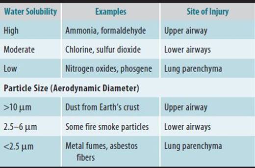
The site of deposition of inhaled materials depends on water solubility for gases and particle size for solids (Table 23–1). Water-soluble gases and particles with a diameter in excess of 10 μm tend to get deposited in the upper airways, whereas insoluble gases and smaller particles penetrate to the lower airways. Subsequent respiratory injury depends on both the site of toxin deposition and the type of cell/structure damaged.
Table 23–1. Site of respiratory tract deposition and effect.
EVALUATION OF PATIENTS WITH OCCUPATIONAL LUNG DISEASE
A careful evaluation can identify and diagnose occupational lung disease successfully in most cases. The following four approaches are recommended: (1) detailed history, including occupational and environmental exposures, (2) thorough physical examination, (3) appropriate imaging studies, and (4) pulmonary function testing.
 History
A detailed history of both the patient’s complaints and environmental/occupational exposures is essential. Work practices should be explored extensively with attention to types and durations of exposures, whether appropriate environmental controls are present, and if respiratory protective gear is used. If available, substance data sheets (SDSs) should be reviewed. These documents profile the important health, safety, and toxicologic properties of the product’s ingredients and under federal law must be furnished by the employer to the worker or to the worker’s health care provider on request.
If available, actual industrial hygiene data on the level of exposure and the agent to which the patient was exposed should be obtained. The history should include the condition of the patient’s home, any hobbies, and social habits because exposures outside the workplace that contribute to or cause the lung injury may be discovered.
Physical Examination
Occupational lung diseases do not present with specific clinical findings. It is difficult, for example, to distinguish asbestosis from idiopathic pulmonary fibrosis or chronic beryllium disease from sarcoidosis. Only in the context of the exposure history will the correct diagnosis be made. A physician who suspects the presence of an occupational lung disease should, nonetheless, perform a complete physical examination rather than focus narrowly on findings suggested by the exposure history. Relevant nonoccupational disease otherwise may be missed.
The physical examination may be helpful if abnormal, but it is, in general, insensitive for detection of mild respiratory tract injury. The vital signs and the level of respiratory distress, if any, should be assessed. The presence of cyanosis and finger clubbing should be noted. Examination of the skin and eyes can yield signs of irritation and inflammation. Oropharyngeal and nasal areas should be inspected for inflammation, ulcers, and polyps. The presence of wheezing, rhonchi, or both is evidence of airways disease, and crackles are suggestive of the presence of parenchymal disease.
Examination of the cardiovascular system for evidence of left ventricular failure is important when crackles are heard. The presence of isolated right ventricular failure suggests the possibility of cor pulmonale as a result of chronic severe lung disease with hypoxemia.
Imaging Studies
A chest radiograph should be part of the workup when lung disease is suspected. However, normal radiographic findings do not exclude significant damage to the lung. Immediately after toxic inhalational injury, the chest radiograph frequently is normal. On the other hand, dramatically abnormal chest radiographs can be seen in individuals without significant lung injury who are exposed chronically to iron oxide or tin oxide. Abnormalities on the chest radiograph do not necessarily correlate with the degree of pulmonary impairment or disability. These are better assessed by pulmonary function testing and arterial blood gas determination.
With dust-exposed persons, chest films should be interpreted according to the International Labor Organization (ILO) classification for pneumoconiosis, in addition to the routine interpretation. The purpose of the ILO classification is to provide a standardized, descriptive coding system for the appearance and extent of radiographic change caused by pneumoconiosis. The classification scheme consists of a glossary of terms and a set of standard radiographs that demonstrate various degrees of pleural and parenchymal change caused by pneumoconiosis. The standard films are now available in digital format from the National Institute for Occupational Safety and Health (NIOSH). The worker’s posteroanterior chest film is scored in comparison with the standard films. In the United States, a certification process for readers using the ILO classification was developed under the auspices of NIOSH. In NIOSH parlance, an “A reader” has taken the American College of Radiology (ACR) pneumoconiosis course but has not passed the certification examination. A “B reader” has taken the ACR course and passed the examination.
Computed tomographic (CT) scanning is a radiographic technique that scans axial cross sections and produces tomographic slices of the organ(s) scanned. Conventional CT scanning of the chest is better able to detect abnormalities of the pleura and the mediastinal structures than is plain chest radiography in large part because it is more sensitive to differences in density. When performed after the administration of intravenous contrast medium, CT scanning is considered to be the imaging study of choice for evaluation of the pulmonary hila.
High-resolution CT (HRCT) scanning incorporates thin collimation (1–2 mm as opposed to 10 mm in conventional CT) with high spatial-frequency reconstruction algorithms that sharpen interfaces between adjacent structures. Studies suggest that HRCT scanning is more sensitive than either conventional CT scanning or chest radiography for assessing the presence, character, and severity of a number of diffuse lung processes such as emphysema and interstitial lung disease (ILD).
Pulmonary Function Testing
Pulmonary function testing is used to detect and quantify abnormal lung function. Measurement of lung volumes and diffusing capacity, gas exchange analysis, and exercise testing need to be performed in a well-equipped pulmonary function laboratory, but spirometry can and should be done in most evaluating centers. There are two different types of spirometers: volume- and flow-sensing devices. Modern computerized versions of both types of spirometers can produce exhaled volume-time and expiratory flow-volume curves. There are advantages and disadvantages to each type of spirometer. Whether a volume- or flow-sensing device is chosen, the best spirometers have comparable accuracy and precision. Performance requirements for spirometers of either type are described in a American Thoracic Society (ATS)/European Respiratory Society (ERS) statement.
The most valuable of all pulmonary function parameters are those obtained from spirometry, namely, forced expiratory volume in 1 second (FEV1), forced vital capacity (FVC), and the FEV1:FVC ratio. These parameters provide the best method of detecting the presence and severity of airway obstruction, as well as the most reliable assessment of overall respiratory impairment. The forced expiratory flow from 25% to 75% of vital capacity (FEF25–75) and the shape of the expiratory flow-volume curve are more sensitive indicators of mild airway obstruction. A simple portable spirometer can be used to obtain the necessary measurements. Lack of patient cooperation, poor testing methods, and unreliable equipment can produce misleading results. The ATS/ERS statement contains criteria for the performance of spirometry, and NIOSH oversees courses for spirometry technicians that lead to their certification. Results of spirometry can be compared with predicted values from reference populations (adjusted for age, height, and sex) and expressed as a percentage of the predicted value. The presence of obstructive, restrictive, or mixed ventilatory impairment then can be determined from the comparison of observed with predicted values. Because the commonly used reference populations consist entirely of whites, there can be problems using predicted values to evaluate patients of nonwhite background. Typically, a 10–15% lowering of the predicted value is done to correct for the generally smaller lungs of nonwhites. A NIOSH study produced separate reference-value equations for whites, African Americans, and Mexican Americans.
Another commonly used single-breath test that reflects the degree of airway obstruction is the peak expiratory flow rate (PEFR). Portable instruments such as the mini-Wright peak-flow meter can be used for its measurement. The major limitation of the PEFR is that patient self-recording of measurements usually is done, and thus there is a potential for malingering. Despite this limitation, the test is useful in detecting changes in airway obstruction over time. In addition, the use of computerized instruments, although more expensive than simple mechanical peak-flow meters, avoids the problems of patient self-recording. Serial peak-flow measurements are especially valuable in the diagnosis of occupational asthma to document delayed responses after the work shift is over.
Because FVC can be reduced as a consequence of disease processes that either restrict airflow into or obstruct airflow from the lungs, differentiation of restrictive from obstructive processes often requires measurement of static lung volumes, that is, total lung capacity (TLC), functional residual capacity (FRC), and residual volume (RV). These lung volumes are measured by inert gas dilution or body plethysmography. Restrictive lung diseases cause a reduction in TLC and other lung volumes, whereas obstructive diseases may result in hyperinflation and air trapping, that is, increased TLC and RV:TLC ratio.
The diffusing capacity of the lung for carbon monoxide (DLCO) is a test of gas exchange in which the amount of inhaled carbon monoxide absorbed per unit time is measured. The DLCO is closely correlated with the capacity of the lungs to absorb oxygen. A reduced DLCO is a nonspecific finding; obstructive, restrictive, or vascular diseases all can cause reductions. Nevertheless, the DLCO is used often in combination with other clinical evidence to support a specific diagnosis or to assess respiratory impairment.
Bronchoprovocation Tests
Bronchoprovocation tests are useful in the diagnosis of occupational asthma. Pulmonary function responses to inhaled histamine and methacholine are relatively easy to measure and give an indication of the presence and degree of nonspecific hyperresponsiveness of the airways. A measure of airway obstruction, such as FEV1, is obtained repeatedly after progressively increasing doses of histamine or methacholine so as to generate a dose-response curve. The test is usually terminated after a 20% fall in FEV1. Patients with asthma typically respond with such a change in lung function after a relatively low cumulative dose of methacholine. Nonspecific challenge testing as described earlier is relatively inexpensive and can be performed on an outpatient basis. A recent ATS statement provides guidelines for the proper conduct of methacholine challenge.
Inhalation challenge testing with specific allergens thought to be causing occupational asthma also can be performed. Bronchoconstriction may occur early (within 30 minutes), late (in 4–8 hours), or in a dual response (Figure 23–1). The occurrence of any of these responses after inhaled allergen is specific and diagnostic of occupational asthma. Unfortunately, specific inhalation challenge tests are both expensive and potentially hazardous. These tests should be performed only at specialized centers.

 Figure 23–1. Potential responses to inhalation of allergen in sensitized workers with asthma.
TOXIC INHALATION INJURY
ESSENTIALS OF DIAGNOSIS
 Inhalational exposure to irritating agents can cause injury along the respiratory tract.
The site of injury depends on the physical and chemical properties of the inhaled agent.
The severity of injury depends on the intensity and duration of the exposure.
Effects can range from transient, mild irritation of the mucous membranes of the upper airways to life-threatening pulmonary edema.
General Considerations
Short-term exposures to high concentrations of noxious gases, fumes, or mists generally are a result of industrial or transportation accidents or fires. Inhalation injury from high-intensity exposures can result in severe respiratory impairment or death.
Details about the exposure in most cases should establish the causative chemical. The more serious exposures generally occur after major spills from industrial or transportation accidents or fires. Early effects depend on the level of exposure and may range from mild conjunctival and upper respiratory membrane irritation in low-dose exposures to life-threatening laryngeal or pulmonary edema in high-dose exposures.
The site of injury depends on the physical and chemical properties of the inhaled agent. The site of deposition of an inhaled gas is determined primarily by water solubility. Other important factors are the duration of exposure and the minute ventilation of the victim. The concentration of an inhaled water-soluble gas such as ammonia is greatly reduced by the time it reaches the trachea because of the efficient scrubbing mechanisms of the moist surfaces of the nose and throat. In contrast, a relatively water-insoluble gas, such as phosgene, is not well absorbed by the upper airways and thus may penetrate to the alveoli.
Pathogenesis
The effects of inhalational exposure to toxic materials can range from transient, mild irritation of the mucous membranes of the upper airways to fatal adult respiratory distress syndrome (ARDS) (Table 23–2).
Table 23–2. Potential effects of inhaled irritants.

The adverse respiratory effects depend on the concentration of the substances inhaled. Low-dose exposure to a water-soluble agent such as ammonia or chlorine usually produces local irritation of conjunctival membranes and the upper airway. Moderate exposure to such an agent can result in hoarseness, cough, and bronchospasm. Acute high-level exposure can cause ARDS. Because of poor water solubility, certain agents, such as phosgene and oxides of nitrogen, are only mildly irritating to the upper respiratory tract. Once inhaled and deposited in the lower respiratory tract, however, these agents are highly irritating to the pulmonary parenchyma and may cause tissue necrosis.
Prevention
The most effective approach to the prevention of acute lung injury from exposure to toxic agents is to substitute less toxic materials in products and processes.
Clinical Findings
The initial focus of the physical examination must be on the airway. If the nose and throat are badly burned, or if there is hoarseness or stridor, chemical laryngitis should be suspected. The presence of early wheezing suggests that the exposure was relatively heavy. Spirometry or peak-flow measurements may demonstrate airway obstruction relatively early after exposure.
The chest radiograph usually will be normal immediately postexposure. Chemical pneumonitis and pulmonary edema (ARDS) may develop within 4–8 hours of heavy exposure. Arterial blood gas measurements may show hypoxemia prior to radiographic evidence of parenchymal injury. Because of the relative lack of immediate signs and frequent delayed reactions to poorly water-soluble agents such as phosgene and oxides of nitrogen, patients exposed to significant concentrations of these agents should be observed for a minimum of 24 hours.
Complications
Long-term sequelae from toxic inhalation injury include bronchiectasis, bronchiolitis obliterans, persistent asthma (see the discussion of irritant-induced asthma later in the text), and pulmonary fibrosis.
Treatment
Management of toxic inhalation injury should include immediate decontamination of exposed cutaneous and conjunctival areas by irrigation with water. If facial cutaneous burns are noted, direct laryngoscopy or fiberoptic bronchos-copy is recommended by some to assess for the presence of laryngeal edema. If present, endotracheal intubation should be considered. However, it is by no means clear who will develop life-threatening upper airway obstruction. A conservative approach of careful clinical monitoring of the victim in an intensive-care unit may be appropriate. If bronchos-copy is performed, evidence of significant inhalation injury includes erythema, edema, ulceration, and/or hemorrhage of the airway mucosa. If particulate material was inhaled, it may be visualized on the airway mucosa.
Simple spirometry or peak expiratory flow measurements to detect early airway obstruction are often quite useful. Flow-volume loops have been used both to diagnose upper airway obstruction and as a more sensitive detector of early lower airway obstruction and they do so better than simple spirometry or peak expiratory flow rates. Supplemental oxygen should be administered if there is any sign of respiratory distress. Wheezing should be treated with an inhaled bronchodilator. Serial periodic clinical examinations, spirometry or peak-flow measurements, chest radiographs, and arterial blood gases are useful in monitoring progression of disease. There is no evidence to support the use of prophylactic antibiotics or the immediate use of corticosteroids in exposed patients.
Vigorous bronchial hygiene measures are required in those who develop severe tracheobronchitis. Drainage of mucus plugs and respiratory secretions should be encouraged by postural drainage, chest physical therapy, deep inspiratory maneuvers, and adequate hydration. If intubated, frequent suctioning of the airways should be performed to remove any adherent soot that may contain irritant and corrosive chemicals. Some authors recommend fiberoptic bronchoscopy to lavage off this adherent material.
Patients who develop pulmonary edema/ARDS require intensive-care-unit management, including mechanical ventilatory assistance. However, if such patients can be supported through the acute phase of the disease process, they may recover with no significant loss of lung function.
Prognosis
Controversy exists, however, about the potential for long-term pulmonary sequelae after toxic inhalation injury. For example, there are well-documented reports of persisting airway obstruction, nonspecific airway hyperresponsiveness, and sequential reduction in residual volume following acute chlorine gas exposure. Until this controversy is resolved, it would seem prudent to follow exposed individuals with periodic clinical examinations and pulmonary function testing for the development of any persistent respiratory impairment. Although there is no controlled experimental evidence to support the practice, a trial of corticosteroids can be considered in a patient who is not recovering promptly. Such a trial may be especially beneficial in a patient with bronchiolitis obliterans following inhalation injury.
OCCUPATIONAL ASTHMA
ESSENTIALS OF DIAGNOSIS
Patients complain of dyspnea, wheezing, and/or cough that correlate with workplace exposures.
Patients often report feeling better in the evenings or during weekends and vacations.
Symptoms may occur 4–8 hours after exposure to the offending antigen. This may occur after the patient has left work or even at night.
The suspected diagnosis should be confirmed with changes in lung function (spirometry or peak flow).
General Considerations
Asthma is characterized by airway obstruction that is reversible (but not completely so in some patients), either spontaneously or with treatment, airway inflammation, and increased airway responsiveness to a variety of stimuli.
In occupational asthma, there is variable airway obstruction and/or airway hyperresponsiveness as a consequence of workplace exposure(s). Work-related variable airway obstruction can be caused by several mechanisms, including type I immune (immediate hypersensitivity) reactions, pharmacologic effects, inflammatory processes, and direct airway irritation. More than 250 agents in the workplace cause asthma, and the list is growing as new materials and processes are introduced. Work-aggravated asthma occurs when workplace exposures lead to exacerbations of preexisting nonoccupational asthma. In the United States, asthma occurs in approximately 5% of the general population. Work-related asthma (ie, both occupational asthma and work-aggravated asthma) has been estimated to be 15–20% of all adult asthma.
There are two major types of occupational asthma. Sensitizer-induced asthma is characterized by a variable time during which sensitization to an agent present in the work site takes place. Irritant-induced asthma occurs without a latent period after substantial exposure to an irritating dust, mist, vapor, or fume. Reactive airways dysfunction syndrome (RADS) is a term used to describe irritant-induced asthma caused by a short-term, high-intensity exposure. Sensitizing agents known to cause occupational asthma can be divided into high-molecular-weight (>1000 Da) and low-molecular-weight compounds (Table 23–3). High-molecular-weight compounds tend to cause occupational asthma via type I immunoglobulin E (IgE)–mediated reactions, whereas the mechanism(s) of low-molecular-weight compounds is (are) unknown. Sensitizer-induced asthma is characterized by specific responsiveness to the etiologic agent. The mechanism of irritant-induced asthma is also unknown, but there is no clinical evidence of sensitization. Irritant-induced asthma involves persistent nonspecific airway hyperresponsiveness but not specific responsiveness to an etiologic agent. While there is no doubt that irritant-induced asthma can be caused by a single intense exposure (eg, RADS), it appears that lower-level exposure over a longer duration of time (months to years) also can cause the disease.
Table 23–3. Some agents causing occupational asthma.

Pathogenesis
Airway inflammation is now recognized as the paramount feature of asthma. Asthmatic airways are characterized by (1) infiltration with inflammatory cells, especially eosinophils, (2) edema, and (3) loss of epithelial integrity. Airway obstruction in asthma is believed to be the result of changes associated with airway inflammation. Airway inflammation is also believed to play an important role in the genesis of airway hyperresponsiveness.
Most of the research on mechanisms that mediate airway inflammation in asthma has focused on high-molecular-weight allergen-induced responses. In a previously sensitized individual, inhalation of a specific allergen allows interaction of the allergen with airway cells (mast cells and alveolar macrophages) that have specific antibodies (usually IgE) on the cell surface. This interaction initiates a series of redundant amplifying events that lead to airway inflammation. These events include mast-cell secretion of mediators, macrophage and lymphocyte activation, and eosinophil recruitment to the airways. The generation and release of various cytokines from alveolar macrophages, mast cells, sensitized lymphocytes, and bronchial epithelial cells are central to the inflammatory process (Figure 23–2). Cytokine networking, with both enhancing and inhibitory feedback loops, is responsible for inflammatory cell targeting to the bronchial epithelium, activation of infiltrating cells, and potential amplification of epithelial injury. Adhesion molecules also play critical roles in the amplification of the inflammatory process. The expression of various adhesion molecules is upregulated during the inflammatory cascade, and these molecules are essential for cell movement, cell attachment to the extracellular matrix and other cells, and possibly cell activation. The mechanism of low-molecular-weight sensitizer-induced asthma is not well understood, although bronchial biopsy studies of affected workers clearly have demonstrated that airway inflammation is present.
Inhalation of the specific etiologic agent in a worker with sensitizer-induced asthma often will trigger rapid-onset but self-limited bronchoconstriction, called the early response (see Figure 23–1). In many sensitized workers, a delayed reaction will occur 4–8 hours later, called the late response. The late response is characterized by airway inflammation, persistent airway obstruction, and airway hyperresponsiveness. In some workers, there is a dual response, and in others, only an isolated late response (see Figure 23–1). Mast-cell degranulation and release of mediators such as histamine are believed to be responsible for the early response. The role of the mast cell in the genesis of the late response is more controversial, but the release of chemoattractant substances such as leukotrienes, chemokines (eg, regulated on activation, normal T-cell expressed, and secreted [RANTES] and interleukin-8 [IL-8]) and cytokines (eg, IL-4, IL-5, and IL-13) may be involved in the influx of neutrophils and eosinophils into the airway epithelium. The eosinophil can release proteins (eg, major basic protein, eosinophilic cationic protein, eosinophil-derived neurotoxin, and enzymes), lipid mediators, and oxygen radicals that can cause epithelial injury. There is increasing evidence that lymphocytes, especially a CD4+ subset known as T-helper 2 (TH2) cells, are involved in the release of cytokines that may activate both mast cells and eosinophils. In IgE-mediated allergic asthma, TH2 cells may be responsible for the maintenance of chronic airway inflammation.
Although the mechanisms by which airway inflammation occurs in irritant-induced asthma are not well understood, neurogenic pathways may be involved (see Figure 23–2). The axonal reflex involving C-fiber stimulation and the release of neuropeptides have been implicated in models of irritant-induced airway inflammation. With high-level irritant exposure, direct chemical injury can lead to an inflammatory response. The important unanswered question is what causes this response to persist in certain individuals.

Figure 23–2. Proposed pathways in the pathogenesis of asthma.
As the sensitizer- or irritant-induced airway inflammatory process proceeds, mucosal edema, mucus secretion, and vascular and epithelial permeability all increase, leading to a reduction of the caliber of the airway lumen and resulting airflow obstruction (Figure 23–3). The level of airway obstruction in patients with asthma is a marker of the severity of disease. With mild asthma, there may be no evidence of obstruction between acute exacerbations, but nonspecific airway hyperresponsiveness is likely to be present. With more severe asthma, there is increased airway hyperresponsiveness, and airway obstruction is present between attacks.
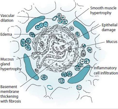
Figure 23–3. Morphologic changes in asthma.
Two other mechanisms by which variable airway obstruction owing to workplace exposure can occur are reflex and pharmacologic bronchoconstriction. In reflex bronchoconstriction, neuroreceptors in the airway are stimulated by agents such as cold air, dusts, mists, vapors, and fumes. The reaction does not involve immunologic mechanisms and does not lead to airway inflammation. In most cases, the patient has a history of preexisting nonoccupational asthma with nonspecific airway hyperresponsiveness so that this is the primary mechanism of work-aggravated asthma. Pharmacologic bronchoconstriction occurs when an agent in the workplace causes the direct release of mediators (eg, cotton dust in textile mills) or a direct effect on the autonomic regulation of bronchomotor tone (eg, organophosphate pesticides inhibit cholinesterase).
Prevention
Prevention of further occupational asthma should be considered in all workplaces where cases are diagnosed. This can be achieved primarily through environmental control of processes known to involve exposure to potential sensitizers and irritants. Protection of workers by substitution of other materials for asthma-inducing agents, the use of appropriate ventilation systems, respiratory protective equipment, and worker education about appropriate procedures is recommended. Avoidance of high-intensity exposures from leaks and spills that may initiate the development of occupational asthma is essential. Medical surveillance for early detection of cases also can contribute to reducing the burden of impairment/disability owing to occupational asthma.
Clinical Findings
The diagnosis of occupational asthma is made by confirming the diagnosis of asthma and by establishing a relationship between asthma and the work environment. The diagnosis of asthma should be made only when both intermittent respiratory symptoms and physiologic evidence of reversible or variable airways obstruction are present. The relationship between asthma and workplace exposure may fit any of the following patterns: (1) symptoms occur only at work, (2) symptoms improve on weekends or vacations, (3) symptoms occur regularly after the work shift, (4) symptoms increase progressively over the course of the workweek, and (5) symptoms improve after a change in the work environment.
At least one of the symptoms of wheezing, shortness of breath, cough, and chest tightness should occur while the worker is at or within 4–8 hours of leaving the workplace. Often the worker’s symptoms improve during days off work or while away from the worker’s usual job. With persistent exposure, the symptoms may become chronic and lose an obvious relationship to the workplace. Concomitant eye and upper respiratory tract symptoms also may be noted. The diagnosis of occupational asthma also should be considered when there is a history of recurrent episodes of work-related “bronchitis” characterized by cough and sputum production in an otherwise healthy individual. While high-molecular-weight sensitizers typically cause early or dual responses, the low-molecular-weight sensitizers tend to induce isolated late responses that may occur hours after the work shift is over.
The evaluation for possible occupational asthma requires a detailed history of the work environment (Figure 23–4). As noted earlier, attention should be given to the agents to which the worker is exposed, the type of ventilation in the work-place, whether respiratory protective equipment is used, and if possible, the level of exposure (ie, whether it is high or low or if accidental exposure through spills ever occurs). A helpful clue to a significant problem in a workplace is the presence of other workers with episodic respiratory symptoms.

Figure 23–4. Algorithm for the clinical investigation of occupational asthma.
The detection of wheezing on chest auscultation is helpful, but the physical examination is frequently normal in asthmatic patients not currently suffering from an exacerbation. Chest radiographs are normal in most individuals with asthma because the disease involves the airways rather than the lung parenchyma. Hyperinflation and flattening of the diaphragms, indicating air trapping, may be seen during exacerbations. Fleeting infiltrates indicating mucus plugging and bronchial wall thickening reflecting chronic inflammation also may be noted.
Spirometry for measurement of FEV1 and FVC is the most reliable method for assessing airway obstruction. However, because asthmatic patients typically have reversible airway obstruction, they may have normal lung function during intervals between acute attacks. The response to inhaled bronchodilator administration has been used as a measure of airway hyperresponsiveness. A 12% improvement in FEV1 of at least 200 mL after inhaled bronchodilator is how the ATS defines a significant improvement indicative of hyperresponsive airways. Across-work-shift spirometry, when available, can provide objective evidence of occupational asthma. A greater than 10% fall in FEV1 across a work shift is suggestive of an asthmatic response.
Serial recording of PEFR over a period of weeks to months is often the best way to document the work-relatedness of asthma. The worker records his or her PEFR at least four times while awake, as well as respiratory symptoms and medication use. When interpreting the worker’s log, attention should be given to any work-related pattern of change. A 20% or greater diurnal variability in PEFR is considered evidence of an asthmatic response (Figure 23–5). The major advantage of serial PEFR measurement over spirometry is the ability to detect late responses that occur after the work shift ends.

Figure 23–5. Serial peak expiratory flow rates (PEFRS) during a 16-day period in a worker with occupational asthma before, during, and after 1 week of exposure to the inciting agent.
Methacholine or histamine challenge can demonstrate the presence of nonspecific airway hyperresponsiveness in a worker suspected of having occupational asthma who has normal spirometry. Such testing can be particularly valuable if it demonstrates an increase in airway responsiveness on returning to work or a decrease when away from work. Specific inhalational challenge testing, that is, challenging the patient with the suspected agent at levels and under conditions that mimic workplace conditions, can be done for medicolegal purposes or to determine the precise etiology in a complex exposure scenario. However, specific challenge testing is time-consuming and potentially dangerous, and usually should be reserved for evaluation of patients in whom there is diagnostic uncertainty.
Allergy skin tests with common aeroallergens can be used to establish whether or not the worker is atopic. Atopy is a risk factor for high-molecular-weight sensitizer-induced asthma. When high-molecular-weight compounds are responsible for occupational asthma, skin tests with the appropriate extracts may help to identify the etiologic agent. Extracts of materials such as flour, animal proteins, and coffee will give positive skin tests in specifically sensitized individuals. Skin testing also may be helpful for a few low-molecular-weight compounds such as platinum salts. IgE antibodies assayed by the radioallergosorbent test (RAST) or by enzyme-linked immunoabsorbent assay (ELISA) may confirm exposure to allergens such as flour, animal proteins, acid anhydrides, plicatic acid, or isocyanates. However, the presence of positive skin reactions and/or specific antibodies is not always correlated with the presence of occupational asthma.
Treatment
Acute asthma attacks requiring emergency management should be treated with supplemental oxygen, beta-agonists, corticosteroids, and if infection is suspected, antibiotics. Hospitalization should be considered in the more severe cases because of the potential for respiratory failure.
Once the diagnosis of occupational asthma is made, the primary intervention is to reduce or eliminate the worker’s exposure to the offending agent. This may be achieved through modifications in the workplace. It may be possible to substitute the offending agent with another safer one. Improved local exhaust ventilation and enclosure of specific processes also may be helpful. With irritant-induced asthma, the use of personal protective equipment may lower exposures to levels that do not induce bronchospasm. Workers who are allowed to continue in the job should have regular follow-up visits, including monitoring of their lung function and nonspecific airway responsiveness. With sensitizer-induced asthma, however, the worker should be precluded from further exposure to the sensitizing agent. It may be necessary to completely remove the worker from the workplace because exposure to even minute quantities of the offending agent may induce bronchospasm. If a worker is required to leave the workplace (eg, a baker with flour-induced asthma), the worker should be considered 100% impaired on a permanent basis for the job that caused the illness and for other jobs with exposure to the same causative agent.
In addition to reduction or elimination of exposure to any specific offending agent, the worker also should avoid exposure to other materials/processes that may exacerbate the worker’s asthma, such as irritating dusts, mists, and vapors. Cessation of smoking and avoidance of exposure to environmental tobacco smoke are also essential.
Prognosis
Once occupational asthma has been diagnosed, an attempt should be made to classify the degree of impairment/disability. An approach to the evaluation of impairment in patients with asthma was developed by the ATS and has been adopted by the American Medical Association. Asthma is a dynamic disease that does not generally result in a static level of impairment. The criteria used for impairment rating are degree of postbronchodilator airway obstruction by spirometry, measurement of airway responsiveness, and medication requirements. Assessment of impairment/disability should be done only after optimization of therapy and whenever the worker’s condition changes substantially, whether for better or worse.
Occupational asthma caused by such diverse agents as diisocyanates, snow crab, and western red cedar show persistence of symptoms and the presence of nonspecific airway hyperresponsiveness for periods up to 6 years after removal from the offending agent. Factors that affect the long-term prognosis of the patient with occupational asthma are the total duration of exposure, the duration of exposure after the onset of symptoms, and the severity of asthma at the time of diagnosis. Those who do poorly have a delayed diagnosis, lower lung function values, and greater nonspecific airway hyperresponsiveness, hence the importance of early diagnosis and early removal from future exposure to the etiologic agent. Treatment with inhaled corticosteroid medications has been shown to improve prognosis for sensitizer-induced occupational asthma.
SPECIFIC AGENTS
1. Diisocyanates
Chemicals of the diisocyanate group are used widely in the manufacture of polyurethane surface coatings, insulation materials, car upholstery, and furniture. The most commonly used diisocyanate is toluene diisocyanate (TDI). Because of its high vapor pressure, the less volatile agent methylene diphenyl diisocyanate (MDI) is used in some production processes. Other diisocyanates, such as hexamethylene diisocyanate (HDI), naphthalene diisocyanate (NDI), and isophorone diisocyanate (IPDI), also have commercial uses. These chemicals are all highly reactive because of the presence of –N–C–O groups, which easily react with biologic molecules and are potent irritants to the respiratory tract. Upper respiratory tract inflammation occurs in almost everyone exposed to TDI levels of 0.5 ppm or more.
Five major patterns of airway response to TDI have been described in humans: (1) occupational asthma of the sensitizer-type, which occurs in 5–10% of exposed workers weeks to months after the onset of exposure, (2) chemical bronchitis, (3) acute but asymptomatic deterioration of respiratory function during a work shift, (4) chronic deterioration of respiratory function associated with chronic exposure to low doses, and (5) persistent asthma or RADS after exposure to high doses.
2. Vegetable Dusts, Including Cotton (Byssinosis), Flax, Hemp, & Jute
Byssinosis occurs in certain workers in the cotton textile industry. The characteristic symptoms are chest tightness, cough, and dyspnea 1–2 hours after the patient returns to work after several days off. The symptoms usually resolve overnight and on subsequent days become milder until by the end of the workweek the worker may become asymptomatic. The prevalence of byssinosis is higher in workers with longer duration of exposure and with greater respirable dust exposure, such as during opening bales and carding, and lowest in those with a shorter exposure history and with lesser dust exposure. The mechanism underlying byssinosis remains unclear. Cotton-dust extracts are capable of causing direct release of histamine and contain endotoxins that can induce a number of inflammatory responses.
3. Metal Salts
Complex salts of platinum used in electroplating, platinum refinery operations, manufacture of fluorescent screens, and jewelry making are known to cause occupational asthma. Specific IgE antibodies to platinum salts conjugated to human serum albumin have been found in sensitized workers by RAST. Rhinitis and urticaria frequently accompany asthma, and this triad is sometimes called platinosis. Nickel, vanadium, chromium, and cobalt are other metals known to cause occupational asthma.
4. Acid Anhydrides
Epoxy resins often contain acid anhydrides as curing or hardening agents. Phthalic anhydride, trimellitic anhydride (TMA), and tetrachlorophthalic anhydride (TCPA) are several of the more commonly used acid anhydrides. Occupational asthma occurs in a small percentage of exposed workers. The serum of affected workers typically contains specific IgE antibodies against acid anhydride–protein conjugates. Trimellitic anhydride exposure can give rise to four clinical syndromes: (1) symptoms of immediate airway irritation, (2) immediate rhinitis and asthma, (3) late asthma with systemic symptoms of fever and malaise, and (4) infiltrative lung disease (hemorrhagic alveolitis) with hemoptysis and anemia.
5. Wood Dusts
A large number of wood dusts are known to cause rhinitis and asthma. Western red cedar is the best studied. This wood contains the low-molecular-weight compound plicatic acid, which is believed to be responsible for causing asthma through an unclear mechanism. Western red cedar asthma falls under the category of low-molecular-weight sensitizer-induced asthma and clinically is much like diisocyanate asthma. There is often a long period between onset of exposure and onset of symptoms, and asthma only develops in a small proportion of exposed subjects. A small dose of plicatic acid can induce a severe asthmatic attack in a sensitized individual, and many workers continue to have persistent asthma years after cessation of exposure.
HYPERSENSITIVITY PNEUMONITIS
ESSENTIALS OF DIAGNOSIS
A link between symptoms and antigen exposure may be obtained from the work or environmental history.
The antigen can be a microbial agent, animal protein, or chemical sensitizer.
The clinical presentation can be acute, subacute, or chronic (insidious onset).
General Considerations
Hypersensitivity pneumonitis, also known as extrinsic allergic alveolitis, refers to an immunologically mediated inflammatory disease of the lung parenchyma that is induced by inhalation of organic dusts that contain a variety of etiologic agents (eg, bacteria, fungi, amebae, animal proteins, and several low-molecular-weight chemicals). Although many different antigens are capable of causing hypersensitivity pneumonitis (Table 23–4), the basic clinical and pathologic findings are similar regardless of the nature of the inhaled dust. The nature of the inhaled antigen, the exposure conditions, and the nature of the host immune response all contribute to the risk for the disease. Hypersensitivity pneumonitis is characterized initially by a lymphocytic alveolitis and granulomatous pneumonitis, with improvement or complete resolution if antigen exposure is terminated early. Continued antigen exposure may lead to progressive interstitial fibrosis.
Table 23–4. Some agents causing hypersensitivity pneumonitis.

Inhalational exposure to antigen in a sensitized individual may result in either an acute or chronic presentation of hypersensitivity pneumonitis depending on the exposure conditions. The acute and more common form of presentation of hypersensitivity pneumonitis usually occurs within 4–6 hours of an intense exposure to the offending antigen. Recurrent low-level exposure to an appropriate antigen may result in the insidious onset of chronic interstitial lung disease with fibrosis.
Pathogenesis
The pathogenesis of hypersensitivity pneumonitis involves repeated inhalational exposure to the antigen, sensitization of the exposed individual, and immunologically mediated damage to the lung. The inflammatory response that results in hypersensitivity pneumonitis appears to involve a combination of humoral, immune complex–mediated (type III), and cell-mediated (type IV) immune reactions to the inhaled antigen. In the presence of excess antigen, immune complexes may be deposited in the lungs. These complexes activate complement, leading to an influx of neutrophils. The local immune response later shifts to a T-lymphocyte-predominant alveolitis, with a differential cell count in bronchoalveolar lavage (BAL) fluid of up to 70% lymphocytes. Examination of BAL lymphocyte subpopulations in patients with hypersensitivity pneumonitis often has revealed a predominance of CD8+ suppressor/cytoxic cells. The peripheral blood and BAL T lymphocytes from patients with hypersensitivity pneumonitis will proliferate and undergo blastogenic transformation with cytokine generation when exposed in vitro to antigen. Animal models also support the role of cell-mediated immunity in the disease. Passive transfer of lymphocytes from sensitized animals to unexposed, nonsensitized animals results in a hypersensitivity pneumonitis–like disease when the latter animals subsequently are exposed to the specific antigen by inhalation. Alveolar macrophages also may play an important role in the pathogenesis of the disease by processing and presenting inhaled antigen to T-helper lymphocytes, as well as by releasing cytokines, which may help to amplify the inflammatory response.
Because only a small number of exposed persons ever develop hypersensitivity pneumonitis, the underlying mechanism of the disease may be a form of immune dysfunction in which a normal host defense response cannot be appropriately downregulated. This immune dysfunction may be, at least in part, genetically mediated. Other environmental factors also may be involved because a number of studies show that hypersensitivity pneumonitis occurs more frequently in nonsmokers than in smokers.
Prevention
Exposure to agents capable of causing hypersensitivity pneumonitis should be avoided. Any area of a workplace or home where there is water damage involves potential risk of exposure to molds that could cause hypersensitivity pneumonitis. Such an area should be thoroughly cleaned and repaired. Moldy silage, compost, and plant material (eg, sugarcane, cork, redwood) as well as areas with heavy bird congregation should be avoided.
Clinical Findings
Symptoms of chills, fever, malaise, myalgia, cough, headache, and dyspnea are noted commonly. Physical examination may reveal a relatively ill-appearing patient with bibasilar inspiratory crackles on chest auscultation. Frequently, acute hypersensitivity pneumonitis is misdiagnosed as an acute viral syndrome or pneumonia because it tends to closely mimic these conditions. Laboratory findings include peripheral blood leukocytosis with increased neutrophils and a relatively decreased lymphopenia. Arterial blood gas values may show hypoxemia.
Chest radiographic findings may be completely normal even in symptomatic individuals. Typically, however, the acute phase is associated with the presence of a reticulonodular pattern. Patchy densities that tend to coalesce also may be seen. These infiltrates usually are bilaterally distributed, but a more focal presentation sometimes occurs.
Pulmonary function testing may reveal a decrease in the FEV1 and FVC with an unchanged FEV1:FVC ratio consistent with a restrictive impairment. A decrease in the DLCO reflecting impaired gas exchange also is typical of the acute presentation. The acute form generally progresses for up to 18–24 hours and then begins to resolve. Recurrence of the syndrome may be seen subsequently with reexposure to the antigen.
Progressive respiratory impairment with symptoms of dyspnea, cough, excessive fatigue, and weight loss may develop without acute episodes. Physical examination may reveal cyanosis, clubbing, and inspiratory crackles. Chest radiographic findings include diffusely increased linear markings and reduced lung size. Findings on HRCT scanning of the chest include centrilobular micronodules, ground-glass opacification, patchy airspace consolidation, and linear densities. Chest CT findings can be suggestive of the diagnosis of hypersensitivity pneumonitis but are not always pathognomonic. Pulmonary function testing usually will show a restrictive impairment with a decreased DLCO, although some patients may be seen with a mixed or obstructive pattern.
The diagnosis of hypersensitivity pneumonitis should be suspected in patients with episodic respiratory symptoms and evidence of fleeting infiltrates on chest radiographs or restrictive impairment on pulmonary function testing. A careful history may elicit the onset of respiratory symptoms with exposure to the offending antigen. The temporal relationship of symptom development after exposure is crucial to the diagnosis. Additional supporting evidence is provided by the remission of symptoms and signs after cessation of exposure to the antigen and their reappearance on reexposure. The home environment also can be a source of the offending antigen. Workplace and home inspections may provide information supportive of the diagnosis (eg, evidence of mold or water damage).
Serologic studies demonstrating specific IgG precipitating antibodies by the traditional double-immunodiffusion technique will be positive in most patients with hypersensitivity pneumonitis if the correct antigen is used, although such antibodies are also detected frequently in exposed individuals who are healthy. False-positive results may be obtained with the use of more sensitive assays for IgG, such as ELISA. False-negative results frequently are a result of the failure to test for the correct antigen. Most commercially available hypersensitivity pneumonitis panels involve only a limited number of common antigens. Inhalational challenge studies with the suspected antigen may assist in the diagnosis of hypersensitivity pneumonitis. Antigen extracts may be administered in an aerosolized form followed by serial pulmonary function testing. Specific challenge testing should be conducted only by a laboratory experienced in the technique. While such challenges provide the “gold standard” method of confirming a direct relationship between a suspected offending antigen and the disease process, workplace studies involving the actual conditions of patient exposure are safer and usually easier to conduct.
Analysis of BAL fluid obtained by fiberoptic bronchoscopy in patients with hypersensitivity pneumonitis often demonstrates an increased percentage of T lymphocytes that are primarily CD8+ suppressor cells. In sarcoidosis, another condition characterized by increased T lymphocytes in BAL, the predominant cells are of the CD4+ helper subtype.
Lung biopsy may be necessary to make the diagnosis in difficult cases, such as those with the chronic form and an insidious presentation of dyspnea. Video-assisted thoracoscopic surgery (VATS) or open lung biopsy is preferred because transbronchial biopsy may not provide adequate tissue for pathologic differentiation of hypersensitivity pneumonitis from other diseases such as sarcoidosis. In acute or early chronic (subacute) hypersensitivity pneumonitis, there is patchy infiltration of predominantly lymphocytes in a bronchocentric distribution, usually with accompanying epithelioid (ie, noncaseating) granulomas. The granulomas are likely what appear as centrilobular micronodules on HRCT scanning. In chronic hypersensitivity pneumonitis, peribronchiolar inflammation remains prominent, and bronchiolitis obliterans is common. Large histiocytes with foamy cytoplasm may be seen in the alveoli and interstitium. Interstitial fibrosis with honeycombing occurs in advanced disease, by which time granulomas no longer may be evident.
Complications
The primary complication of hypersensitivity pneumonitis is the development of irreversible lung fibrosis.
Treatment
The key to successful treatment of hypersensitivity pneumonitis is avoidance of the offending antigen. As described for occupational asthma, this may be achieved by product substitution or institution of effective engineering controls. Respiratory protective equipment also may be appropriate in situations where possible exposure is only occasional. If persistence of symptoms occurs despite engineering control measures and respiratory protective equipment, complete removal of the worker from exposure is necessary.
Corticosteroids remain the mainstay of treatment of patients with severe or progressive hypersensitivity pneumonitis, despite the lack of controlled data regarding the effect of these agents on the disease process. An empirical trial of prednisone (1 mg/kg per day), with monitoring of chest radiographic and pulmonary function changes 1 month after starting the trial, is a reasonable approach. Therapy should be continued until there is significant clinical improvement. If bronchospasm is present, beta-agonists should be administered. Supplemental oxygen should be given to patients with hypoxemia, and intensive-care-unit support may be needed in particularly severe acute cases.
Prognosis
Workers with a diagnosis of hypersensitivity pneumonitis should have frequent follow-up, especially if continued exposure to antigen is possible. If further exposure to the offending agent is avoided, the prognosis is good. Significant pulmonary morbidity may occur if persistent exposure is allowed.
INHALATION FEVERS
ESSENTIALS OF DIAGNOSIS
Inhalational exposure to organic dusts, polymer fumes, and certain metals can cause a flu-like illness.
The illness is usually self-limited.
Bilateral infiltrates on chest X-ray are usually present.
General Considerations
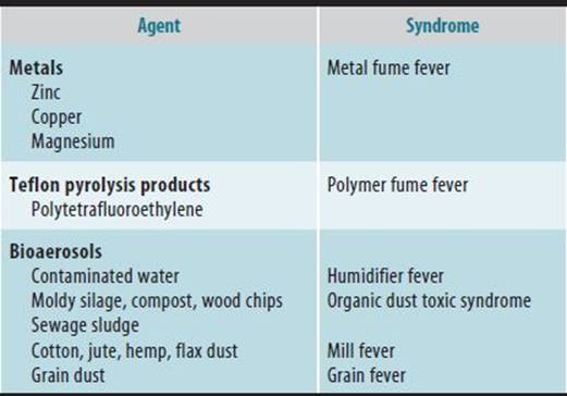
Inhalation fever refers to several syndromes that are characterized by short-term but debilitating flulike symptoms after exposure to organic dusts, polymer fumes, and metal fumes (Table 23–5). In addition to fever, the symptoms include chills, myalgia, headache, malaise, cough, and chest discomfort.
Table 23–5. Some agents causing inhalation fever.
In contrast to occupational asthma and hypersensitivity pneumonitis, which require susceptibility and/or sensitization, the attack rate for the inhalation fevers is high; that is, most people will experience symptoms as a result of high-level exposure to the etiologic agents.
SPECIFIC SYNDROMES
1. Metal Fume Fever
General Considerations
Inhalation of certain freshly formed metal oxides can cause metal fume fever, an acute self-limiting flulike illness. The most common cause of this syndrome is the inhalation of zinc oxide, which is generated from molten bronze or welding galvanized steel. The oxides of only two other metals, copper and magnesium, have been proven to cause metal fume fever. When zinc is heated to its melting point, zinc oxide fumes are generated. The particle size of the generated fumes ranges from 0.1 to 1 μm in diameter, although aggregation with the formation of larger particles occurs readily. The underlying pathogenesis of metal fume fever is incompletely understood. However, there is evidence from controlled human exposure studies that zinc oxide fume inhalation induces a leukocyte recruitment to the lungs with an associated release of cytokines, which causes systemic symptoms.
It is estimated that more than 700,000 workers in the United States are involved in welding operations, so the potential for inhalational exposure and metal fume fever is great. The clinical syndrome begins 3–10 hours after exposure to zinc oxide. The initial symptom may be a metallic taste associated with throat irritation and followed within several hours by the onset of fever, chills, myalgia, malaise, and a nonproductive cough. Occasionally, nausea, vomiting, and headache are noted. Physical examination during the episode may reveal a febrile patient with crackles on auscultation of the chest. Laboratory evaluation frequently reveals a leukocytosis with a left shift and an elevated serum lactate dehydrogenase level. The chest radiograph, pulmonary function tests, and arterial blood gas measurements usually are normal. Transient chest radiographic infiltrates and reduced lung volumes and DLCO have been reported in severe cases. Signs and symptoms generally peak at 18 hours and resolve spontaneously with complete resolution of abnormalities within 1–2 days.
Treatment of metal fume fever is entirely symptomatic. Control of elevated body temperature by antipyretics and oxygen therapy for hypoxemia may be required. There is no evidence that steroid therapy is of any benefit. Prevention relies on appropriate engineering controls and/or personal protective equipment to reduce exposure. There are no good data on the long-term sequelae of repeated exposures.
2. Polymer Fume Fever
A syndrome similar to metal fume fever may occur after inhalation of combustion products of polytetrafluoroethylene (Teflon) resins. The properties of Teflon—strength, thermal stability, and chemical inertness—make it a widely used product in the manufacture of cooking utensils, electric appliances, and insulating material. When Teflon is heated to temperatures greater than 300°C (572°F), numerous degradation products are formed that appear to cause the syndrome. Exposure to such combustion products can occur during welding of metal coated with Teflon, during the operation of molding machines, and while smoking cigarettes contaminated with the polymer.
Exposure to a high concentration of polymer fumes causes a fever to develop within several hours. Often this occurs toward the end of the work shift or in the evening after work. The symptoms, signs, and laboratory findings of polymer fume fever are essentially the same as those of metal fume fever. The syndrome is self-limiting and resolves within 12–48 hours. Exposure to very high concentrations of polymer fumes may lead to the development of severe chemical pneumonitis with pulmonary edema. In such cases, the symptoms, signs, and laboratory features are similar to pulmonary edema from other causes.
3. Organic Dust Toxic Syndrome
Inhalation of various bioaerosols contaminated with fungi, bacteria, and/or endotoxins can cause an acute febrile syndrome known as organic dust toxic syndrome (ODTS). Exposures to moldy silage, moldy wood chips, compost, sewage sludge, grain dust (grain fever), cotton dust (mill fever), animal confinement building environments, and contaminated humidifier mist (humidifier fever) are associated with the development of inhalation fever. The clinical syndrome of ODTS is essentially identical to that described earlier for metal or polymer fume fever. Severe pulmonary inflammatory reactions have been described with massive exposures, but these are rare.
METAL-INDUCED LUNG DISEASE
ESSENTIALS OF DIAGNOSIS
Inhalational exposure to several metals can cause immune-mediated interstitial lung disease (ILD). the clinical presentation is similar to that of other types of ILD.
General Considerations
Metal-induced ILD appears to be due to cell-mediated sensitization to the offending agent. While greater exposure is associated with increased risk, likely genetic predisposition plays an important role.
SPECIFIC METALS
1. Hard Metal
Hard metal is a cemented alloy of tungsten carbide with cobalt, although other metals such as titanium, tantalum, chromium, molybdenum, or nickel also may be added. These cemented carbides have found wide industrial use because of their properties of extreme hardness, strength, and heat resistance. Their major use is in the manufacture of cutting tools and drill-tip surfaces.
Workers exposed to hard metal are at risk for developing interstitial lung disease, the so-called hard-metal disease, and occupational asthma. The putative cause of both these disease processes is cobalt. Some workers may present with features of both hard metal–induced airway and parenchymal diseases. Workers at risk for these diseases are those engaged in the manufacture of the alloy, grinders and sharpeners of hard metal tools, and diamond polishers and others who use disks containing cobalt and metal coaters who use powdered hard metal. Occupational asthma caused by cobalt in hard-metal workers is similar to that caused by other low-molecular-weight sensitizer agents.
Workers with hard-metal disease typically complain of symptoms of dyspnea on exertion, cough, sputum production, chest tightness, and fatigue. Physical examination may reveal evidence of crackles on chest auscultation, reduced chest expansion, clubbing, and in advanced cases, cyanosis. Chest radiographs may show bilateral rounded and/or irregular opacities with no pathognomonic features. Pulmonary function tests tend to show both a restrictive ventilatory impairment and a decreased DLCO. The diagnosis of hard-metal disease often is made on the basis of pathologic examination of lung tissue rather than by clinical evaluation. The histologic findings are those of interstitial pneumonitis, frequently of the giant-cell type (eg, giant-cell interstitial pneumonia), and interstitial fibrosis. Characteristic multinucleated giant histiocytes may be seen in BAL fluid as well.
The primary treatment of hard-metal disease is removal of the affected worker from further exposure. Relatively rapid progression to impairment is not infrequent, and resolution after cessation of exposure may not occur. Complete removal from cobalt exposure is advisable because a case has been reported of a worker who developed rapidly fatal lung disease with continued exposure. Because hard-metal disease is often progressive, empirical therapy with corticosteroids may be required.
2. Beryllium
Beryllium is a light-weight, tensile metal that has a high melting point and good alloying properties. It has a wide range of applications in modern industrial processes. Although beryllium is no longer used in the manufacture of fluorescent light tubes, it is used commonly in the ceramics, electronics, aerospace, and nuclear weapons/power industries. Workers at risk are those involved in processes that generate airborne beryllium, including melting, casting, grinding, drilling, extracting, and smelting of beryllium. Acute beryllium-induced pneumonitis can occur after high-intensity exposure but has largely disappeared owing to improved workplace control of exposures. Chronic beryllium disease, which involves sensitization to the metal through a cell-mediated (type IV) mechanism, still occurs after lower-level exposures in susceptible workers. Beryllium can be phagocytosed by macrophages that present beryllium antigen to lymphocytes, resulting in sensitization and proliferation of beryllium-specific CD4+ T cells. Beryllium-activated T cells may release various cytokines and other inflammatory mediators, resulting in granuloma formation. Latency from time of initial beryllium exposure to the development of clinically manifest disease ranges from months to many years.
Why only a small percentage of an exposed population becomes sensitized to beryllium is not well understood. Recent studies have found a genetic marker of risk for beryllium sensitization, a glutamic acid substitution in residue 69 of the beta chain of the major histocompatibility complex molecule HLA-DP.
Chronic beryllium disease is a granulomatous inflammatory disorder that is very similar to sarcoidosis. In fact, the histologic findings in chronic beryllium disease are identical to those of sarcoidosis; that is, epithelioid (nonca-seating) granulomas with mononuclear cell infiltrates and varying degrees of interstitial fibrosis. Chronic beryllium disease usually affects only the lungs, but involvement of skin, liver, spleen, salivary glands, kidney, and bone may occur. Extrapulmonary involvement is less common than in sarcoidosis.
Workers with chronic beryllium disease commonly present with insidious onset of dyspnea on exertion, cough, and fatigue. Anorexia, weight loss, fever, chest pain, and arthralgias also may occur. Physical examination findings usually are confined to the lungs, with crackles being the most common, but they may be absent with mild disease.
Chest radiographic findings are ill-defined nodular or irregular opacities and hilar adenopathy. The latter is seen somewhat less frequently than in sarcoidosis and rarely occurs in the absence of parenchymal changes. The small nodular opacities sometimes are more prominent in the upper lung zones and may coalesce into more conglomerate masses. High-resolution CT scanning is more sensitive than plain chest radiography, but histologically confirmed cases occur with normal scans.
Pulmonary function testing may be normal with mild disease, but there is usually a restrictive, obstructive, or mixed pattern of impairment and a reduced DLCO. Resting arterial hypoxemia and further desaturation with exercise are common with more severe disease.
Often a meticulously obtained occupational history is required to suggest beryllium as the causative agent. Because of the similarity between chronic beryllium disease and sarcoidosis, demonstration of beryllium sensitization is necessary to confirm the diagnosis. A relatively specific blood lymphocyte proliferation test (LPT) is available in which the beryllium-specific uptake of radiolabeled DNA precursors by the patient’s lymphocytes cultured in vitro is quantitated. The sensitivity of the LPT for chronic beryllium disease is greater than 90% when using peripheral blood lymphocytes and can be increased if lung lymphocytes obtained from BAL are used. The blood LPT also can be used to screen for sensitization among beryllium-exposed workers.
The current criteria for the diagnosis of chronic beryllium disease are (1) a history of beryllium exposure, (2) a positive peripheral blood or BAL LPT, and (3) the presence of epithelioid granulomas and mononuclear infiltrates, in the absence of infection, in lung tissue. This approach relies on the LPT to confirm sensitization to beryllium and trans-bronchial biopsy of lung tissue to confirm the presence of disease.
Because the disease process involves a type of hypersensitivity, a worker with chronic beryllium disease should be completely removed from further beryllium exposure. A trial of corticosteroids is warranted in symptomatic workers with documented pulmonary physiologic abnormalities because this may induce a remission in some. If steroid therapy is initiated, objective parameters of response such as chest radiographs and pulmonary function test results should be monitored serially in order to adjust appropriately the dose and duration of treatment. Chronic beryllium disease has the propensity to develop into chronic irreversible pulmonary fibrosis, so careful monitoring of affected workers is necessary.
3. Other Metals
Inhalation of relatively high concentrations of cadmium, chromium, or nickel fumes or mercury vapor can cause toxic pneumonitis. Occupational exposure to certain metals (eg, antimony, barium, iron, and tin) can lead to deposition of sufficient radiodense dust that chest radiographs demonstrate opacities in the absence of lung parenchymal inflammation and fibrosis.
PNEUMOCONIOSES
ESSENTIALS OF DIAGNOSIS
Chronic exposure, usually over years, to mineral dusts can cause fibrotic ILD.
Symptoms are typically progressive dyspnea and dry cough.
Diagnosis is usually made on the basis of radio-graphic abnormalities, which may proceed lung function impairment.
General Considerations
The pneumoconioses are a group of conditions resulting from the deposition of mineral dust in the lung and the subsequent fibrotic lung tissue reaction to the dust. The diagnosis is usually made based on chest imaging. Radiographically evident interstitial opacities may appear before impairment of pulmonary function or symptoms.
The risk of disease is clearly associated with level of exposure. Chronic exposure (ie, years) is required for most types of pneumoconiosis. Typically, a long latent period (>5 years) between onset of exposure and clinical manifestation of disease is also required
SPECIFIC PNEUMOCONIOSES
1. Silicosis
Silicosis is a parenchymal lung disease that results from the inhalation of silicon dioxide, or silica, in crystalline form. Silica is a major component of rock and sand. Workers with potential for exposure are miners, sandblasters, foundry workers, tunnel drillers, quarry workers, stone carvers, ceramic workers, and silica flour production workers.
Exposure to silica can lead to one of three disease patterns: (1) chronic simple silicosis, which usually follows more than 10 years of exposure to respirable dust with less than 30% quartz, (2) subacute/accelerated silicosis, which generally follows shorter, heavier exposures (ie, 2–5 years), and (3) acute silicosis, which is seen often following intense exposure to fine dust of high silica content over a several-month period.
Chronic silicosis is characterized by the formation of silicotic nodules in the pulmonary parenchyma and the hilar lymph nodes (Figure 23–6). The lesions in the hilar lymph nodes may calcify in an “egg shell” pattern that, while only occurring in a small proportion of cases, is virtually pathognomonic for silicosis. Lung parenchymal involvement tends to have a predilection for the upper lobes. The coalescence of small silicotic nodules into larger fibrotic masses, called progressive massive fibrosis (PMF), may complicate a minority of cases. PMF tends to occur in the upper lung fields, may obliterate blood vessels and bronchioles, causes gross distortion of lung architecture, and leads to respiratory insufficiency.

Figure 23–6. Radiographic changes of simple silicosis.
Accelerated silicosis is similar to chronic silicosis except that the time span is shorter and the complication of PMF is seen more frequently. Acute silicosis is a rare condition seen in workers who are exposed to very high concentrations of free silica dust with fine particle size. Such exposures occur frequently in the absence of adequate respiratory protection. The characteristic findings differ from chronic silicosis in that the lungs show consolidation without silicotic nodules, and the alveolar spaces are filled with fluid similar to that found in pulmonary alveolar proteinosis. Acute silicosis leads to death in most cases.
Alveolar macrophages play an important role in the pathogenesis of silicosis because these cells ingest inhaled silica and then release cytokines that recruit and/or stimulate other cells. Although crystalline silica can be cytotoxic secondary to direct chemical damage to cellular membranes, the primary effect of inhaled silica on macrophages is activation. The silica-activated macrophages recruit and activate T lymphocytes, which, in turn, recruit and activate a secondary population of monocytes-macrophages. The activated macrophages produce cytokines, which stimulate fibroblasts to proliferate and produce increased amounts of collagen.
There are few symptoms and signs of chronic simple silicosis. The diagnosis usually is made by chest radiographs, which frequently reveal small round opacities (<10 mm in diameter) in both lungs, with a predilection for the upper lung zones. If an adequate occupational history is obtained from the patient along with a thorough review of the chest radiographs, the diagnosis of silicosis should not present any great difficulty. Pulmonary function testing in patients with simple silicosis is usually normal but occasionally may demonstrate evidence of a mild restrictive ventilatory defect and decreased lung compliance. In addition, a mild obstructive impairment is found occasionally in patients with simple silicosis, often as a consequence of chronic bronchitis caused by nonspecific dust effects and/or smoking. With complicated silicosis involving progressive fibrosis (nodules >10 mm in diameter), increasing dyspnea is noted, initially with exertion and then progressing to dyspnea at rest. Complicated chronic silicosis is associated with greater reductions in lung volumes, decreased diffusing capacity, and hypoxemia with exercise. Progressive massive fibrosis is the end-stage of complicated chronic silicosis.
There is an increased incidence of mycobacterial disease, both typical and atypical, in silicosis. Fungal diseases (especially cryptococcosis, blastomycosis, and coccidioidomycosis) are also seen with greater frequency. The mechanism by which the immune-inflammatory responses to inhaled silica lead to the increased incidence of mycobacterial and fungal infections is not clearly understood.
Because no treatment for silicosis is currently known, management is directed toward the prevention of progression and the development of complications. Continued exposure should be avoided, and surveillance for tuberculosis should be instituted. Tuberculin-positive persons with silicosis have an approximately 30-fold greater risk for developing tuberculosis and should be treated for latent tuberculosis with a regimen proven to be efficacious. In acute silicosis, therapeutic whole-lung lavage has been employed to physically remove silica from the alveoli.
The prognosis for patients with chronic silicosis is good, especially if they are removed from exposure. Mortality remains high, however, in those who develop PMF.
2. Asbestosis
Asbestos is the name for the fibrous forms of a group of mineral silicates. The types of asbestos that have been used commercially are chrysotile, amosite, crocidolite, anthophyllite, tremolite, and actinolite, with chrysotile being the most commonly used. The durability, heat resistance, and ability to be woven into textiles of asbestos led to a wide variety of industrial applications. Major occupational exposures occurred with asbestos mining and milling, manufacture or installation of insulation for ships or buildings, manufacture of friction materials for brake linings and clutch facings, asbestos cement manufacture, asbestos textile manufacture, and asbestos-containing spray products for decorative, acoustical, and fireproofing purposes.
Asbestosis refers to the diffuse interstitial pulmonary fibrosis caused by inhalation of asbestos fibers. The inhaled fibers are deposited primarily at the bifurcations of conducting airways and alveoli, where they are phagocytosed by macrophages. The initial injury is characterized by damage to the alveolar epithelium, incomplete phagocytosis by and activation of alveolar and interstitial macrophages, and release of proinflammatory cytokines as well as cytotoxic oxygen radicals by activated macrophages. A peribronchiolar inflammatory response ensues involving fibroblast proliferation and stimulation, which eventually may lead to fibrosis. Many factors are felt to play a role in disease initiation and progression, including the type and size of fiber, the intensity and duration of exposure, history of cigarette smoking, and individual susceptibility. A dose-response relationship exists such that asbestosis is more common in workers with a higher exposure level. Once asbestosis begins, it may progress irrespective of removal from continued exposure. Finally, there is a considerable latency period (usually at least 20 years) between onset of exposure and development of clinically apparent disease. The diagnosis of asbestosis is made by a thorough exposure history, clinical examination, appropriate imaging studies, and pulmonary function testing. The symptoms of asbestosis are indistinguishable from those of any other gradually progressive interstitial pulmonary fibrosing disorder, with progressive dyspnea and nonproductive cough being the most prominent. Bibasilar crackles with a “Velcro” quality can be auscultated over the posterolateral chest in the middle to late phase of inspiration. The crackles of asbestosis are unaffected by coughing.
Imaging studies that are helpful in the evaluation of asbestos-exposed patients are the chest radiograph and HRCT scan. The chest radiograph shows characteristic small, irregular or linear opacities distributed throughout the lung fields but more prominent in the lower zones. There is loss of definition of the heart border and hemidiaphragms. The most useful radiographic finding is the presence of bilateral pleural thickening, which does not occur commonly with other diseases-causing interstitial pulmonary fibroses (Figure 23–7). Diaphragmatic or pericardial calcification is almost a pathognomonic sign of asbestos exposure. The ILO classification system is often used in the United States to rate the degree of profusion of small, irregular opacities and of pleural thickening on the chest radiograph. Conventional chest CT scanning is more sensitive than chest radiography for the detection of pleural disease but not for parenchymal disease. HRCT scanning is the most sensitive imaging method for detecting early asbestosis.

Figure 23–7. Radiographic changes of asbestosis.
Depending on the severity of disease, pulmonary function testing will show varying degrees of restrictive impairment and decreased DLCO. Because asbestosis begins as a peribronchiolar process, reduced flow rates at low lung volumes, indicative of small airways obstruction, may be seen.
As for silicosis, there is no known treatment for asbestosis. Fortunately, only a minority of those exposed are likely to develop radiographically evident disease, and among these, most do not develop significant respiratory impairment. Workers with asbestosis should be removed from further asbestos exposure because the risk that parenchymal scarring will progress appears to increase with cumulative asbestos exposure. Any other factors that may contribute to respiratory disease should be reduced or eliminated. This is especially true of cigarette smoking because there is some evidence that it may contribute to the initiation and progression of asbestosis.
The substitution of other fibrous materials for asbestos and the institution of strict environmental controls where it is still present have led to a dramatic reduction in occupational exposures to asbestos. Medical surveillance of all currently exposed workers in the United States is required by Occupational Safety and Health Administration (OSHA) regulation.
3. Coal Workers’ Pneumoconiosis
Coal workers’ pneumoconiosis is the term used to describe parenchymal lung disease caused by the inhalation of coal dust. Miners who work at the coal face in underground mining and drillers in surface mines are at greatest risk of contracting this disease. A heavy coal dust burden is required to induce coal workers’ pneumoconiosis, and the condition is seen rarely in those who have spent fewer than 20 years underground.
The coal macule is the primary lesion in coal workers’ pneumoconiosis. It is formed when the inhaled dust burden exceeds the amount that can be removed by alveolar macrophages and mucociliary clearance. This leads to retention of coal dust in the terminal respiratory units. Prolonged retention causes lung fibroblasts to secrete a limiting layer of reticulin around the dust collection, or macule, near the respiratory bronchiole. Progressive enlargement of the macule may weaken the bronchiole wall to create a focal area of centrilobular emphysema; coalescence of small macules into larger lesions may occur. Initially, there is a predilection for the upper lung lobes, but with progression of the disease, the lower lobes become involved. As for silicosis, coal workers’ pneumoconiosis can be characterized as simple (radiographic lesions <10 mm in diameter) or complicated (lesions >10 mm in diameter). Only a small proportion of miners (<5%) develop complicated or progressive fibrotic disease. Progressive massive fibrosis, identical to that described earlier for silicosis, may occur.
The symptoms of cough and sputum production are common among coal miners and often are the result of chronic bronchitis from dust inhalation rather than coal workers’ pneumoconiosis. As with silicosis, simple coal workers’ pneumoconiosis is often asymptomatic. The symptoms and signs associated with complicated disease are the same as those described earlier for silicosis. Progressive massive fibrosis almost invariably leads to respiratory insufficiency and death.
The chest radiograph in simple coal workers’ pneumoconiosis shows the presence of small, rounded opacities in the lung parenchyma. Often seen first in the upper lung zones, these opacities may involve the lower zones in the later stage of the disease. Calcification of the hilar lymph nodes is not seen unless there is concomitant silica exposure. Complicated coal workers’ pneumococcosis/PMF is diagnosed when large parenchymal opacities are present.
Caplan syndrome may occur in coal miners with rheumatoid arthritis and is characterized by the appearance of rapidly evolving rounded densities on chest radiographs. These have a propensity to cavitate and histologically are composed of layers of necrotic collagen and coal dust. The pulmonary manifestations of Caplan syndrome may precede or coincide with the onset of arthritis.
Pulmonary function findings vary with the stage of disease in a manner similar to that described for silicosis. In simple disease, there are usually no significant pulmonary function abnormalities. In complicated disease, either a restrictive or mixed restrictive and obstructive pattern may occur with a decreased diffusing capacity and abnormal arterial blood gases. It is important to remember that an obstructive ventilatory impairment in a coal miner may be a result of chronic bronchitis, coal workers’ pneumoconiosis, or both.
Simple coal workers’ pneumoconiosis usually follows a benign course. Unlike silicosis, no increase is seen in either pulmonary tuberculosis or fungal infections of the lung. In complicated disease, the affected worker may have mild to severe respiratory symptoms and significant impairment. In such cases, depending on the degree of impairment, the worker should be removed from continued dust exposure. In the United States, underground miners are able to participate in a federally run medical surveillance program that provides free periodic chest radiographs. If coal workers’ pneumoconiosis is evident on the chest radiograph, the affected miner has the right to work in a low-dust job in the mine without loss of pay. In addition, personal dust exposure is monitored to confirm that exposures remain low.
Prevention of coal mine dust-related respiratory disease depends primarily on effective control of exposure to coal mine dust. In the United States, good progress has been made in reducing the incidence and prevalence of coal workers’ pneumoconiosis since the passage in 1969 of the Coal Mine Health and Safety Act, which established programs to monitor dust levels in mines and to provide radiographic surveillance of miners.
4. Other Pneumoconioses
Other mineral dusts capable of causing pulmonary parenchymal fibrosis include graphite (which causes disease similar to coal workers’ pneumoconiosis), kaolin and diatomaceous earth (which cause silicosis-like disease), and talc and mica (which cause disease that has features of both silicosis and asbestosis). A metal dust that can cause pneumoconiosis is aluminum oxide, which can form fibers under certain conditions.
A new cause of ILD was reported involving a series of cases of ILD from a single nylon flock manufacturing plant. Finely cut nylon, called flock, is used to make fabric for upholstery, clothing, and automobiles. Nylon flock fibers are 10–15 μm in diameter, but respirable-size particles are generated during cutting operations. Lung biopsies from patients with nylon flock–related ILD have shown lymphocytic bronchiolitis and peribronchiolitis with lymphoid hyperplasia.
CHRONIC OBSTRUCTIVE PULMONARY DISEASE
Chronic obstructive pulmonary disease (COPD) is typically divided into two main categories, chronic bronchitis and emphysema, although many patients with COPD have features of both. Work-related COPD is usually of the chronic bronchitis category, although cadmium and coal dust have been associated with emphysema.
CHRONIC BRONCHITIS
ESSENTIALS OF DIAGNOSIS
History of chronic exposure to inhaled irritants at work is necessary for the diagnosis of occupational COPD. there may or may not be a history of coexistent cigarette smoking.
Chronic cough and sputum production are required for the diagnosis of chronic bronchitis.
Airflow limitation as evidenced by a decreased FEV1:FVC ratio that does not improve with inhaled bronchodilator is another essential feature of CoPD.
General Considerations
Chronic bronchitis is characterized by inflammation of the bronchial tree and is manifested by persistent cough productive of sputum on most days for at least 3 months of the year for at least 2 successive years. The inhalation of irritant dusts, fumes, and gases can cause chronic simple bronchitis, that is, persistent sputum production without airflow obstruction (Table 23–6).
Table 23–6. Some agents causing chronic bronchitis.
Minerals
Coal
Oil mist
Silica
Silicates
Synthetic vitreous fibers
Portland cement
Metals
Osmium
Vanadium
Welding fumes
Organic dusts
Cotton
Grain
Wood
Smoke
Tobacco smoke
Fire smoke
Engine exhaust
The diagnosis of chronic bronchitis is straightforward and based entirely on whether the worker’s history is consistent with the definition given earlier. Once chronic bronchitis has been diagnosed, establishing a causal role for an occupational exposure is also based on the history obtained from the worker. Symptoms of cough and sputum production that are temporally associated with workplace exposure should suggest the diagnosis. Whether workers with chronic simple bronchitis are at risk for the development of chronic airflow obstruction and permanent respiratory impairment is an area of controversy that has yet to be completely resolved. The development of permanent respiratory impairment may depend on a variety of host factors such as preexisting nonspecific airway hyperresponsiveness, protease-antiprotease activity, and whether there is concomitant cigarette smoking. The population attributable risk is approximately 15% for occupational factors in the etiology of COPD. Smoking workers are at greater risk of developing respiratory symptoms with exposure to other irritants, and a work-related contribution to their symptoms should be considered.
Pathogenesis
Airway inflammation and lung injury are key features of COPD. Exposure to irritating agents in the workplace is hypothesized to cause airway inflammation by activating epithelial cells and macrophages to release chemokines, prostanoids, and proinflammatory cytokines. Activated macrophages and recruited neutrophils release various proteases, including matrix metalloproteases (MMPs) that can damage lung tissue, stimulate mucus hypersecretion, and lead to airway remodeling. In addition, there is increasing evidence that CD8+ T lymphocytes are also recruited as part of the irritant-induced inflammatory response and play a role in the pathogenesis of COPD.
Prevention
Reduction of exposure to irritants in the workplace can prevent cases of chronic bronchitis through the application of the hierarchy of control strategies as described for occupational asthma. Protection of workers by substitution of nonirritating materials, the use of appropriate ventilation systems, respiratory protective equipment, and worker education about appropriate procedures is recommended.
Clinical Findings
Upper respiratory tract inflammatory symptoms, eye irritation, and an increased incidence of symptoms among coworkers all are features that support a work-related problem. Physical examination may demonstrate no evidence of pulmonary abnormality. Spirometry and expiratory flow-volume curves may or may not show evidence of airway obstruction. A nonsmoking worker exposed to high concentrations of an irritant at the workplace who has evidence of airway obstruction and no history of asthma should be suspected of having occupationally induced chronic bronchitis.
Treatment
Because chronic bronchitis often has a multifactorial etiology, a multifocal approach to management should be taken. If the worker smokes, cessation should be encouraged. Work exposure to the suspected agent should be reduced or eliminated. Pharmacologic agents of benefit are the beta2-agonists, inhaled steroids, and inhaled anticholinergic agents. Periodic follow-up with particular attention to symptoms and worsening airway obstruction on serial spirometry is warranted.
Prognosis
The prognosis of workers with chronic irritant-induced bronchitis has not been well described. Some data, however, suggest that accelerated loss of ventilatory function can occur. In light of this, it may be prudent to assume that all workers with chronic work-related bronchitis are at risk of developing permanent respiratory impairment. Those with worsening symptoms or lung function abnormalities should be considered for removal from further exposure.
BRONCHIOLITIS OBLITERANS
ESSENTIALS OF DIAGNOSIS
The clinical presentation is usually insidious onset of cough and dyspnea.
Irreversible airflow limitation is present on pulmonary function testing.
Minimal changes are found on chest radiographs.
General Considerations
There is a history of a relevant exposure to a toxic agent. Bronchiolitis is inflammation of the small airways, and when the inflammatory response leads to obstruction of bronchiolar lumens, the term bronchiolitis obliterans is used. The bronchiolar obstruction is caused by intraluminal polyps of organizing connective tissue (proliferative-type) and/or airway remodeling and smooth-muscle hypertrophy (constrictive-type). The most common occupational cause is irritant gas inhalation (eg, oxides of nitrogen, chlorine, phosgene, ozone, hydrogen sulfide, and sulfur dioxide) (see discussion of toxic gas inhalation above). Bronchiolitis obliterans also has been reported in nylon-flock workers, battery workers (exposed to thionyl chloride), and textile workers exposed to polyamide-amine dyes. A new cause of bronchiolitis obliterans was reported in a group of workers from a single microwave popcorn plant, exposure to buttery flavoring (putative agent, diacetyl). A NIOSH survey of other workers in the plant found a high prevalence of obstructive-type spirometric abnormalities with a diacetyl exposure-response relationship. Cases of bronchiolitis obliterans have been identified in other food production facilities where diacetyl is used as a flavoring.
PLEURAL DISORDERS
ESSENTIALS OF DIAGNOSIS
Pleuritic pain, that is, sharp pain on inspiration, may accompany pleural disease caused by occupational exposures, but the diagnosis is usually made on the basis of chest imaging.
General Considerations
The pleura is the serous membrane that lines the lungs, the mediastinum, the diaphragm, and the rib cage. It is divided into the visceral pleura, which lines the lung surface, and the parietal pleura, which lines the remaining structures. The primary cause of occupationally induced pleural disease is asbestos, although talc and mica can cause benign pleural disease and zeolite can cause mesothelioma.
BENIGN PLEURAL EFFUSIONS
Pleural effusions resulting from asbestos exposure may occur in up to 3% of exposed workers. The risk of developing an effusion is greater in those with heavy exposure. Benign asbestos effusions tend to develop within 5–20 years of the onset of exposure.
A pleural effusion can be attributed to asbestos if the following criteria are met: (1) a significant history of occupational exposure with an appropriate latent period since onset of exposure, (2) exclusion of other known causes of pleural effusion, and (3) a repeat evaluation of the effusion within a minimum of 2 years confirms that it is benign.
The majority of workers who have pleural effusions from asbestos exposure are asymptomatic. Physical examination in those with large effusions may show diminished rib cage expansion, dullness to percussion, and decreased breath sounds on the side of the effusion. Chest radiographs typically show small to moderately large, unilateral pleural effusions. Bilateral involvement occurs in approximately 10% of cases of benign asbestos effusions. Pleural thickening may be noted, although often the effusion is the first manifestation of asbestos-induced disease. Diffuse pleural thickening involving both pleural surfaces and obliteration of the costophrenic angle may develop in the wake of benign asbestos effusions. Thoracentesis will obtain pleural liquid that is a sterile exudate with no specific findings, although increased eosinophils are suggestive of an asbestos etiology.
It is essential to exclude other etiologies of pleural effusion, especially tuberculosis and malignancy. Regular follow-up with repeat thoracentesis if pleural fluid persists is essential. There is no known treatment. Recurrences occur, but in most cases the effusion clears spontaneously within a year without any obvious residual pleural disease.
PLEURAL PLAQUES
Pleural plaques are circumscribed areas of pleural thickening that are the most common radiographic findings as a result of chronic asbestos exposure. Plaques usually involve the parietal pleural surface and tend to occur over the central portions of the hemidiaphragm and along the inferior posterolateral aspect of the lower ribs.
Bilateral pleural plaques almost invariably are a result of past asbestos exposure, and their prevalence is related to both the intensity of exposure and the duration since onset of exposure. Workers with a greater exposure have a higher chance of developing plaques. In workers without asbestosis, plaques rarely cause signs and symptoms. The diagnosis usually is made from a routine chest radiograph. When plaques lie parallel to the beam, they appear as slightly to moderately protuberant linear or ovoid opacities along the costal or diaphragmatic margins. If calcified, they have an irregular, unevenly dense appearance. Although oblique radiographic views are recommended by some, chest CT scanning provides the most sensitive and specific technique for confirming the presence of plaques. Pathologically, the plaques are composed mainly of collagen with little accompanying inflammation. Asbestos fibers can be demonstrated in plaque tissue by electron microscopy, although this is not required for routine clinical diagnosis.
A worker with a past history of asbestos exposure and pleural plaques on chest radiograph should be evaluated for the presence of asbestosis. Even if no evidence of parenchymal disease is found, the worker should be monitored periodically for the possible development of this condition.
Although workers with pleural plaques and no parenchymal disease typically do not develop respiratory impairment, there is evidence that heavily exposed workers with radio-graphic evidence of plaques, but no asbestosis, tend to have decreased lung function in comparison with workers with similar exposure histories whose chest radiographs are normal.
Because of the risk of development of bronchogenic carcinoma with asbestos exposure, cigarette smoking should be discouraged. The increased risk of lung cancer is not due to the plaques but to the cumulative dose of asbestos, the plaques merely acting as a marker of exposure.
Diffuse pleural thickening involving both visceral and parietal pleura also can result from past asbestos exposure. Such thickening occasionally is associated with a restrictive-type respiratory impairment even in the absence of asbestosis. Neither circumscribed plaques nor diffuse pleural thickening is believed to undergo malignant transformation to mesothelioma.
LUNG CANCER & MESOTHELIOMA
Lung cancer and mesothelioma are discussed in Chapter 19.
REFERENCES
Blanc PD: Occupation and COPD: a brief review. J Asthma 2012;49:2 [PMID: 21895566].
Cain JR: Diagnosing metal fume fever—an integrated approach. Occup Med (Lond) 2010;60:398 [PMID: 20407044].
de Lange DW: Do corticosteroids have a role in preventing or reducing acute toxic lung injury caused by inhalation of chemical agents? Clin Toxicol (Phila) 2011;49:61 [PMID: 21370942].
Eisner MD: Novel risk factors and the global burden of chronic obstructive pulmonary disease. Am J Respir Crit Care Med 2010;182:693 [PMID: 20802169].
Hines SE: The role of lymphocyte proliferation tests in assessing occupational sensitization and disease. Curr Opin Allergy Clin Immunol 2012;12:102 [PMID: 22306552].
Lacasse Y: Recent advances in hypersensitivity pneumonitis. Chest 2012;142:208 [PMID: 22796841].
Leung CC: Silicosis. Lancet 2012;379:2008. [PMID: 22534002].
Marcon A: Can an airway challenge test predict respiratory diseases? A population-based international study. J Allergy Clin Immunol 2014;133:104 [PMID: 23683511].
NIOSH: B reader program: http://www.cdc.gov/niosh/topics/chestradiography/breader.html.
Rosenman KD: HLA class II DPB1 and DRB1 polymorphisms associated with genetic susceptibility to beryllium toxicity. Occup Environ Med 2011;68:487 [PMID: 21186201].
Seidler A: Progression of beryllium sensitization to chronic beryllium disease. Occup Med (Lond) 2012;62:506 [PMID: 22705916].
Selman M: Hypersensitivity pneumonitis: insights in diagnosis and pathobiology. Am J Respir Crit Care Med 2012;186:314 [PMID: 22679012].
Tarlo SM: Diagnosis and management of work-related asthma: American College of Chest Physicians Consensus Statement. Chest 2008;134:1S [PMID: 18779187].
Myers R. Asbestos-related pleural disease. Curr Opin Pulm Med 2012;18:377 [PMID: 22617814].
Silverman DT: The Diesel Exhaust in Miners study: a nested case-control study of lung cancer and diesel exhaust. J Natl Cancer Inst 2012;104:855 [PMID: 22393209].
 SELF-ASSESSMENT QUESTIONS
Select the one correct answer for each question.
Question 1: The diffusing capacity of the lung for carbon monoxide (DLCO)
a. is the amount of inhaled carbon monoxide returned in exhaled air
b. is closely correlated with the capacity of the lungs to absorb oxygen
c. is increased with obstructive, restrictive, or vascular diseases
d. is often misused to assess respiratory impairment
Question 2: Bronchoprovocation tests
a. are useful in the diagnosis of occupational asthma
b. should be done in a hospital environment
c. give an indication of the presence and degree of inflammation of the airways
d. are usually terminated after a 40% fall in FEV1
Question 3: The site of deposition of an inhaled gas
a. depends on the duration of exposure and its concentration
b. is determined primarily by water solubility
c. such as phosgene is the moist surfaces of the nose and throat
d. such as ammonia is likely the alveoli
Question 4: Occupational asthma
a. may be caused by such diverse agents as diisocyanates, snow crab, and western red cedar
b. resolves rapidly after removal from the offending agent
c. is predicted by a history of childhood asthma
d. treatment with inhaled corticosteroids does not improve prognosis
Question 5: Hypersensitivity pneumonitis
a. should be distinguished from extrinsic allergic alveolitis
b. is an immunologically mediated inflammatory disease of the airway
c. is induced by inhalation of organic dusts that contain a variety of etiologic agents
d. has different clinical and pathologic findings for each etiologic agent
Question 6: Silicosis
a. is a thoracic pleural disease
b. results from the inhalation of silicon dioxide, or silica, in crystalline form
c. is primarily the result of cytotoxic effects on lymphocytes
d. produces small round opacities (<10 mm in diameter) in both lungs, with a predilection for the lower lung zones
Question 7: Coal workers’ pneumoconiosis
a. is, unlike silicosis, often asymptomatic
b. is seen often in those who have spent more than 2 years in underground mining
c. may lead to progressive massive fibrosis, identical to that of silicosis
d. is confirmed by biopsy of the coal macule
Question 8: In reflex bronchoconstriction
a. neuroreceptors in the airway are stimulated by agents such as cold air, dusts, mists, vapors, and fumes
b. the reaction involves immunologic mechanisms and leads to airway inflammation
c. the patient has no history of preexisting nonoccupational asthma
d. there is no history of nonspecific airway hyperresponsiveness