
Dennis J. Shusterman, MD, MPH
The upper airway contributes to respiratory function by providing air conditioning, filtering, and sensory monitoring of the ambient environment. These same structures are vulnerable to the effects of inhaled irritants and allergens. A growing body of evidence links the development of rhinitis with that of asthma, making the prevention (and early recognition) of upper airway inflammation a priority.
FUNCTIONAL ANATOMY OF THE UPPER AIRWAY
Anatomy of the Upper Airway
The upper airway extends from the nares to the larynx (Figure 22–1). The surface area of the nasal cavities is increased by the presence of the nasal turbinates enhancing the nose’s air conditioning and filtering ability. The anterior nasal cavity is lined with a squamous epithelium; posterior to the tip of the inferior turbinate, it transitions to a ciliated epithelium, complete with secretory cells, submucous glands, and venous capacitance vessels. The nasal vasculature responds to a variety of humoral and neural factors which by changing the nasal mucosal thickness affect upper airway patency. These stimuli also affect glandular secretion, giving rise to the two main symptoms associated with nasal disease: rhinorrhea and airflow obstruction. An area at the top of each nasal cavity is dedicated to the olfactory (cranial nerve I) neuroepithelium, the only portion of the central nervous system exposed directly to the environment, and which continuously regenerates throughout one’s lifespan. The entire nasal and oral cavities (as well as conjunctivae) are also innervated by the trigeminal nerve (cranial nerve V), which gives rise to sensations of temperature, mechanical stimulation, and chemical irritation (Figure 22–2).
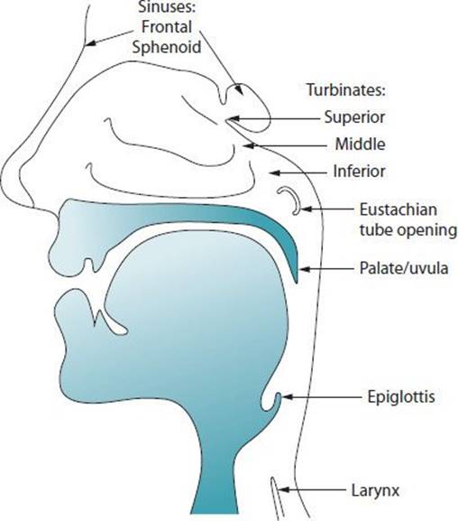
 Figure 22–1. Anatomy of the upper airway.
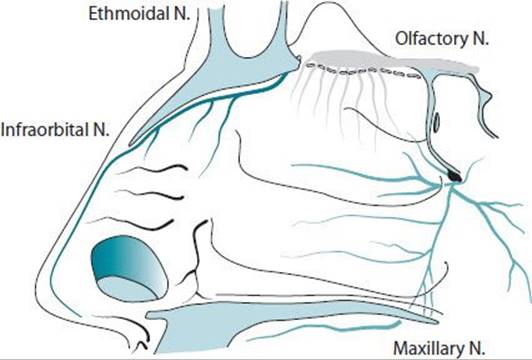
Figure 22–2. Innervation of the nasal cavity. The olfactory epithelium connects, via perforations in the cribriform plate, with the olfactory bulbs. The ethmoid and infraorbital nerves arise from the ophthalmic (first) division of the trigeminal nerve; the maxillary nerve constitutes the second division of the trigeminal.
Functions of the Upper Airway
The upper respiratory tract performs several essential physiologic functions. These include air conditioning, filtering, microbial defense, sensation, and phonation (Table 22–1). During the fraction of a second that inspired air travels through the upper airway, its temperature is adjusted to near body temperature, and its relative humidity regulated to between 75% and 80%. These physical alterations to inspired air help minimize thermal and osmotic stresses on the tracheobronchial tree. The major fraction of particulate matter larger than 1 μm in diameter is deposited in the upper airway (Figure 22–3). The majority of impacted material—captured in the mucous blanket—is transported posteriorly via ciliary action until it empties into the nasopharynx and then is swallowed (a smaller fraction being transported anteriorly to the nasal vestibule). The high surface area of the turbinates and the high water content of nasal mucus further provide a “scrubbing” mechanism for water-soluble air pollutants (Figure 22–4). Thus, depending upon the concentration and duration of exposure, water-soluble gases and vapors may have their initial (or principal) effect on the mucous membranes of the nose, throat, and conjunctivae.
Table 22–1. Functions of the upper airway.
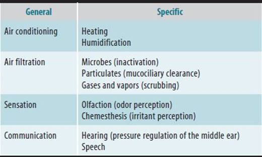
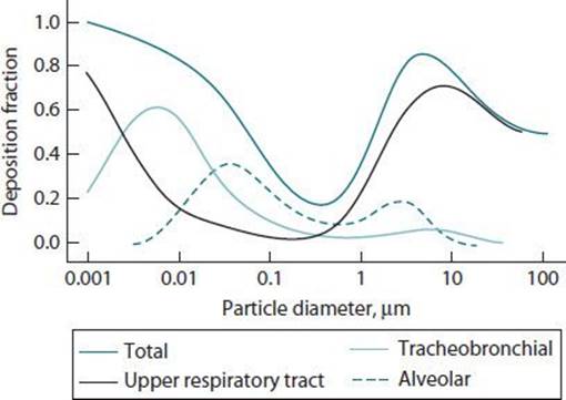
Figure 22–3. Fractional deposition of particulate matter in the upper respiratory tract, tracheobronchial tree, and alveoli, by particle diameter.
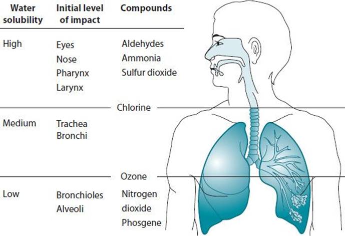
Figure 22–4. Water solubility and site of initial impact of airborne irritants. Highly water-soluble pollutants dissolve quickly in mucous membrane water, and alert the individual to the presence of the pollutant via trigeminal irritation.
The sensory functions of the upper airway are twofold: olfaction and irritant perception. Odor perception, mediated by the olfactory nerve, contributes to quality of life—allowing one to appreciate fragrances, as well as augmenting the primary tastes in the appreciation of food. In addition, olfaction has a safety function. Individuals lacking odor perception (anosmics) cannot distinguish fresh from spoiled food, tell that a gas pilot light has gone out in their kitchen, or sense that a respirator filter has become saturated with an odorous vapor against which they are to be protected. Upper respiratory tract irritation (conveyed by the trigeminal nerve) can be protective, in that nose and throat (as well as eye) irritation triggers escape behavior during an industrial mishap, at times before chemical injury to the lung can occur. With lower-level exposures, trigeminal (eye, nose, and throat) irritation (collectively referred to as “sensory irritation”) may be the primary health endpoint of concern, and indeed is a major symptom complex in so-called “sick building syndrome.”
OCCUPATIONAL & ENVIRONMENTAL CONDITIONS OF THE UPPER AIRWAY
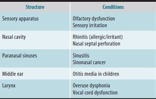
A variety of exposure-related health effects involve the upper airway. Structures potentially affected include the nasal cavity, paranasal sinuses, sensory nerves, Eustachian tubes/ middle ear, and larynx (Table 22–2).
Table 22–2. Upper airway health effects associated with occupational and environmental agents.
Occupational & Environmental Allergic Rhinitis
ESSENTIALS OF DIAGNOSIS
 Rhinorrhea, nasal airflow obstruction, nasal pruritus, and sneezing.
Symptoms may occur seasonally (“intermittent”) or perennially (“persistent”).
Common aeroallergens encountered in the general environment include pollens, mold spores, and animal-related allergens.
Allergens responsible for occupational allergic rhinitis are identical to those producing occupational asthma, and include both high- and low-molecular-weight substances.
Diagnosis involves confirmatory allergy testing (either epicutaneous skin prick testing or in vitro measurement of antigen-specific IgE).
 General Considerations
An estimated 20% of the population suffers from allergic rhinitis, and another 5% suffers from various forms of nonallergic rhinitis. As a result of exposure to common aeroallergens, individuals may experience: (1) seasonal pollinosis; (2) perennial allergy to common indoor allergens (eg, dust mite, molds, or pet allergens); or (3) a mixed pattern. Both conditions produce symptoms of nasal pruritus, sneezing, rhinorrhea, and nasal congestion, although perennial allergic rhinitis frequently adapt to their symptoms to the point that additional prompting may be necessary to elicit a complete history. The terms “seasonal” and “perennial” allergic rhinitis are increasingly being replaced by the terms “intermittent” and “persistent” allergic rhinitis. Seasonal allergens vary geographically, and some areas have already shown changes consistent with long-term climate change. Dust mites require a minimum of approximately 40% relative humidity to survive, and therefore are rarely found in far northern latitudes.
Occupational & Environmental Exposure
Workplace allergens producing allergic rhinitis may be either commonly encountered allergens, exposure to which may be incidental to the work environment (eg, grass pollen exposure in a landscaping gardener), or unusual agents encountered only in industrial environments (eg, trimellitic anhydride exposure in a plastics worker). As is the case with asthma, occupational allergic rhinitis may either be work-induced or work-exacerbated. Table 22–3 lists representative agents producing occupational allergic rhinitis; the reader will recognize that these same agents can produce occupational asthma (and, indeed, many sensitized individuals suffer from both conditions). Figure 22–5depicts a classification of work-related rhinitis, based upon that of work-related asthma.
Table 22–3.Some agents associated with occupational allergic rhinitis.
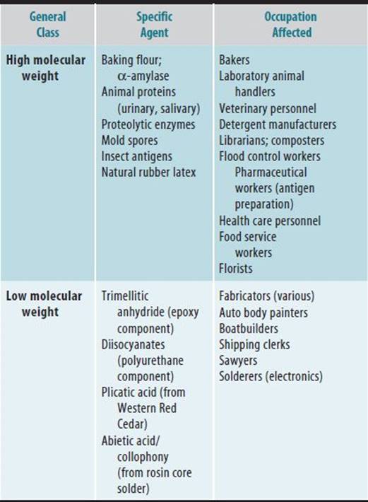
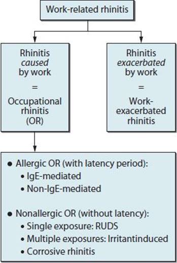
Figure 22–5. Classification of occupational rhinitis, analogous to that for occupational asthma, as proposed by the European Academy of Allergy and Clinical Immunology.
Metabolism & Mechanisms of Action
In sensitized individuals, specific antigen can initiate mast cell degranulation in the nasal mucosa, resulting in immediate release of such preformed mediators as histamine, heparin, tryptase, and leukocyte chemotactic factors. A “late-phase” reaction, occurring 2–6 hours later, releases leukotrienes, prostaglandins, and cytokines. The effects of these mediators include glandular secretion (rhinorrhea), nerve stimulation (nasal pruritus and sneezing), vasodilation (congestion), and chemotaxis (inflammation). Of importance, the mucous membranes of the nose and conjunctivae are contiguous, and are often affected by the same antigen exposures (hence, “rhinoconjunctivitis”).
Clinical Findings
A. Symptoms & Signs
Allergic rhinitis is typically manifest by symptoms of rhinorrhea, nasal airflow obstruction, nasal pruritus, and sneezing. Symptoms may occur seasonally (“intermittent”) or perennially (“persistent”). Signs include swollen, pale nasal turbinates, copious, watery nasal secretionsm, and mucous stranding.
B. Laboratory Findings
• Eosinophilia on nasal cytology
• Positive epicutaneous skin prick testing 1
• Allergen-specific IgE on in vitro testing (RAST or ELISA)1
• Increased total serum IgE (variable finding)
• Peripheral eosinophilia (variable finding)
C. Imaging Studies
• Increased thickness of nasal turbinates may be observed on CT scanning.
D. Special Tests
• Nasal inspiratory peak-flow measurements. This ambulatory, self-administered test may provide objective validation of cross-shift symptoms and can be employed during adjacent periods of allergen avoidance and normal work routine to help establish an occupational etiology.
• Sensory testing. Qualitative and quantitative tests of olfactory function can help document the response to allergen avoidance and medical therapy.
E. Special Examinations
• Rhinolaryngoscopy. Flexible (fiberoptic) rhinolaryngos-copy allows the examining physician to visualize the sinus ostia, larynx, and olfactory cleft, as well as to assess for the presence of nasal polyps.
Differential Diagnosis
• Irritant rhinitis
• Nonallergic rhinitis
• Viral upper respiratory tract infection
Prevention
Allergen avoidance should be an important component of therapy, both to control nasal symptoms and to prevent the progression of allergic rhinitis to asthma. In terms of environmental aeroallergens, the major exposures within the realm of control of patients are perennial allergens encountered in the home (or office) environment.
In the industrial workplace, engineering controls or personal protective equipment may be sufficient to control antigen exposures. However, some individuals may require reassignment, particularly if chest symptoms are coincident. In some cases, substitute chemicals or processes have been effective in ameliorating the risk of occupational sensitization. After a peak incidence of sensitization of health care workers to natural rubber latex in the mid-1990s, for example, the increased use of nonlatex gloves (and lower-allergen, non-powdered latex gloves) resulted in a dramatic reduction of new cases.
Treatment
Medical therapies for allergic rhinitis include oral medications (antihistamines and leukotriene inhibitors) and topical medications (nasal corticosteroids, cromolyn sodium, antihistamines, and cholinergic blockers). Topical saline flushes have also been employed as an adjunct to traditional medications.
Of the oral antihistamines, “second generation” mediations that may enable patients to control symptoms while simultaneously staying productive and alert. These include fexofenadine, loratadine, desloratadine, cetirizine, and levocetirizine. As many as 2 weeks of therapy may be necessary before an optimal response is observed from topical anti-inflammatory medications (corticosteroids; cromolyn sodium; or the combined antihistaminic/anti-inflammatory agents, azelastine and olopatadine).
Topical nasal decongestants are to be avoided except for very brief control of acute symptoms. Continuous therapy with topical decongestants poses a risk of tachyphylaxis and rebound congestion (rhinitis medicamentosa). For patients complaining of prominent secretions, a trial of ipratropium bromide nasal spray (a cholinergic blocker) may be indicated.
The efficacy of desensitization therapy (“allergy shots”) has been better evaluated for common aeroallergens than for specific occupational sensitizers. Patients electing to use saline flushes should either use commercial products or be cautioned regarding the hazards of microbial contamination of home preparations.
Prognosis
Assuming the practicality of allergen avoidance (and absent progression to rhinosinusitis), allergic rhinitis has an excellent prognosis. Untreated occupational allergic rhinitis may presage the subsequent development of occupational asthma.
Limited data link allergic rhinitis to obstructive sleep apnea. High-grade nasal obstruction predisposes to oral breathing, bypassing the filtration and air-conditioning functions of the upper airway. This may be one of the mechanisms whereby rhinitis and asthma severity are linked. Nasal mucosal swelling may also occlude the ostia of the paranasal sinuses and/or middle ear (Eustachian tubes). Ostial occlusion leads to pressure imbalance, effusion, and eventually infection (sinusitis or otitis media).
Occupational and Environmental Irritant Rhinitis
ESSENTIALS OF DIAGNOSIS
Nasal irritation, dryness, stinging, burning, rhinorrhea, nasal obstruction.
Facial pressure and decreased olfaction.
Mucosal erythema is a common sign.
Punctate erosions of the nasal septum.
Septal perforation.
Irritant rhinitis occurs in the absence of specific sensitization (although it may occur coincident with allergic rhinitis).
Irritant rhinitis is dose related. Thus, in contrast to allergy, symptoms may be present in a substantial fraction of coworkers in an industrial setting.
Cytologic changes consistent with irritant rhinitis have been documented among urban dwellers whose primary exposure is to high levels of photochemical oxidants (smog).
General Considerations
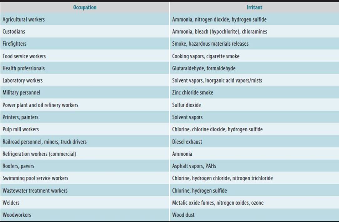
The eyes, nose, and throat are sensitive to chemical irritants (including gases, vapors, dusts, and smokes), with sensory irritation being the most commonly reported symptom complex in problem work environments. Types of chemical irritants in home or office air include (1) combustion products (from tobacco smoke and malfunctioning appliances) and (2) volatile organic compounds (VOCs; from cleaning products, office supplies and machines, building materials and furnishings, and microbial sources). Industrial environments present workers with an even wider range of airborne irritants, with the majority of permissible exposure levels (PELs) being based on the irritancy of the compound in question. Extreme forms of industrial irritant rhinitis (“corrosive rhinitis”) occur in electroplaters and others exposed to chromic acid who may develop nasal mucosal ulcerations and even septal perforation. Ambient exposure to photochemical air pollution can produce objective inflammatory changes in the upper airway, including squamous metaplasia. Representative environmental and occupational irritants appear in Tables 22–4 and 22–5, respectively.
Table 22–4. Environmental irritants.
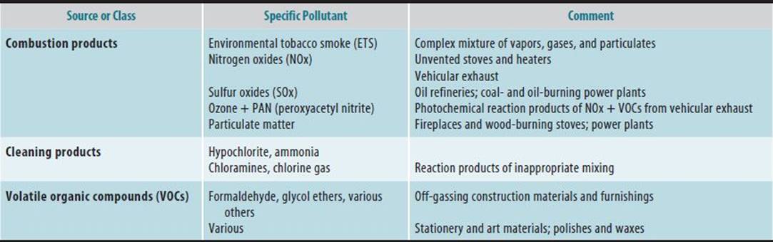
Table 22–5. Selected occupational irritants.
Persistent rhinitis symptoms and signs after a one-time high-level irritant exposure has been termed reactive upper airways dysfunction syndrome (RUDS). This diagnosis is analogous to the lower airway condition referred to as irritant-induced asthma or reactive airways dysfunction syndrome (RADS). However, in contradistinction to RADS, RUDS lacks objective diagnostic criteria (ie, physiologic changes on provocation testing), making the diagnosis one based on clinical criteria alone.
Metabolism & Mechanisms of Action
“Irritation” encompasses a spectrum of effects, including: (1) subjective sensory irritation, (2) stimulation of neurogenic reflexes, and (3) actual tissue damage. Neurogenic reflexes triggered by physical or chemical stimuli are also prominent in a subset of nonallergic rhinitis referred to as “vasomotor rhinitis” (see discussion below). Stimulation of trigeminal nerve afferents—which are sensitive to low pH, endogenous inflammatory mediators (such as bradykinin), and various chemical irritants—results in two major types of reflex response: (1) parasympathetic reflexes, conveyed by the facial nerve (cranial nerve VII), and (2) the axon reflex, an antidromic response involving neuropeptides released from afferent branches of the trigeminal nerve.
Two familiar examples of parasympathetic reflexes are: (1) gustatory rhinitis (a copious, watery rhinorrhea that occurs with the ingestion of spicy foods) and (2) “skier’s nose” (watery rhinorrhea in response to cold, dry air). Verifying their mechanism, both of these conditions can be blocked by the topical anticholinergic agent, ipratropium bromide.
The axon reflex, through the release of substance P, also acutely triggers glandular secretion and vascular dilatation. Subacutely, substance P potentiates the response of mast cells to antigens, forming one of several known links between the allergic response and chemical irritation. In another such link, both diesel exhaust particles and second-hand tobacco smoke enhance allergic sensitization (act as adjuvants) and intensify the allergic response(priming). In return, preexisting nasal allergies increase an individual’s sensitivity to chemical irritants (neuromodulation). Thus, our understanding of the immunologic and neurogenic systems in the airway has come to include reciprocal modulatory effects elicited by allergens and chemical irritants.
Clinical Findings
A. Symptoms & Signs
Irritant rhinitis is marked by subjective irritation (often expressed as “dryness,” “stinging,” or “burning”). Pruritus and sneezing are not typical symptoms. Rhinorrhea and nasal congestion (airflow obstruction) are secondary (reflex) symptoms that occur variably among individuals affected by irritant rhinitis. Nasal erosions (as well as septal perforation) can occur with concentrated and protracted exposures to airborne irritants. This has been termed “corrosive rhinitis.”
All the symptoms of irritant rhinitis are variable. They include nasal irritation (dryness, stinging, burning), rhinorrhea, nasal obstruction, facial pressure, and decreased olfaction. Mucosal erythema is a common sign. There may be punctate erosions of the nasal septum. Nasal erosions (as well as septal perforation) can occur with concentrated and protracted exposures to airborne irritants. This has been termed “corrosive rhinitis.”
B. Laboratory Findings
Irritant rhinitis yields a negative allergy workup. Polymorphonuclear leukocytes (neutrophils) predominate on nasal smear.
C. Imaging Studies
If reflex congestion is present, turbinate hypertrophy may be apparent on CT scanning.
D. Special Tests
• Nasal inspiratory peak-flow measurements. If subjective congestion is prominent in response to workplace or environmental exposures, exposure-related changes in nasal patency may be documentedutilizing a nasal peak inspiratory flow meter.
• Sensory testing. Qualitative and quantitative tests of olfactory function can help document the response to irritant avoidance and medical therapy.
E. Special Examinations
Flexible (fiberoptic) rhinolaryngoscopy allows the examining physician to visualize the sinus ostia, larynx, and olfactory cleft, as well as to assess for the presence of nasal polyps.
Differential Diagnosis
• Allergic rhinitis
• Nonallergic rhinitis
• Viral upper respiratory infection
Prevention
The majority of occupational permissible exposure limits have been set for the avoidance of chemical irritant effects, in particular sensory irritation. Similar logic underlies several ambient air quality standards, as well as statutory restrictions on smoking in public spaces.
High-grade nasal obstruction predisposes to oral breathing, bypassing the filtration and air-conditioning functions of the upper airway. This may be one of the mechanisms whereby rhinitis and asthma severity are linked. Nasal mucosal swelling may also occlude the ostia of the paranasal sinuses and/or middle ear (Eustachian tubes). Ostial occlusion leads to pressure imbalance, effusion, and eventually infection (sinusitis or otitis media).
Treatment
• Reduction of exposure.
• Nonspecific supportive measures (eg, saline nasal lavage).
• Topical steroids (of questionable value).
• Topical cholinergic blockers (ipratropium bromide) for prominent rhinorrhea.
• In atopic patients, control of intercurrent allergic rhinitis—whether occupational or nonoccupational—may decrease reactivity to chemical irritants.
Prognosis
With the exception of corrosive rhinitis with nasal septal perforation, the prognosis for irritant rhinitis after exposure reduction is excellent. However, some individuals with RUDS may show persistent nasal hyperesthesia and hyperreactivity, despite therapy.
Occupational & Environmental Nonallergic Rhinitis
Nonallergic rhinitis encompasses a variety of entities, including “vasomotor” rhinitis, endocrine rhinitis (including rhinitis of pregnancy), rhinitis medicamentosa, nonallergic rhinitis with eosinophilia (“NARES”) syndrome, rhinitis of granulomatous disease (Wegener granulomatosis), immotile cilia/Kartagener syndrome, rhinitis in cystic fibrosis. The mechanism(s) underlying nasal hyperreactivity are poorly understood.
ESSENTIALS OF DIAGNOSIS
Symptoms of vasomotor rhinitis are variable, including rhinorrhea, nasal obstruction, facial pressure, and decreased olfaction.
There are no characteristic physical findings in vasomotor rhinitis.
Negative allergy workup.
Lack of inflammatory cells on nasal smear.
General Considerations
Vasomotor rhinitis, a subcategory of nonallergic rhinitis, is a term that is often used to describe increased nasal reactivity to nonspecific physical and chemical stimuli. Symptoms of rhinorrhea and/or congestion tend to predominate, with neither subjective irritation nor nasal pruritus being prominent. Relevant physical stimuli include low humidity, extremes in or rapid changes of temperature, and excessive air motion. Possibly linked to this diagnosis are gustatory rhinitis (rhinorrhea in response to the ingestion of spicy foods) and bright-light rhinitis (self-explanatory). Roughly 40% of individuals with allergic rhinitis also complain of reactivity to nonspecific physical and chemical stimuli. Problematic occupations include outdoor work, biotechnology and food processing (cold rooms), and office work. The American Society of Heating, Refrigerating, and Air-Conditioning Engineers (ASHRAE) has promulgated guidelines for temperature and humidity control in indoor air; these parameters should be assessed as part of any “problem building” investigation.
High-grade nasal obstruction predisposes to oral breathing, bypassing the filtration and air-conditioning functions of the upper airway. This may be one of the mechanisms whereby rhinitis and asthma severity are linked. Nasal mucosal swelling may also occlude the ostia of the paranasal sinuses and/or middle ear (Eustachean tubes). Ostial occlusion leads to pressure imbalance, effusion, and eventually infection (sinusitis or otitis media).
Metabolism & Mechanisms of Action
The pathogenesis of vasomotor rhinitis is unclear. In some studies, parasympathetic overactivity appears to be responsible for hypersecretion. Other studies have identified a subset of patients with local mucosal allergy (ie, sensitized mucosal mast cells in an absence of systemic allergy). The concept of local mucosal allergy (or “entopy”) has been more extensively studied with common aeroallergens than with occupational agents.
Clinical Findings
A. Symptoms & Signs
Symptoms in vasomotor rhinitis are all variable. They include rhinorrhea, nasal obstruction, facial pressure, and decreased olfaction. There are no characteristic physical findings in vasomotor rhinitis
B. Laboratory Findings
• Negative allergy workup
• Lack of inflammatory cells on nasal smear
C. Imaging Studies
• If reflex congestion is present, turbinate hypertrophy may be apparent on CT scanning.
D. Special Tests
• Nasal inspiratory peak-flow measurements. If subjective congestion is prominent in response to workplace or environmental exposures, exposure-related changes in nasal patency may be documentedutilizing a nasal peak inspiratory flow meter.
• Nasal provocation with cold, dry air. When exposed to cold, dry air, individuals with vasomotor rhinitis, on average, congest more than do normal controls. Because of high inter-individual variability, however, this test does not provide for reliable clinical diagnosis.
• Histamine challenge. Histamine has been used, in titrated doses, to document nonspecific nasal reactivity. The concentration is increased by a fixed ratio until a predetermined increase in nasal airway resistance is documented (analogous to the methacholine challenge test). However, there is considerable overlap in response among diagnostic groups.
• Nasal allergen challenge. As indicated above, some individuals with rhinitis symptoms react to local instillation of antigen in the nose, while simultaneously maintaining negative skin test reactivity and a lack of antigen-specific IgE in the serum.
E. Special Examinations
• Rhinolaryngoscopy. Flexible (fiberoptic) rhinolaryngos-copy allows the examining physician to visualize the sinus ostia, larynx, and olfactory cleft, as well as to assess for the presence of nasal polyps.
Differential Diagnosis
• Allergic rhinitis
• Irritant rhinitis
• Viral upper respiratory infection
Prevention
There is no known primary prevention for vasomotor rhinitis. Avoidance of extremes of temperature and humidity, as well as avoidance of chemical irritant exposure, may provide symptomatic relief in some individuals.
Treatment
Approved therapies for vasomotor rhinitis include selected topical steroids (fluticasone propionate and beclomethasone dipropionate), topical antihistamines (azelastine and olopatadine), and topical cholinergic blockers (ipratropium bromide).
Prognosis
The prognosis for resolution of vasomotor rhinitis is very guarded. Some referral centers claim long-lasting relief after capsaicin desensitization, considered an experimental procedure at this time.
PARANASAL SINUS DISEASE
Sinusitis
ESSENTIALS OF DIAGNOSIS
Sinusitis may affect single or multiple sinuses, unilaterally or bilaterally.
Sinusitis is classified as acute (up to 4 weeks duration); intermediate (4–12 weeks); and chronic (>12 weeks).
Primary symptoms are nasal congestion, facial pressure, purulent nasal discharge, decreased olfaction, and systemic symptoms (such as fatigue and, occasionally, fever).
Findings on CT scanning may include mucoperiostial thickening, air-fluid levels, and obstruction of the osteo-meatal complex. Nasal polyposis may be an associated finding.
The role of sinusitis in the genesis of headaches is controversial.
Sinusitis has been linked to asthma incidence and severity.
General Considerations
Both allergic and irritant rhinitis can progress to rhinosinusitis. Epidemiologically, active smokers are at higher risk for developing acute (and chronic) sinusitis. Evidence for a link between sinusitis and second-hand tobacco smoke exposure appears to be mounting, as well. Relatively few studies have systematically examined the endpoint of sinusitis and occupational exposures. Surveys of furriers, spice workers, vegetable picklers, hemp workers, and grain and flour workers all include increased prevalence rates for sinusitis; however, these studies are based on self-report. More recently, cohort studies of World Trade Center responders have suggested increased rates of upper airway disorders, including sinusitis, compared to unexposed individuals.
Metabolism & Mechanisms of Action
Irritant- and allergen-induced nasal mucosal swelling can compromise the patency of the paranasal sinus ostia, thereby producing pressure imbalance, effusion, and impaired clearance of secretions, and leading to the development of sinusitis. Most bouts of acute sinusitis result from viral upper respiratory tract infections, and are self-limited. In acute bacterial sinusitis, the most common organisms involved include Streptococcus pneumoniae, Haemophilus influenzae, and Moraxella catarrhallis. Less frequently, Staphylococcus aureus, anaerobes, or gram-negative organisms are present. Invasive fungal sinusitismay be seen with immune suppression (eg, in the presence of diabetes mellitus). Noninvasive allergic fungal sinusitis has also been described, in which affected sinuses are colonized by one or more fungal species (such as Schizophyllum commune), which in turn attract eosinophils. Considerable tissue damage can occur from the inflammatory mediators released by these mast cells.
Chronic rhinosinusitis, with or without polyposis, involves noninfectious chronic inflammation, the mechanisms of which have yet to be fully elucidated. Inflammation in the upper and lower respiratory tracts appears to be linked, in that active sinusitis typically augments nonspecific bronchial reactivity in asthmatics. Postulated mechanisms include: upregulation of neurogenic and humoral responses; loss of air conditioning and filtration functions due to chronic oral breathing; and aspiration of biochemical-mediator-laden nasal secretions into the lower respiratory tract.
Clinical Findings
A. Symptoms & Signs
Symptoms include nasal airflow obstruction(congestion), facial pressure, impaired olfaction, and systemic symptoms of fatigue and variable fever. Signs include sinus tap tenderness (frontal/maxillary), and mucopurulent nasal secretions visible on routine examination.
B. Laboratory Findings
Abnormalities of the complete blood count (leukocytosis), as well as elevations of the erythrocyte sedimentation rate or C-reactive protein are both nonspecific and insensitive measures in acute or chronic sinusitis.
C. Imaging Studies
Potential findings on CT scanning include mucoperiosteal thickening, air-fluid levels, and obstruction of the osteomeatal complex. Polyposis and bony erosions may also be found.
D. Special Tests
• Nasal nitric oxide sampling. Because the sinuses serve as a reservoir for nitric oxide (NO), nasal NO levels tend to be higher than levels measured in exhaled breath. However, nasal NO trends down with increasing obstruction of the osteomeatal complex (ie, obstruction due to sinusitis and/or nasal polyposis).
E. Special Examinations
• Rhinolaryngoscopy. Mucopurulent discharge per sinus ostia is a common sinusitis-related finding on rhinolaryngoscopy.
Differential Diagnosis
• Viral upper respiratory tract infection
• Odontogenic (dental) pain
• Migraine headache
• Sinus neoplasm
• Nasal foreign body
• Invasive fungal sinusitis
• Allergic fungal sinusitis
• Underlying immune deficiency or mucociliary disorder (cystic fibrosis; immotile cilia syndrome)
• Underlying granulomatous process (Wegener)
Prevention
• Irritant and allergen avoidance
• Effective medical therapy for allergic rhinitis
• Nasal hygiene (saline flushes) for those working in dusty environments
Treatment
Acute and uncomplicated sinusitis lasting 10 or fewer days should be treated as a self-limited condition, and presumed to be of viral origin. Acute sinusitis symptoms lasting greater than 10 days may be a candidate for antibiotic therapy. Given the potential role of β-lactamase producing organisms, recommended empiric antibiotic therapy is evolving.
Therapy for chronic rhinosinusitis (with or without polyps) emphasizes topical corticosteroids, at times augmented by oral leukotriene antagonists. Therapy for invasive fungal sinusitis consists of systemic antifungal agents. Therapy for allergic fungal sinusitis consists of oral steroids. Clinical trials have shown no consistent benefit from the administration of antifungal agents. When exposure controls and medical therapy fail to yield expected improvement, patients may benefit from otolaryngologic consultation. In some cases, functional endoscopic sinus surgery may be indicated to promote effective sinus drainage.
Prognosis
The prognosis for resolution of acute sinusitis is good. The prognosis for resolution of chronic sinusitis is guarded.
Sinonasal Cancer
A number of occupations and imputed exposures have been linked with the development of malignant neoplasms of the paranasal sinuses. The strongest (and most consistent) findings pertain to formaldehyde-exposed workers and to leather- and woodworkers, although some studies also have found nickel- and chrome-refining and chrome-plating workers to be at risk.
Laryngeal Pathology
Symptoms referable to phonation (typically, hoarseness) can also occur in work settings. Temporary and reversible hoarseness may occur either from exposure to inhaled chemical irritants or from overuse of the voice. Although overuse is most widely recognized in lecturers and singers, it also occurs among industrial employees who need to shout in order to communicate in noisy environments. The most ominous condition heralded by hoarseness—squamous cell carcinoma of the larynx—has been associated with a number of exposures/occupations, including: polycyclic aromatic hydrocarbon exposure (cigarette smoking, metalworking fluids, work in aluminum reduction plants, use of coal as a cooking fuel indoors), as well as exposure to asbestos and personal consumption of ethanol.
Two other occupational/environmental conditions deserve mention. After significant smoke inhalation injury laryngeal strictures may occur, resulting either from the initial chemical/thermal insult or secondary to prolonged intubation. In addition, laryngeal papillomatosis has been described in a case report of a physician whose apparent exposure was human papillomavirus aerosolized during laser surgery.
A functional laryngeal condition of note is vocal cord dysfunction (VCD). VCD involves episodic hoarseness, shortness of breath, stridor (often confused with wheezing), and globus (a pressure sensation in the throat or upper chest). Coughing is also common in VCD. Because of overlapping symptoms with asthma, VCD may be misdiagnosed as the latter. Predisposing conditions include postnasal drip and gastroesophageal reflux. In the occupational setting, VCD has been documented after acute irritant exposures, giving rise to the diagnosis of “irritant-associated VCD.” Most recently, this condition has been documented among a subset of individuals exposed to alkaline dust as World Trade Center responders.
Diagnosis of VCD requires documentation of paradoxical vocal cord motion (adduction during inspiration as visualized during rhinolaryngoscopy). Alternatively, the finding of variable extrathoracic obstruction during the inspiratory phase of the flow-volume loop is highly suggestive for this condition. After ruling out more serious conditions (eg, neoplasms, vocal cord paralysis, and spasmodic dysphonia), treatment consists of voice rest, hydration, and biofeedback/voice training under the supervision of a qualified speech pathologist.
Otitis Media in Children
An increased incidence of otitis media with effusion has been reported among children exposed to environmental tobacco smoke, typically in the home. Postulated mechanisms center on Eustachian tube dysfunction, with secondhand tobacco smoke producing ciliostasis and mucous membrane congestion, resulting in impaired pressure equalization, middle ear effusion, and reduced drainage of middle ear secretions. Because of the strength and consistency of this finding, the workup of recurrent otitis media in young children always should include questions about parental smoking.
Sensory (Olfactory) Alterations
Both temporary and long-lasting alterations in olfactory function have been reported among workers exposed to a variety of industrial chemicals. Chemically induced olfactory dysfunction may include (1) quantitative defects, including hyposmia (reduced odor acuity) and anosmia (absent odor perception), and (2) qualitative defects, including olfactory agnosia (decreased ability to identify odors) and various dysosmias (distorted odor perception). Occupational groups and exposures for which defects in odor detection or identification have been identified include alkaline battery workers and braziers (cadmium ± nickel exposure), tank cleaners (hydrocarbon exposure), paint formulators (solvent ± acrylic acid exposure), and chemical plant workers (ammonia and sulfuric acid exposures). Of note, olfactory deficits have also been identified among World Trade Center responders, compared to age-, sex-, and smoking status-matched controls. At high concentrations (exposure at levels in excess of approximately 50 ppm), hydrogen sulfide is known to produce profound and reversible olfactory fatigue.
Chemical irritants may cause hyposmia via nasal obstruction, or alternatively, may produce direct damage to the olfactory neuroepithelium. Experimentally, at least one study has shown the olfactory equivalent of a temporary threshold shift (reversible olfactory deficit) after several hours of controlled exposure to solvents (toluene or xylene); subjects recovered olfactory acuity within about 2 hours of cessation of exposure. Of note, no perceptual deficit was evident for a test compound unrelated to the exposure (methylphenyl carbinol). This reversible and specific phenomenon might therefore be thought of as an extension of the familiar process of odor adaptation, in which odors lose their intensity during continuous exposure.
Other causes of olfactory impairment not directly related to chemical exposures include head trauma, chronic nasal obstruction from rhinosinusitis, postinfectious inflammation, neurodegenerative disorders (Alzheimer and Parkinsonism), endocrine disorders, hepatic and renal disease, neoplasms, various drugs, ionizing radiation, selected psychiatric conditions, and congenital defects (eg, Kallmann syndrome).
DIAGNOSTIC TECHNIQUES
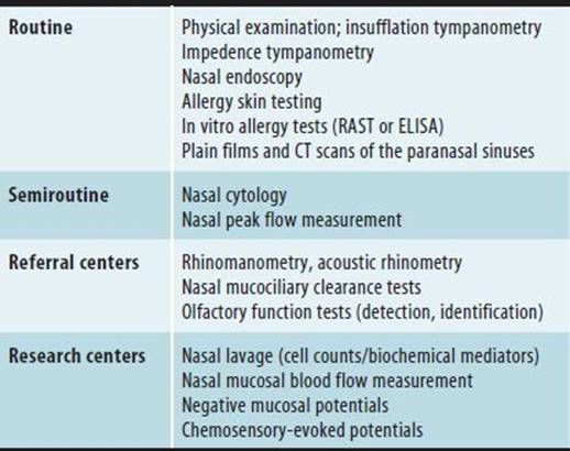
A number of diagnostic tools are useful in the study of nasal responses to environmental agents; these have been classified here as routine, semiroutine, and techniques used in clinical referral centers or research centers (Table 22–6).
Table 22–6. Diagnostic tools for the upper airway.
Semiroutine Methods
A. Nasal Cytology
Nasal smears for cytologic analysis are used to provide information regarding the types of inflammatory cells in nasal mucus and/or the superficial mucosal layers. Samples are taken from the medial surface of the inferior turbinate using a curette, and are done under direct visualization. Typically, eosinophils predominate in allergic inflammation, whereas neutrophils predominate with viral and bacterial infections. Neutrophils also predominate in nasal smears taken from individuals with irritant rhinitis, whereas inflammatory cells may absent altogether in patients with nonallergic rhinitis.
B. Peak Nasal Flow Measurement
Nasal inspiratory peak-flow measurement is listed here as semiroutine not because of any technical challenges involved, but because the technique and equipment are unfamiliar to many health care providers. Commercially available nasal inspiratory flow meters have become more compact and rugged than in the past (Figure 22–6). To take a measurement, the patient breathes out maximally (to residual volume), places the mask over his or her nose and mouth, and then inhales forcefully through the nose to total lung capacity. Three replicate measures are normally taken, with the highest value being taken as representative.
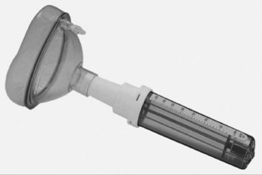
Figure 22–6. Commercial nasal inspiratory flow meter.
A diary of nasal peak-flow measurements (along with nasal symptoms) can be kept, with the patient recording peak flow before, during, and after a work shift. If possible, recordings should be taken over a full work week, along with adjacent weekends. Interpretation of these data is analogous to the process of interpreting peak expiratory flow data in the diagnosis of occupational asthma, although no consensus standards exist for “significant” work-related decrements in peak flow.
Techniques Used in Referral Centers
A. Rhinomanometry
Rhinomanometry, or the measurement of nasal airway resistance (NAR), involves simultaneously measuring airflow and pressure between the nasopharynx and anterior nares. With posterior rhinomanometry, the individual being tested breathes nasally with an anesthesia mask applied over the nose and mouth, and with a small plastic pressure tap held between the tongue and palate. In anterior rhinomanometry, one nostril at a time is occluded with a pressure tap while the subject breathes slowly through the opposite nostril with a flow meter applied.
Anterior rhinomanometry is particularly useful for documenting fixed anatomic pathology that may be unilateral in distribution (eg, deviated septum or polyposis). Posterior rhinomanometry gives a more stable estimate of total nasal airway resistance than does the anterior technique and is therefore of particular utility in documenting the response of the nose to challenge agents (allergens or irritants).
NAR has been used as the endpoint for various pharmacologic challenge protocols, principally to document the so-called non-specific nasal hyperreactivity. This procedure requires the use of serially increasing concentrations of histamine or methacholine, with the endpoint being the concentration necessary to induce a predetermined percentage increase in NAR. Using this method, allergic rhinitics studied in and out of season show systematic differences in nonspecific nasal reactivity (greater during allergy season). Rhinomanometry can also be used as an objective endpoint after nasal allergen or cold air challenge.
B. Acoustic Rhinometry
Another technique designed to measure nasal airway patency is acoustic rhinometry (AR). The apparatus consists of a tube with an acoustic pulse generator (and microphone) at one end and a nasal adaptor at the other; the instrument alternately sends and receives sound pulses. By measuring the intensity of reflected sound waves at various time intervals from the initial pulse, an acoustic rhinometer produces a map of total nasal cross-sectional area as a function of distance from the nares. Like rhinomanometry, AR is often used to document the response to pharmacologic, irritant, cold air, or allergen challenge. The relationship between cross-sectional area and nasal airway resistance, however, is a complex one, rendering the physiologic and symptomatic interpretation of acoustic rhinometry somewhat difficult.
C. Sensory Testing
Olfactory sensory testing focuses on alternative endpoints: qualitative or quantitative. Qualitative odor testing uses panels of test odorants to assess odor identification ability. Typically, such tests are administered as a multiple-choice task in order to prevent the patient’s personal experience from having undue influence on testing results. One commercially available qualitative test, the University of Pennsylvania Smell Identification Test (UPSIT), takes the form of scratch-and-sniff panels on a paper base; the test has been well standardized with extensive population norms. The advantage of this test is its portability, and the fact that results generally correlate well with quantitative endpoints.
In the clinical setting, quantitative olfactory testing consists of olfactory threshold testing. The simplest clinical screening test is the alcohol sniff test. The alcohol sniff test utilizes commonly available packaged isopropanol swabs, opened at the top and held below the breathing zone of a patient whose eyes are closed and who is breathing nasally. The swab is advanced vertically by one centimeter with each breath, until the patient reports perceiving an odor. With normal olfactory acuity, the patient should be able to detect an odor by the time the stimulus reaches 20 cm below the nose. More formally, odor threshold testing can be carried out utilizing a forced-choice discrimination task using a series of squeeze bottles with matching blanks. A threshold so obtained is an odor detection threshold. Alternate systems utilize a series of pen-like devices whose wicks are saturated with test odorants.
D. Mucociliary Clearance Tests
Mucociliary clearance tests include both invasive and noninvasive procedures. The best-standardized test is the observation of ciliary beat frequency in vitro. This method is often employed as a screening step (prior to electron microscopy) in the diagnosis of disorders involving ultrastructural abnormalities in epithelial cilia (eg, primary ciliary dyskinesia/Kartagener syndrome). Specimens typically are obtained either by scraping or biopsy of the inferior turbinate; ciliary beat frequency is normally in the range of 9–15 Hz. In addition to frequency, trained observers can note the degree of spatial coordination of adjacent ciliary units, an important component of intact function.
The saccharine test is the simplest measure of nasal mucociliary dysfunction. In this procedure, a small grain of saccharine is placed on the anterior portion of the inferior turbinate, and the time interval before the subject tastes the saccharine is recorded. A prolonged test—defined as greater than 30 minutes—indicates impaired mucociliary function.
Mucociliary clearance is important because of its essential function in microbial defense. Patients with impaired mucus formation (cystic fibrosis) or impaired ciliary function (primary ciliary dyskinesia) experience repeated episodes of bronchitis, otitis, and sinusitis, often with ultimate cardiopulmonary complications (bronchiectasis and corpulmonale). Environmental factors that have been noted to impair mucociliary clearance include viral infection, antigen challenge, cigarette smoke, and sulfur dioxide exposure.
REFERENCES
Altman KW: Odor identification ability and self-reported upper respiratory symptoms in workers at the post-9/11 World Trade Center site. Int Arch Occup Environ Health 2011;84:131 [PMID: 20589388].
de la Hoz RE: Occupational rhinosinusitis and upper airway disease: the world trade center experience. Curr Allergy Asthma Rep 2010;10:77 [PMID: 20425498].
Eccles R: A guide to practical aspects of measurement of human nasal airflow by rhinomanometry. Rhinology 2011;49:2 [PMID: 21468367].
Feng CH: The united allergic airway: connections between allergic rhinitis, asthma, and chronic sinusitis. Am J Rhinol Allergy 2012;26:187 [PMID: 22643942].
Jones LL: Parental smoking and the risk of middle ear disease in children: a systematic review and meta-analysis. Arch Pediatr Adolesc Med 2012;166:18 [PMID: 21893640].
Kenn K: Vocal cord dysfunction: what do we know? Eur Respir J 2011;37:194 [PMID: 21205712].
Quirce S: Noninvasive methods for assessment of airway inflammation in occupational settings. Allergy 2010;65:445 [PMID: 19958319].
Shusterman D: The effects of air pollutants and irritants on the upper airway. Proc Am Thorac Soc 20118:101 [PMID: 21364227].
Sin B: Pathophysiology of allergic and nonallergic rhinitis. Proc Am Thorac Soc 2011;8:106 [PMID: 21364228].
Slavin RG: Update on occupational rhinitis and asthma. Allergy Asthma Proc 2010;31:437 [PMID: 21708054].
 SELF-ASSESSMENT QUESTIONS
Select the one correct answer for each question.
Question 1: Occupational allergic rhinitis
a. is work exacerbated, but not work induced
b. is linked with occupational asthma
c. may result in release of mediators such as antigens
d. is associated with a negative epicutaneous skin prick test
Question 2: Allergens responsible for occupational allergic rhinitis
a. are distinct from those producing occupational asthma
b. include only low-molecular-weight substances
c. include only high-molecular-weight substances
d. may include common aeroallergens, such as grass pollen
Question 3: Irritant rhinitis
a. produces definitive symptoms and signs distinct from those of allergic rhinitis
b. is diagnosed by a positive allergy workup
c. predisposes to oral breathing via reflex nasal congestion
d. via reflex nasal congestion
Question 4: Vasomotor rhinitis
a. is a subcategory of allergic rhinitis
b. is defined by reactivity to specific chemical stimuli
c. causes rhinorrhea, with nasal pruritus being definitive
d. also occurs in many individuals with allergic rhinitis
Question 5: Sinusitis
a. has been linked to asthma incidence and severity
b. may follow allergic but not irritant rhinitis
c. spares active smokers because they develop resistance
d. always involves bacterial infections
1If neither an in vitro test system nor a skin test reagent is available for a given occupational allergen, response to allergen avoidance or workplace challenge may provide the best clue to the specific diagnosis.